

- The cell membranes; structure and functions
- Exchange of molecules, signals and ions
- Membrane transport mechanisms
- Receptors and receptor functions
- Ion exchange mechanisms
- Cell-cell-matrix interactions
- Cell adhesion molecules (CAMs)
- Integrins
- Selectins
- Cadherins
- Immunoglobulin superfamily
- Cytokines
- Growth factors
- Cell adhesion molecules (CAMs)
- The cell junctions
- Intracellular organelles of the cell
- Cytoskeletal structures
- Microtubules, micro and intermediate filaments
- Mitochondria
- Lysosomes
- Endoplasmic reticulum, ribosomes
- Golgi network
- Cytoskeletal structures
- Nucleus, DNA and genes, telomeres
- The response of cell to stimuli
- The Protein molecule
- Structure and chains
- Functions
- The Synthetic process in the cell
- Intracellular functional proteins
- Heat shock proteins
- Ubiquitin
- The extracellular matrix
- Collagen, elastin
- Adhesion proteins-laminin, fibronectin
- Proteoglycans
- Basement membranes
- Relation between cell and extracellular matrix
Each cell is a complete living organism on its own but as compared to a unicellular organism (like the amoeba) a cell in the human body lives in a “society of cell”. For this community life each cell has to have at least the following common biological attributes:
- A biological system for communication with the neighbouring cells as well as cells at a distance. This is achieved by the existence of cell junctional molecules, cell adhesion molecules, cytokines, including growth factors, interleukins, secretory products including neuro-secretory molecules and hormones. These molecules function at three levels with some overlaps:2
- Autocrine: Cell functions for its own growth and existence
- Paracrine: Cell functions that influence the neighbouring cells
- Endocrine: Cell functions that influence cells at a distance.
- A biological set up within the cells to perform the essential life functions including respiration, digestion, detoxification and excretion of waste products and above all manufacture and supply of energy for all these functions. These functions are dependent on the presence of
- Cell membrane systems
- Cell micro-organelles namely mitochondria, lysosomes, endoplasmic reticulum, golgi apparatus, etc.
- A biological set up to safeguard its life and structures by a defense mechanism against foreign as well internal agents. This exists as a part.
- The cell membrane and organelle membrane functions.
- The protection provided to all cells by the immune system in the body assured by an efficient communication set up referred to in I above.
- A biological system to direct and control all the functions listed in I, II and III above and to control the cell reproduction for the continuation of the species. This function rests in the nucleus in the form of the genetic apparatus or the cell genome which possesses a programme acquired during its evolution.
The four sets of life function are more or less common to all cells, in addtion cells have specialized functions depending upon the organ/system to which they belong.
In this chapter a brief description of the first two items (cell communication systems and cell organelle systems) is given, the others will follow in respective chapters.


Fig. 1.1: The phospholipid trilaminar cell membrane
Phpl | : Phospholipid molecules with heads and tails |
Prt | : Protein molecule for ion exchange gate |
RCP 1 | : Receptor molecule intramembranous |
Rcp 2 | : Receptor molecule in outer layer of membrane |
Chl | : Cholesterol molecule |
Car | : Carbohydrate molecule |
The subject of modern molecular biology is bound to be difficult for the students of morphology and average medical graduates, but a basic understanding of the disease processes is not possible without this understanding. This has become more important, since many of the newer therapeutic strategies are beginning to be based on these molecular interactions. Presented here is a highly simplified version avoiding chemical formulae and chemical reactions in the descriptions wherever possible. Students who are interested in further details of the molecular basis of medicine must consult other texts for this purpose.
Cell Membrane—Structure and Functions
Phospholipid Molecules (Fig. 1.1)
The cell cytoplasm and all its contents including the nucleus are enclosed within the three layered (trilaminar) cell membrane or the plasma membrane. It is not like a polythene or plastic sheet but a gel like arrangement of phospholipid and protein molecules in a constant state of flux. This structure is according to the Danielli-Davson model as modified by Singer and Nicolson (1972). The membrane is composed of two layers of phospholipid molecules which are pictured in the shape of tennis rackets placed parallel to each other and alternating in position of their blades. One layer of the racket blades faces outwards and the other inwards. The racket handles are aligned in parallel row in the substance of the membrane. The heads of the phospholipid molecules form the hydrophilic (water wettable) and tails are the water repellant (hydrophobic) components of the membrane. The inner layer of molecules consists of phosphatidyl inositol, phosphatidyl ethanolamine and serine, while the outer layer is composed of phosphatidyl choline, sphingomyelin and glycolipids. Other lipid molecules in the membrane are glycolipids and cholesterol.
Other Molecules and Components
All along the membrane are distributed a variety of molecules including cholesterol, carbohydrates and proteins. The membranes are the media in which large number of enzyme based metabolic activities go on. The energy manufacture by the ATP-ADP, cycle and the exchanges of ions/electrolytes like sodium, calcium, and potassium (ion channel) take place in the membrane. In addition to the phospholipid cell membrane some specialized cells contain additional cell surface structures in association with the plasma membrane. For example, the red cells have a meshwork of protein molecules called spectrin. These molecules are twisted around each other and linked to the plasma membrane. Similarly, the skeletal muscle plasma membrane is supported by a 3dystrophin-glycoprotein complex. This complex connects the sarcoplasma membrane to actin fibrils inside the cytoplasm and to the laminin molecule in the extracellular matrix through a transmembrane glycoprotein receptor. This link supports the membrane allowing contraction and relaxation while maintaining its position and shape. Genetic absence of such additional structures creates congenital diseases.
Most of the protein molecules involved in the exchange of ions and serving as receptors are intrinsic proteins that are enclosed within the plasma membrane while those protein molecules that take part in the cell-to-cell or cell -to-matrix interactions and adhesions are partly extrinsic in location.
Similar phospholipid membranes enclose the internal organelles of the cell, including the nuclear membrane. These internal membranes may only be one-layer of the phospholipid molecules, as described below.
Functions of the Cell Membranes
- Maintenance of cell structure, keeping all the internal structures in specifically defined locations.
- Protection of cell constituents against damage by external agencies.
- Maintenance of communication with the surrounding cells, connective tissue (extracellular matrix) as well as cells and organs in other parts of the body through the intermediation of biomolecular and ion exchanges is made possible by a number of mechanisms operating in the cells. These mechanisms do not disturb the rest of the cell's functions, while the exchanges take place.
There are two properties of the membranes which keep the external molecules away, one is the fact that the phospholipid molecules are hydrophobic that is water repellant and second is the electrical charge on the surface which repels the molecules bearing the same charge. But the unique property of the membrane is the instant modification of both these functions to permit the attraction of molecules. With the help of protein molecules and receptor proteins, the membrane will allow the watery molecules to pass through and with the reversal of its electrical charge (process of depolarization and repolarization) the membrane will attract ions. This is expressed as the dynamic nature of the cell membranes.
Membrane Exchanges, Transfer and the Transport Mechanisms
The cells communicate with each other and exchange material between the inside and the outside by the following mechanisms:
- Direct cell-to-cell contact at sites of membrane adhesions and cell junctions (described below).
- Simple diffusion and filtration. Smaller molecules, fluids move across the membrane because of its semi-permeable nature and the differences in the concentrations of the fluids and molecules inside and outside the cell. The fluid molecules are taken in by the process of pinocytosis.
- Ion exchange based on electrical charge on the membrane and the incoming/outgoing ions.
- Carrier mediated transport. Insoluble molecules ride on the back of protein molecules to enter in (endocytosis) and out (exocytosis) of the membrane. The molecule with or without the carrier protein gets wrapped up in a fragment of the cell membrane to form a vacuole as it moves across the membrane. The gap in the membrane immediately closes over by the adjoining phospholipid molecules.It is important to learn some basics of these processes, since genetic disorders of these mechanisms underlie a number of disesaes.
- Intercellular communication channels:
- Receptor mediated exchange
- Ion exchange
- Cell adhesion molecules (CAMs)
Cell Membrane Receptors
“Receptor mediated exchanges are like the ‘receptionist’ in offices. Some receptionists are available at the entry, others inside the offices. They receive a visitor, identify him or her, identify the visitor's purpose and take him to the office and officer as required. Some receptionists personally escort the visitor and return to their post after delivery, others do not leave their position but convey the message through a signal (telephone), and get the visitor's work done. Still others, hand the visitor over to a second messenger for the work. This is exactly how membrane receptors work.”
In the biology of cells, receptors are surface molecular units, mostly protein but may be glycoprotein, lipoprotein molecules. They may be located, (a) in the outer cell membrane or (b) inside the cell or (c) the receptor is ‘transmembranous’, that is, it has an extramembranous subunit, a membranous coupling and an intracellular subunit in contact with one or more specific cell organelle. Receptors are molecule specific, that is, for each type of substance there may be a specific receptor. Further depending upon the need of the cell and the quantity of molecules that reach the membrane, the cell may increase or decrease the number of receptors, since the cell manufactures the receptor molecules. This process is known as up regulation or down regulation of receptors. These molecular receptors are different from structural or anatomic receptors like pressure receptors, volume receptors, touch receptors, temperature receptors, etc.4
Based on function there are three families of membrane receptors:
- Enzyme linked receptors These receptors are involved in activation of cell growth and manufacture of other molecules by the cells. For example, the Tyrosine kinase class of enzyme linked receptors respond to most growth factors to control growth of a cell. The tyrosine kinase associated receptors take part in the activation of synthesis and secretion of hormones like the growth hormone, erythropoietin, prolactin and cytokines like the interferon. Threonin kinases and serine kinase receptors are other examples.
- Ion channels which are the targets for molecules sodium, potassium and calcium and certain peptide hormones. The activated receptors arranges for the movement of the particular ion in or out of the cell.
- G-protein linked receptors are transmembranous (that is they have an extramembranous component, a membranous and an intramembranous component) and generate a second messenger molecule like the cyclic—monophosphate system (c-GMP) for many neuroendocrine agents. These receptors activate the phosphorylating enzymes required for supply of energy for metabolic functions as well as for synthetic activities within the cells. These include the ATP-ADP-ATP cycle and the GDP-GTP-GDP cycle activated either directly or through cyclic adenyl phosphates (c-AMP) systems. This activity is also known as the “second messenger activation” (described below).
Mechanism of Receptor Action (Table 1.1)
An extracellular stimulus in the form of a cell or bacteria or chemical mediator, molecule or ion or electron comes to the cell surface and seeks out the specific receptor for it. The receptor action takes place in 4 steps to transmit the signal to its destination; the process is called signal transduction and the 4 steps are; recognition, receptor activation, transportation and transfer activation. Recognition of the stimulus is the first step. The site on which the receptor attaches itself to the molecule is called the ligand of the molecule. The contact results in activation of the receptor into transporting either the molecule across the membrane or the message or signal to its destination. A step of transfer activation follows in which the message is transferred to its destination in one of the two mechanisms:
- Direct activation of the system to synthesize proteins or the other required substance.
- Activation of another intermediary which in turn activates the target destination. This is known as the second messenger mechanism.
|

G-proteins and the Second Messenger System of Signal Transduction
Among the most important signal system is the activation of the adenosine monophosphate/phosphatase cycle (c-AMP) by the G-proteins or the Guanosine nucleotide binding regulatory proteins. The transmembrane G-protein is activated by a stimulus (like various hormones—see below) binding to its special ligand. This results in the GDP to GTP diosephosphate to triosephosphate conversion. The G-protein subunits then activate either a membrane located ion exchange channel or membrane bound enzyme or activate a “second messenger”, the most common second messenger is the cyclic-AMP. This activated second messenger then regulates other intracellular activities.
A number of different hormones act through the G-protein linked second messenger system like glucagon, ACTH, thyroid hormone, epinephrine, histamine.
The discovery of the G-protein system earned the 1994 Nobel Prize in Physiology for Drs. Alfred G Gilman, and Martin Rodbel; (though it would be quite appropriate but the G-protein is not named after the G in Dr. Gilman's name!). The action of the G-protein has been compared 5to that of an electric switch. The switch knob is the receptor for the finger, the contact point inside the switch is the second messenger that activates the flow of the electric current. Both the outer knob and the contact point themselves are not affected by the action.
Apart from the normal metabolic pathways, the G-protein is also involved in abnormal situations. In infections by the cholera bacillus, the secretion of water and electrolytes out of the intestinal epithelium is mediated by the G-protein, c-AMP complex.
Actions of the Cyclic Nucleotide Systems
The c-AMP and the GTP and other regulatory proteins that are activated by the receptor activation, lead to gene activation followed by the entire process of RNA mediated transcription and translation leading to the synthesis of proteins. The energy for the enzyme activity is derived from the activation of the ATP-ADP cycle of oxidative phosphorylation and other mitochondrial based systems involving the phosphokinases referred to later. Hormones like insulin and thyroxine are produced in their receptive cells through this system and act on their target cells in the same way. Further, the two kinases, c-AMP and GTP oppose each other in their activities. It is this which allows cells to carryout opposing actions simultaneously. For example, the conversion of glucose to glycogen and glycogen to glucose, the activation of mitosis to multiply and to stop mitosis by contact inhibition.
Ion Exchange Channels
The other process of cell communication is by ion exchange. Ions are electrically charged molecules and this channel is used by electrolytes like sodium, calcium, potassium in a fluid (watery) medium. This is the permeability function of the cell membrane. Ions cannot penetrate the hydrophobic (water repelling) phospholipid membrane. The traffic of ions is made possible by protein molecules produced by the cell itself, these protein molecules form the ion channels enclosing a water molecule. The channels are like gates which may be open, or closed-resting or closed-activated. Different proteins (different in their peptide chain composition) line the channels for different ions. As an ion approaches the cell membrane, it slides along the membrane surface, till it finds the specific gate meant for it (a process called gating). The ion channels work like pumps to push fluids and electrolytes from a higher electrogradient towards a lower gradient, across the cell membrane. Ions may however, be pumped against a gradient too. This process changes the electrical charge on the membrane to make it either positive or negative—a process known as depolarization and repolarization of the membrane charges.
Two examples of important cell functions that are directly based on the ion channel are:
- Muscle contraction is induced by rapidly alternating changes in the intracellular levels of calcium through the calcium channels. The calcium ions enter and exit by this activation (described later).
- The transfer of a nerve impulse is affected by the change of sodium levels through the sodium ion channel.
Such channels are a very common method for treatment of certain diseases with the use of calcium or sodium channel blockers.
As has been referred to already, the membranes of the cell organelles like mitochondria, lysosomes and nucleus also act through the same mechanisms to carryout their physiological and pathological functions.
Molecular Interaction between Cells
No cell in the body is isolated from its environment, even the free floating red cells and platelets are constantly exchanging information with their surroundings. This state of interaction is based on the existence of a vast number and varieties of chemical agents which are variously called as molecules, chemical mediators, or factors. Other than the specifically secreted substances like hormones or mucin or enzymes, there are some molecular agents that are common to most cells in the body:
- Intercellular communication molecules known as Cell Adhesion Molecules or CAMs.
- Molecules secreted from cells, specially leukocytes and vascular cells in response to activating stimuli. These are grouped as cytokines.
- Molecular signals between cells by a system of cell membrane receptors.
- Specialized function molecules differing with each type of cells.
Nomenclature and Identification of Chemical Mediators
Cellular interactive molecules are being discovered at a rapid rate by research scientists. In many of the chemicals, neither the chemical structure nor the full functional details are known yet. Therefore, a large number of well- recognized chemical mediators are simply known by the function they perform—like growth factors, colony stimulating factors. Many of these factors and cytokines are known by the name of the cell from which they were first discovered and even though later the same factor has been detected in other cells the original cell name continues. An example is the platelet derived growth Factor, or the PDGF, one of the oldest known growth promoting factor discovered by activated platelets. The PDGF is known to be elaborated by a large number of cells—macrophages, lymphocytes, endothelial cells, fibroblasts, but it is still known as the PDGF. Another 6common system of naming the chemical molecules is by numbering the cell clone to which they belong. the clonality of the cells (that is the genetic origin) of the cells, is determined by such molecules which they carryon their surface or in their cytoplasm. These factors are identified by immunological reactions using specific monoclonal antibodies against the agent. These factors are then known by the cluster of designation or the CD system. For example, T lymphocytes are classified into a very large number of CD groups like CD4+, CD8+, myeloid series of cells are characterized by the CD43 number, smooth muscle cells by CD34 and so on. The designations are decided by periodic international conferences of experts and by now nearly 250 CD numbers have been allotted. In addition manufacturers of molecular biological reagents give their own code numbers or names to the molecules that are used in immunocytochemical investigations. An example is CA 125, a glycoprotein secreted by ovarian cancer cells.
Cell Adhesion Molecules—CAMs (Fig. 1.2)
Right from the early embryogenesis, the cells start elaborating chemicals that not only govern their own growth (autocrine effect) but also the growth and functions differentiation of neighbouring cells and tissues (paracrine effect). The growth of cells in number, size and differentiation (that is development of specific functional and structural types) is subject to such regulation and interaction between the cell and its surrounding environment. This environment of the supporting connective tissue in common parlance is biologically known as the extra cellular matrix or ECM and includes not only the formed elements of connective tissue like collagen, elastic fibers, adhesive proteins, basement membranes but also the gel like compounds that bind the fibers together. This is what was commonly known as the ground substance and consists of proteoglycans. It has been established that these two basic components of all organs and tissues—the cells and the ECM, greatly influence each other during their growth and development throughout life. Disturbance in their interaction causes disturbances in growth, resulting sometimes in cancer too. This interaction is mediated through the CAMs (Fig. 1.2).
Cell adhesion occurs when a plasma membrane adhesion receptor reacts with a molecule in the extracellular matrix or on a neighbouring cell and the ligand-receptor forms a connection with the cell's own cytoskeleton. The process is reversible, and repetitive. The process is controlled by expression and functions of adhesion receptors and by contact with corresponding ligands.
Examples of cell- cell, cell-matrix, interactions involving adhesion molecules are:
- Fertilization of the ovum by sperm
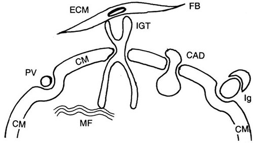
Fig. 1.2: Cell adhesion molecules (CAMs)
CM | : Cell membrane | Ig | : Immunoglobulin |
PV | : Pinocytic vacuole | CAD | : Cadherin |
MF | : Microfilament | IGT | : Integrin |
FB | : Fibroblast | ECM | : Extracellular matrix |
- Embryogenesis and morphogenesis with different tissue cells responding to growth stimuli. The cell adhesion molecules participate in regulation of differentiation, proliferation and cell death by apoptosis.
- Tissue repair, haemostasis.
- Immune response, inflammatory response.
The CAMs are named after the cell of origin as for example:
ICAM—1, 2: Intercellular adhesion molecules also known as CD54
VCAM: Vascular cell adhesion molecule also identified as CD106
PECAM: Platelet-endothelium adhesion molecule or CD31
ELAM: Endothelium-leucocyte adhesion molecule also known as E-selectin
LCAM: Leucocyte cell adhesion molecule or L-selectin
These CAMs have corresponding ligands on other cell surfaces where they attach. The most common interaction is the one between leucocytes and endothelial cells.
The cell adhesion molecules may be detected on the surface of cells as well as free in the circulating blood by appropriate techniques and can aid in identification of cells.
TYPES OF INTERACTIVE CELLULAR MOLECULES
There are at least 5 groups of CAMs:
Integrins Molecules that connect the functional or parenchymal cells with the ECM.
Cadherins They are the calcium dependent adhesion molecules.
Selectins These agents contain the protein molecules called lectins or lectin like components which bind with 7the carbohydrate (glyco-) ligands and determine the interaction of leukocytes with vascular cells.
Immunoglobulin super family These molecules consist of a vast array of molecules, by which the cells communicate with immunocompetent cells and which form intermediary links (ligands) for the above listed CAMs.
A 5th group of cell adhesion molecules is recognized as the CD44 molecules and their isoforms which take part in the immune system. CD44 bind to hyaluronic acid and are expressed on lymphocytes, monocytes, neutrophils and are involved in both cell-to-cell and cell-to-matrix interactions.
Some details of these cell adhesion molecules are now described. In later chapter it will become clear that these agents are involved in a variety of cell functions, both in the normal and in wide range of diseased states specially in immune functions and in disturbances of growth including cancer (See Table 1.2).
Integrins (Ruoslahti et al 1994)
Integrins are a family of membrane glycoprotein surface receptors which are primarily concerned with cell to extracellular matrix (ECM) contact and communication. The integrin molecule has an α-subunit and a β-subunit. Each subunit has a larger trans-and extramembrane domain and a smaller cytoplasmic domain. The extramembrane portion is in contact with corresponding receptor or specific ligands on the ECM components like collagen, laminin, fibrinogen, fibronectin. The major ligands for this contact are provided by the immunoglobulin superfamily of surface receptors. The intracellular subunits contact the cytoskeleton. The integrins are named after the subunit groups like α1/β1 known as CD 11 and β-subunit as CD 18 also.
Functions of Integrins
These molecules have an important function of determining and maintaining the integrity of the cells and to integrate the cells into their respective environments.
- During embryonic development, the integrin connections influence the development of cells by a close interaction between the cells and the ECM.
- The integrins anchor the cells to the basement membrane. This contact with basement membrane is not just meant to be a physical support but also determines the orderly growth and development of the cells. Without this contact, the cells seem to lose their polarity, (described later), and their functional maturation. In this role, not just the integrins but other CAMs are also involved. Loss of the integrins leads to loss of cell polarity and its position which has been recognized as one of the features of a cancer cell.
- In different organs the functions of integrins differ. In the renal glomeruli for example, the integrins determine the adhesion of glomerular cells to the laminin component of the glomerular basement membrane and the mesangium. The proper growth of the glomerular epithelial cells is the function of the glomerular basement membrane while the epithelial cells in turn maintain the basement membrane through the CAMs.
- Integrins play an important role in the movement of leukocytes and platelets over endothelial cells to reach a focus of inflammation. This interaction between leukocytes and endothelial cells depends upon the recognition by the cells of surface CAMs on each other and to use them to move.
- Integrins have been found to be implicated in wound healing, as the surrounding epithelium regenerates and moves to cover the healing wound. It uses the integrin mediated communications with the ECM.
- It will be explained in the Chapter 9 that loss of or abnormalities of integrins (and other CAMs) is an important feature of cancer cells. This defect helps the cells to move away from the parent growth, thus spreading across basement membranes into the ECM and across vascular membranes into capillaries to spread to distant sites. The establishment of a secondary colony of such cancer cells is also dependent on the interaction between the surface integrins/CAMs on the cancer cells and the cells of the host organ.
Selectins
This family of CAMs is named after lectins. Lectins are protein molecules in seeds with the property of binding with glycoproteins and glycolipids on the surface of animal cells to cause agglutination or clumping. Three types of selectins have been identified and named after the cells on which first detected:
P-selectin from platelets also known as CD-62
E-selectin from endothelial cells (ELAM-1)
L-selectin from leukocytes (LCAM-1)
Functions of Selectins
- Movement and identification of the destination of leukocytes and platelets: As will be described in details in the chapter on inflammation (chapter 5), leukocytes and platelets are attracted to the site of inflammation/injury and they recognize the site by identifying the selectins on the endothelial cells at the site and the endothelial cells recognize the leukocytes. This mutual recognition is possible due to the presence of specific selectin binding receptors on the cells.
- “Homing” of lymphocytes: Lymphocytes wander all over the body and circulate through the lymph nodes and spleen etc, where each type of lymphocyte (T cell, B cell and their subsets) have specific locations to reach. This destination of the lymphocytes is recognized mutually by the lymphocytes and the lymph-nodal venular endothelial cells by the interaction of selectins on their surface. The selectins are therefore, also known as the “homing receptors” on lymphocytes. (The term “homing” refers to the trained courier pigeons that recognize their destination when sent up in the air).
Cadherins (Takeichi 1990)
Calcium dependent cell adhesion molecules are important surface receptors that bind adjacent cells together. These are also glycoprotein complexes and have their greatest role in the embryological development of cells and organs. An essential step in the development of the embryo is the continual migration of cells of one type to another site to set up an organ (like the migration of neural crest cells all over the body). This migration and reclustering of cells is influenced largely by the cadherins.
The cadherin glycoprotein is a transmembrane receptor. It connects on the inside in the cytoplasm with the actin filaments through the catenin molecules (see below) generally at the sites of the cell junction called zonula adherans. The extracellular component (domain) is sensitive to calcium ion contact. An important role for cadherins is to prevent invasion of extracellular matrix by cancer cells. The following types of cadherins are known: E-cadherins: Epithelial cell, N-Cadherins: Nerve cell, myocardial cells, M-Cadherins: Muscle fibres, R-Cadherin: Retinal cadherin and P-Cadherin from placenta.
Catenin molecules are intracellular proteins that have β, γ subunits for providing contact with the specific cadherins.
Immunoglobulin Superfamily of CAMs
They are a wide variety of immunoglobulin molecules located on almost all cells in the body and serve as essential ligands for cell-to-cell contact through the other CAMs and cytokines. They are of course, of paramount importance in the functioning of the immune system. They are involved in the following reactions:
- Recognition of immune cells—different types of lymphocytes, macrophages.
- Recognition and binding of the major histocompatibility complexes (MHCs).
- Distinction of body's own cells and proteins from those that come from outside (recognition of self from non-self).
- Neural development as parts of the N-CAMs (neural cell adhesion molecules).
- Serving as receptors/ligands for other CAMs, cytokines, especially endothelial growth factor and platelet derived growth factor molecules.
- Serving as CAMs between leukocytes and endothelial cells as L-CAMs, V-CAMS (vascular cell adhesion molecules).
- Family of glycoproteins liberated by embryonic cells and by certain cancer cells known as carcinoembryonic antigen or CEA. This serves as a well-known diagnostic maker in the serum of patients of cancers of colon, lungs, etc.
- Linkages between laminin of basement membranes and elastin of the ECM.
- Identification in the laboratory of the cell type. For example, the recognition of helper T cells and suppressor T cells is based on the identification of specific CD antigens on their surfaces. Leukocyte, especially lymphocyte LCAM also known as CD44 is a useful marker for certain lymphocytic malignancies.
Some of these CAMs have been separated into the 5th category of cell adhesion molecules under the name of CD44 in which the surface ligand is a constituent of the proteoglycans of the ECM, namely hyaluronic acid.
Cytokines
Cytokines are soluble proteins produced by haemopoietic and nonhaemopoietic cells and include agents involved in activation of immune functions. These include:
- The activation of specific lymphocytes (T cells, B cells and their subsets) into recognizing and acting against invading bacteria, abnormal cells, and other antigens.
- The sequential activation and progression of the inflammatory reaction from the first signals to the regeneration and repair of damaged tissue.
- The responses of cells to stimuli like activation of growth of cells in the bone marrow.By now, over 50 different cytokines have been identified and belong to 6 categories, in each of which there are subtypes named as α, β or by numericals:Interferons (IFN -)Interleukins (IL -)Tumour necrosis factors (TNF)-also called “cachectin”Transforming growth factors (TGF)Colony stimulating factors (CSF)Other growth factors.
Growth Factors
A variety of growth promoting and inhibiting factors, like the platelet derived growth factor (PDGF), the epidermal growth factors (EGF), the fibroblast growth factor (FGF), endothelial derived growth factor (EDGF) and the transforming growth factor (TGF), are all cytokines liberated by cells on stimulation. These factors have been named after the cell from which they were originally detected and isolated. Though subsequently the factors have been identified in other cell systems also, the same name continues. An example is the platelet derived growth factor (PDGF) which is liberated by macrophages, fibroblasts, endothelial cells, in addition to platelets. These factors attach to receptors on target cells that have protein (tyrosine) kinetic activity and thereby, lead to the activation of the genetic mechanism in the cells for synthesis of proteins and enzymes required for the cell growth—either to stimulate or to inhibit it according to the stimulus and thereby, influence the cell metabolism. The differentiation and maturation of cells is also activated by these signals, they act through or on the enzymic pathways. The two pathways are the energy generation system of ATP-ADP cycle and the protein synthetic pathway of the cAMP and cGMP, involving the second messenger and the regulatory G-proteins. An example is the leukopoiesis and erythropoiesis in the bone marrow, under the influence of colony-stimulating-factors (CSF) for granulocytes and monocytes (CSF-GM). Erythropoietin secreted by the kidney is one such agent (a hormone) that stimulates the erythropoietic colonies and blast formations.
Among the important growth factors is the transforming growth factor-beta or TGF-β of which three isoforms are recognized, TGF-β, 1,2 and 3. This factor is involved in a wide variety of functions at the cellular and molecular level. For example, it is a chemotactic factor that attracts leukocytes and platelets. It stimulates other cells to secrete more cytokines, and stimulates the growth of cells including the cells of the ECM. It is an important factor in the repair of the tissues after injury and inflammation. It will be appreciated that deficiency of a growth factor (deficient production or inhibition after production) or its excess (excess secretion or defective degradation) can be responsible for a diseased state.
These factors will be referred to again and again in the pathogenesis of diseases in various chapters of the book.
These cytokines may be present in the cells in either an inactive form or in small quantities. The stimulation for more production leads to the release and activation of the inactive form, which then activates its own synthesis in larger quantities by an autocrine action and stimulates other cells to do so by a paracrine action.
The cytokines that are involved in the leukocyte-endothelial interactions are also known as chemokines, (Furie et al 1995) while the growth factors and other stimulating cytokines have recently been called the crinopectins (crino means secretion and pectin refers to adhesion) (Feige et al 1995).
These and other functions of cytokines are described in greater details in the Chapters on Immunology (4), inflammation (5) and disturbances of growth (10).
Arrangement of cells in an organ: All cells in organs are arranged in a specified pattern which is determined by the nature of the function of the organ. The position is fixed with respect to the basement membrane and/or the lumen or the vascular channels and, most important, with respect to the neighbouring cells. This fixed position is called the polarity of the cells—the cell has an upper pole, a lower or basal pole or surface and possibly three or four lateral surfaces in touch with the neighbouring cells. The exception to this fixed pattern are the circulating and wandering cells, the red cells, the leukocytes and macrophages, platelets etc. The polarity of the fixed tissue cells does not change, in fact as we shall see later the change in polarity may be one of the first feature of a cancer cell in the organ. The maintanence of polarity is the function of the cadherins or calcium mediated cell adhesion molecules already described. These molecules are distributed in the cell at strategic locations for this purpose. The polarity determines the cell functions, both within it and with the surroundings. The functions towards the surface pole are different from those at the base. For example, the cilia or villi are placed in cells at the exposed surface and the golgi network is located towards the lumen in the secreting cells. The secretions from the endocrine cells are directed at the base of endocrine cells to be passed into the capillaries along the base, the phosphorylating enzyme systems for the ADP-ATP generation is located in the surface membrane.10

Fig. 1.3: Polarity of cells in glands
Different cells in a gland (intestinal, respiratory) have different functions with corresponding polar positions. Cell bases along the basement membrane (BM), secreting cells (1) have microvilli at the surface towards the lumen. Mucus secreting goblet cell (2) has its globular part towards the lumen. Endocrine secreting cell (3) has the endocrine granules (for example, serotonin) towards the capillary at the base and nucleus towards the lumen. Leukocyte in the capillary is in contact with endothelial cell through CAMs (M). Other CAMs (II) located at the cell membrane.
BM: Basement membrane, ECM: Extracellular matrix, LEUCO: Leukocyte, XX: Integrins, II: CAMs; MC(m), cadherins placed in appropriate sites in the cell regulate the polarity

Fig. 1.4: Polarity of cells in liver chord
The adjoining cell surfaces and the surfaces facing the sinusoids are different. Bile canaliculi (BC) are placed in adjoining cells. KP: Kupffer cell, EC: endothelial cell
Cell Junctions and Adhesions
The fixed cells in an organ form continuous masses of cells in an orderly fashion. This is possible only, because the cells are joined to each other and the supporting connective tissue by special adhesion. These adhesions are of different types so that not only do they fix the cells but also serve as the sites for receiving the signals as explained in the preceding paragraphs. This allows a whole organ or a mass of cells to react identically and simultaneously as a single unit to a single stimulus. In most epithelial or parenchymal cells, four types of intercellular junctions are described but the type and number differ with type of cells; they are visible clearly in the electron microscope.
Types of Cell Junctions (Fig. 1.5)
Tight Junction or Zona Occludens
This is the site of fusion of cell membranes of adjacent cells to each other along a short zig-zag point—looks almost like a tailor's stitching or a welded point. This junction extends all-around the cell walls, forming a belt. At this site the cell membranes are impermeable.

Fig. 1.5: Cell adhesion junctions (Cell junction)
1. Occluded zone | 2. Adherant zone |
3. Desmosome | 4. Gap junction |
MF: Microfilament | N: Nucleus |
IF: Intermediate filaments |
Adhesion Junction or Zonula Adherance
Here, the membranes of adjacent cells come very close to each other but do not fuse, leaving a minute gap. There is a zone of condensation of some filamentous proteins along the membranes of opposite cells. This zone is in contact with the actin microfilaments underneath the cell membrane and is one of the points of exchange of signals and molecules between cells.
Desmosome or macula densa This is a non-occluded zone and is again formed by condensation of protein molecules at a short point in the opposing cell membranes. This junctional site is in contact with the intermediate filaments (like desmin) inside the cells. The desmosome junctions are visible under the light microscope in the epidermal cells of skin and mucous membranes as the prickles (also known as tonofila-ments) between adjacent cells. It gives the appearance of sheets of cells knitted together like a lace or tapestry.
Gap Junction or Nexus
This too is formed by protein molecules condensed in the cell membrane at opposing sites in adjacent cells. This junction allows exchange of molecules and is also known as the “electronic coupling” of cells.
The end-to-end intercellular junction in myocardial fibres is formed by intercalated discs which join the adjacent cells in a wavy stitch from cell-to-cell at their long axis, thus forming continuous sheets or the so called syncitium. These discs make it possible for the cardiac muscle contraction impulses to be transmitted to a whole mass of cells simultaneously.
The various systems of exchange of material, signals, and information described above are summarized in the Figure 1.6 and the accompanying Table 1.3.
INTRACELLULAR COMPONENTS OF THE CELLS—THE CELL ORGANELLES (Fig. 1.7)
The Cytoskeleton
The functional structures within the cell are suspended in a gel like medium called the cell sap or protoplast or cytosol and the cell maintains a three dimensional structure by filaments stretching between the cell walls on all sides. These filaments are both rigid and elastic so that the cells can modify their shape without breaking. Along these filaments, there is a network of microtubules which carry molecules all over the cell. Together these structures form the cytoskeleton. Suspended between all this network are the clusters of cell organelles, the mitochondria, the lysosomes, the nucleus, the endoplasmic reticulum, ribosomes and the golgi network. There are two types of filaments, the microfilaments and the intermediate filaments that are composed of contractile proteins. These filaments keep on pulsating, contracting and relaxing so that they set up currents of flow in the cytosol to move the floating particles and molecules.
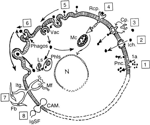
Fig. 1.6: Membrane exchange mechanism (Read together with Table 1.3)
Pnc: Pinocytosis | Phago: Phagocytosis |
Cp: Carrier protein | Itg: Integrin |
Rcp: Receptor | Mf: Microfilament |
Vac: Endocytic vacuole | Fb: Fibroblast |
Ich: Ion channel | IgSf: Immunoglobulin superfamily |
Phls: Phagolysosome | CAM: Cell adhesion molecule |
N: Nucleus | LS: Lysosome |
|

Microfilaments
These are the actin filaments 8 mµ in diameter. Though actin filaments are present in the submembranous position in all cells as well as the nuclei, they are maximum in smooth muscle and skeletal muscle fibres, since the actin is responsible for muscle contractions. As referred to above, the actin contractions also generate currents within the cytosol for a directed flow of cytosolic molecules. The actin filaments are in contact with the junctional complexes of the cell membranes and through the cell adhesion molecules like the intergrins with the extracellular matrix. Actin filaments help to move the leukocytes and to move parts of the cytoplasm and cell membrane during phagocytosis.
Intermediate Filaments
They are slightly larger than the microfilaments measuring 10 mµ in diameter and are the main skeletal support of the cell shape. Different types of cells have different types of intermediate filaments. Identification of these filaments helps to identify the cells. The technique of identification of the intermediate filaments is widely used for identification of origin of cancer cells. It utilises specific antibodies against the filaments by the immunohistological technique.
A list of intermediate filaments is provided later in Chapter 9; the important ones are:
- Keratin of different types are present in epithelial cells.
- Desmin is a feature of connective tissue fibres, specially muscle fibres.
- Vimentin is another filament of epithelial cells
- Glial fibrillary acidic protein (GFAP) is found in neuroglial cells.
Lamins: The nuclear membrane also possesses intermediate filaments called lamins.
Microtubules
These are fine hollow tubes composed of protein molecules called tubulin and run criss-cross in the cytosol. They are like the water, electricity conduits and drain pipes in a building carrying molecules, ions and fluid from one site to the other. In the myocardial fibres the job is done by the sarcoplasmic reticulum and the complex network of transverse tubular system to bring calcium molecules in and out of the cell to initiate contraction and relaxation of the muscle. The microtubules are an important component of the mitotic spindle.
Functional Cellular Organelles
So far, the framework—the skeleton, the transport and traffic systems of the cell have been described. The functions of the cells are carried out by its respiratory system that gives oxygen based energy in the mitochondria, its protein and enzyme manufacturing system (like the liver in the body) in the endoplasmic reticulum which together with the Golgi network is also the circulatory system. The excretory system is in the form of the lysosomes. These functions are governed and supervised under the control of the genetic make-up of the cells in the nucleus. All these organelles are provided with their own membrane enclosures so that they not only function independently of other cytosolic components, but can contain powerful synthetic and lytic (cell destroying) enzymes in an enclosed fashion, thus protecting the cell. These internal cell membranes too are basically phospholipid but may be single or double layered.
Mitochondria
The mitochondria are the energy manufacturing components and carry out many enzymatic processes. Mitochondria (single: mitochondrion, derived from Greek mitos meaning thread and chondros is grain or granules, abbreviated to Mc) are spherical or oval structures and vary from a few (as in a smooth muscle cell) to hundreds, in a single cell (as in the liver cell and cardiac cell). They may be irregularly dispersed in a cell or may be clustered in any one site. These structures increase their number by a self-replicating binary fission. Since they possess apparatus for respiration; protein manufacturing enzymes as well as DNA and RNA, it is believed that the mitochondrion might have been an independent, unicellular organism that evolved into an intracellular unit of a more complex animal and plant cell.13
Structure
The mitochondria are oval or spherical structures each enclosed within a double membrane. The outer membrane is smooth while the inner membrane is thrown into folds that form shelf like structures in the lumen known as cristae. On these cristae are placed molecules called elementary particles or inner membrane spheres. The cavity of the mitochondria is filled with matrix in which are located various enzymes and DNA. The mitochondrial DNA is derived exclusively from the mother's DNA.
Functions of Mitochondria
The mitochondria are the power plants of the cell, they supply high energy phosphates and oxygen in the oxidative phosphorylation cycle. The adenosine di-phosphate (ADP) is oxidized to adenosine tri-phosphate (ATP). The ATP gives away its oxygen for the cell and reverts back to ADP. This is re-cycled with oxygen to continue the system. This system is now known as OXPHOS SYSTEM, it utilises four enzyme complexes called oxphos I to oxphos IV located on the innermembrane of the mitochondria. The system is under the control of the nuclear as well as the local mitochondrial DNA. Mutations in the mitochondrial DNA in muscles cause the mitochondrial myopathies.
Metabolism of carbohydrates (glycolysis), lipids (fatty acid oxidation) and proteins (tri-carboxylic acid or TCC cycle) liberate metabolic products that are taken up by the different enzymes in a sequential manner in the oxphos system. These enzymes include NADH-CoQ (ubiquinone co-enzyme Q-10), succinic dehydrogenase and cytochrome-C in the complexes I to IV and ATP-synthetase in the final step (complex V) which drives the synthesis of ATP. The importance of recognizing the oxphos system lies in the fact that genetic mutations in the mitochondrial and/or nuclear DNA may disturb the enzyme systems leading to a group of diseases known as the oxphos diseases.
Mitochondria also liberate other enzyme systems like, for example those that are concerned with cell death, as well others that activate the release of oxygen free radicals (detailed later in the book).
Mitochondria also possess their own DNA, (referred to as mtDNA) the only DNA to exist outside the nuclear chromosomes. This DNA is exclusively of maternal origin and is also known as the non-genetic DNA. It does not replicate in cell division. It controls a number of enzymic reactions that originate in the mitochondria.
Lysosomes
Lysosomes are the other important cell constituent, serving as the cell's excretory and detoxifying organelle. As contrasted to the mitochondria the lysosome is the centre for degradation and destruction or lysis of unwanted metabolites and molecules or metabolic end products. The name lysosome comes from Greek lysein meaning to dissolve or decompose and soma means body.
Structure
These globules are composed of a large number of hydrolytic enzymes in inactive form enclosed within a monolayer membrane. The enzymes include acid phosphatases, acid esterases. The leukocyte granules are lysosomes so are the granules in osteoclasts.
Functions
The lysosomes are instrumental in three vital functions:
- To dissolve out and degrade or detoxify the end products of metabolism within the cells or products brought into the cells. This degrading system is a very important function and proceeds in step wise fashion. At each step, one or more of the catabolic enzymes comes into activity. If any one enzyme is deficient in this chain, the metabolic process will stop at that level and the semi-finished product will accumulate in the lysosome or in the cell with no possibility of its removal. This defect forms the basis for what are called the lysosomal storage diseases (see Chapter 7).
- The disposal of particulate matter like bacteria by leukocytes. The leukocytes phagocytose (engulf) the bacterium or the particle which enters the cell surrounded by a vacuole of the cell membrane by the process of endocytosis. This vacuole or vesicle is called the phagosome. The phagosome on coming in contact with one or more lysosome, fuses with it to form a larger vacuole called the phagolysosome. Within this enclosed space the lysosomal enzymes are released to digest the bacterium—the lysosome enjoying a meal in private. This way the hydrolytic enzymes are not released into the cell cytosol (thus protecting the cell from their destructive action). After the bacteria have been destroyed, the product is excreted out of the cell by exocytosis again taking a fragment of the cell membrane with it.One of the results of the two processes of lysosomal activity described above is that, over the years the lysosomes may go on accumulating some material representing the end stages of the metabolic or detoxifying actions. These accumulations appear as what are known as the wear and tear pigments or lipochrome pigments and are best seen in the cardiac muscle, in the testicular-Leydig cells and the liver cells. This pigment indicates an ageing phenomenon.
- The lysosomes carryout an important function of self-destruction—autolysis of the cell when it has lost its life support. When cells die, they have to be dissolved and the debris removed. This job of dissolution is carried out by the activation of the cell's lysosomes by the falling pH and the anoxia and release of the enzymes. The leukocytes that come into the area around the dead cells, perform the same function with their lysosomal enzymes.
Endoplasmic Reticulum
The endoplasmic reticulum or ER is a system of tubes and cisterns enclosed in monolayer membrane and forms a network or lace work (reticulum) in the cell. These flat tubes are continued into the space between the two layers of the nuclear membrane, into the Golgi network and the cell membrane. Two types of ER are recognized, the rough endoplasmic reticulum or RER which has rows of ribosome granules along its surface and smooth endoplasmic reticulum or SER which has no such granules. The endoplasmic reticulum is involved in the vital function of manufacture of proteins, enzymes, hormone molecules and other secretions from the cell. This process will be explained shortly.
Ribosomes
They are composed of RNA and proteins of different types according to their composition and functions namely the ribosomal RNA or rRNA, the messenger RNA or the mRNA, the transfer RNA or the tRNA. There are others also. The ribosomes are assembled from amino acids as per the genetic codes in the nucleus and then carryon the function of transcription and translation and assembly of enzymes and proteins and secretions. Apart from being arranged along the membranes of the rough endoplasmic reticulum, there are small clusters of ribosomes in the cytosol, known as polyribosomes. The functions of the ribosomes and the endoplasmic reticulum will be described shortly.
Golgi Network
It is a cluster of flattened saucer like membrane bound channels which receive the products prepared by the endoplasmic reticulum and have the function of refining finishing, packing the hormones, the secretory material, proteins and enzymes before secreting them out to the cell surface or to the mitochondria or to the nucleus.
Cytoplasmic Granules
Other than the above organelles there are secretory and neurosecretory granules and glycogen particles, distributed randomly in the cytoplasm in specialized cells. The neurosecretory and hormonal granules are membrane bound with darker cores; different types have different structure as seen under the electron microscope.
Nucleus
The term nucleus is derived from the Latin meaning the nut kernel. The nucleus is the nerve centre or the “master computer” of the cell. It keeps all the information or programmes required for the cell functions in coded form in the genes. The genes are arranged in an orderly and fixed fashion on the chromosomes and the chromosomes are clumped into the chromatin, the basophilic material of the nucleus. The nucleus is enclosed within a double layered nuclear membrane with a space between the two layers.
Genes (from the Greek genein meaning to produce)
The genes are composed of deoxyribonucleic acid protein complex (DNA), though genetic information is carried also in the ribonucleic acid or RNA in certain viruses. Both these nucleic acids, the DNA and the RNA are acidic compounds of complex sugars which is ribose in RNA and deoxyribose in DNA, pyrimidine and purine bases and phosphates. The purine bases are adenine (A) and guanine (G) in both DNA and RNA and the pyrimidine bases are thymidine (T) and cystine (C) in DNA and Uracil (U) and cystine in RNA. The bases plus sugars is the unit called nucleoside, while the bases plus sugar plus phosphorus is the unit called the nucleotide.
Structually, the DNA is composed of multiple nucleotide (polynucleotide) chains twisted or coiled up in a double stranded spiral manner similar to a winding stair cases. The twisted arrangement is known as a double helix (which in Greek means coil). The frame of the helical stair case is composed of the sugar plus phosphate moieties while the rungs or steps of the staircase are formed by the bases stretching across the spiral bars. The bases are arranged in a predetermined sequence with the two strands of the DNA running in opposite directions parallel to each other. The bases form pairs in which thymidine (T) is always complimentary to adenine (A) and cystine (C) is complimentary to guanine (G). Thus, in one strand if the base arrangement is TACT, its opposite number in the other strand will be ATGA.
- One unit length of DNA is the base pair (bp)
- 1000 bp make a kilobase (kb)
- One million bp make one megabase (mb)
- 3000 mb form the total length of a half set of human chromosome.
This helical structure is according to the model described by Watson and Crick in the 1950s. (For details consult Connors and Ferguson 1993).15
Each base unit or the step is composed of three pairs of the bases listed above. This unit is therefore called a triplet. The arrangement of bases in the triplet are distinctive for each gene and distinguishes it from others. This triplet arrangement is the codon. The code is the manual of instructions that comes with an instrument or a complex toy (the animal and plant body is after all a toy for the Maker!) and lays down the instructions on how to assemble the different components of the entire instrument and individual parts of the instrument. In the same way, the DNA code directs the cell's manufacturing machinery on how to assemble the amino acids to form a particular peptide and protein structure. This then determines the structural components of the entire body. The code is inherited from the parents and therefore, many features in the offspring are the reproductions of the parents. The portions of the gene that carry genetic information is called the exons while inbetween the exons are the non-genetic DNA proteins known as introns, which are involved in the arrangements of the exons.
RESPONSE OF NORMAL CELL TO PHYSIOLOGICAL STIMULI
As already defined life consists of constant responses to stimuli. All cells take part in this process all the time. These response may be triggered by external stimuli or by the internal genetic programme inbuilt into the cells. Each organ has its own specific and distinctive functions. Some of the general responses of cells to external stimuli may be as follows:
- Movement of the cell: Leukocytes, platelets are constantly on the move. Spermatozoa move towards the ovum. These movements are activated by cytokine stimuli.
- Activation of cell growth: This may take the form of:
- Activation of mitosis
- Activation of increase in cell size by increasing the protein and other components in the cells
- Activation of cell differentiation—by the output of new types of cell molecules and cell organelles.These processes are best seen all the time in organs like the bone marrow and the intestinal mucosa.
- Stimulation of secretions and other products from the cell: These products including mucins are the hormones, cytokines, enzymes, immune molecules, chemical mediators etc.
- Stimulation of metabolic activities in the cells—like the production of glucose, proteins, etc.
Many of the above listed activities will be described in later chapters. One common response in majority of these actions or responses is the activation of the synthetic functions of the cell—be it secretion of mucus, or output of a hormone, or of albumin or immunoglobulin or glycogen, or glucose-the cell's synthetic apparatus comes into activity. A basic understanding of the process of synthesis is essential to understand disturbed functions in pathologic states. Since protein synthesis is the fundamental function of cells some details of the manufacture of proteins is described here starting with a brief outline of what is protein and what are the various types of proteins.
Protein molecule
The student is reminded of the important role of proteins in the structure and functions and a few important aspects are mentioned here since reference will be made to these features repeatedly in the book. The term protein comes from the Greek word, proteios meaning of the first rank “—a reference to its importance.” Proteins are composed of amino acids arranged in a specific pattern. Of the total 100 proteins in nature 20 amino acids make up the body proteins, in a variety of combinations. The arrangement has been referred to in terms of the way in which the chains of the amino acids are arranged.
The alpha (α) chain is a rod like innercore, surrounded by a second layer of coiled or helical chain around it. The chain is stabilised by hydrogen bonds.
The beta (β) chain structure on the other hand, is a sheet and not a rod. The amino acids form a pleated or knitted structure and the chains are not folded or coiled but fully extended. These chains are also stabilised by hydrogen bonds placed differently from the alpha chains.
Similarly, there are delta (δ) and gamma (γ) chains. In a given protein, there may be only alpha chains or only beta chains or both the chains are present.
The chain strucutre is examplified by the rope like coiling of alpha chains in the collagen chains to be described later in this chapter.
The other feature of amino acid chains is their pattern of folding by which some amino groups are exposed on the surface, while others are hidden in the folds. This folding pattern is also typical of each protein. The exposed groups take part in the bonds that the molecule forms with other radicals—like the carbon bonds, hydrogen bonds, electrostatic bonds. Some of the chains have what are called the globular domains or ends. This refers to a compact folded part of the chain (like pearls on a string) and is again distinctive for a particular protein molecule.
Functions of Proteins
All enzymes, and there are thousands of them, are protein (enzyme protein). Transport of other molecules in the body and within the cells, specially of insoluble molecules is 16carried out by proteins (transport proteins). Examples are the carriage of oxygen by the haemoglobin molecule, the transport of bilirubin by serum albumin. Many molecules are stored in the cells in combination with storage proteins like the iron is transported by transferrin in the plasma to the liver cell where it is stored by ferritin (storage proteins). Movement of a cell or molecules within a cell is conducted by protein molecules. The muscle contraction is due to the sliding motion of actin and myosin protein fibrils also known as the microfilaments and intermediate filaments (contractile proteins). The structure of the organs and tissues is maintained by proteins like the collagen (structural proteins). Proteins are responsible for the immunefunctions or defence reactions of the body (immunoglobulins, cytokines and mediator proteins). Proteins convey impulses and signals across cell membranes like the transmission of nerve impulse (receptor proteins, signal proteins). Proteins control the growth and differentiation of cells (growth factors or proteins, building blocks and cytokines). Proteins are responsible for the interactions between cells and the connective tissue or the extracellular matrix (adhesion proteins, cell adhesion molecules, integrins).
Then there are some types of complex proteins. These include the lipoproteins (proteins-lipids complex) that act as carriers for a number of lipids and cholesterol. Glycoproteins are proteins complexed with polysaccharides (including the glycosylated proteins) and form the components of the extracellular matrix.
SYNTHETIC PROCESS IN THE CELL (FIG. 1.8)
Given below is a highly simplified and anecdotal explanation of this highly complex biomolecular system.
There are two main steps in the manufacture of protein or other molecules:
Transcription or the preparation of a copy of the code on RNA ribosomes which lays down the sequence in which the amino acids are to be joined or assembled in the particular molecule.
Translation is the actual assembly of the amino acids in that sequence. This job is carried out by another set of RNA.
Both types of RNA are assembled from amino acids in the nucleus, according to their own code. The double helix of the DNA chain (that is the two sides of the winding staircase) opens up like the splitting of the staircase longitudinally down its centre and into the space so created move in the messenger RNA or mRNA molecules to be arranged as per the codes in the DNA. This process is aided by the enzyme polymerase. The split DNA helix closes on the RNA (This step is like the printing platter in a printing press or the sliding platform in a photocopy machine). After “printing” the sequence of amino acids, the helix reopens to allow the mRNA to move out of the nucleus. The mRNA now moves into the endoplasmic reticulum in the cytoplasm. In these complex tubules, other RNA compounds like rRNA and tRNA assemble the various amino acids from the cell sap and introduce them into the endoplasmic reticulum. This is where the analogy to a conveyor belt in a factory is applied. The endoplasmic reticulum is the conveyor belt on which the parts of an instrument or the toy are placed and the ribosomes on its sides are the factory workers or robots, each one adding and arranging the particular part of the instrument according to the instruction manual (the transcript).
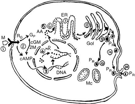
Fig. 1.8: Protein synthesis in a cell: Steps in the process:
M: Activating molecule (hormone, cytokine, etc)
R: Receptor RG: G-protein receptor cAMP: Cyclic AMP
AA: Amino acids 2M: Second messenger system
ER: Endoplasmic reticulum GP: G-protein
Mc: Mitochondria MR: mRNA GOL: Golgi tubules
PR: Protein molecule
- Activation of the membrane receptor/G-protein receptor
- Activation of phosphokinase direct through cAMP or/and through a second messenger including G-proteins
- Activation of the concerned genes
- Transcription of RNA-mRNA, tRNA, rRNA by the opening up of the DNA double helix.
- The mRNA moves to the endoplasmic reticulum
- The tRNA and others assemble the required amino acids into the ER tubules.
- Formation of chains of amino acids into peptides, polypeptides.
- Passage of the unfinished chains into Golgi tubules
- Folding of chains to form the protein molecule
- Secretion of the finished product by exocytosis outside or transfer to internal cell organelles
As the compound travels along the endoplasmic reticulum, the molecule takes shape first as a peptide, then as a polypeptide chain and finally as a protein. Through the endoplasmic reticulum the rather crude or unfinished molecules are transferred to the Golgi apparatus for further finishing, refining and packaging (like the packaging unit in a factory) and finally to be conveyed to their destination.
The entire process of switching on the gene and the mRNA is activated by the system of signal transduction, the receptor mediated instructions described above. The signals end up by activating the kinetic enzymes (manufacturing enzymes) like the phosphokinases in the cell.
In the cell, the energy manufacture and storage for use in the metabolic processes is the function of the nucleotides. These are complexes of ribose and deoxyribose sugars, purine and pyrimidine bases and phosphates. The metabolically important nucleotides are those in which the bases are adenosine, guanosine and inosine. These form mono, di and tri phosphate linkages. For example, adenosine monophosphate or AMP, adenosine diphosphate or ADP and adenosine triphosphate or ATP.
These nucleotides serve as carriers of activated intermediary products and receptors to growth factors and kinetic hormones for the synthesis of carbohydrates, proteins, and lipids. This process has been explained above with the receptor mechanisms.
Recycling of Cell Membranes
The constant traffic of molecules and other material across the different membranes would naturally take bites out of the membrane, requiring immediate closing of the ‘gap’ in the membrane. Normally this happens instantly. Under pathological conditions the membrane damage is more severe. The fragments of the cell membrane are recycled within the cell by a system of degradation and resynthesis. Under abnormal conditions when the cells die or are otherwise stimulated, there is an active system of release of highly active biomolecules known as eicosanoids from the enzymic degradation of the phospholipids. Enzymes like the phospholipase, lipo-oxygenases and cyclo-oxygenase are activated and release substances including the prostaglandins, leukotrienes as metabolities of arachidonic acid. These molecules are of great importance in the regulation of cell functions and synthesis, particularly in the reactions of cells to injury.
Intracellular Carrier Proteins
It will be appropriate to introduce two more proteins here that help to move molecules within the cell cytoplasm. These are the “heat shock protein” and “ubiquitin”.
Heat Shock Proteins—HSP, also known as Stress Proteins:
HSP are present in most cells specially the renal tubular cells. These molecules act as chaperones for other molecules. They normally direct and guide metabolic molecules to the sites of the metabolic activity, for example the metabolites that are brought into the cell for disposal are guided by the HSP to lysosomes so that the metabolites do not stray around. Whenever the cell is subjected to adverse conditions—toxins, metabolic poisons, drugs, etc—the cell secretes more of the HSP to cope up with the extra load of metabolites coming to the cell—hence the name, stress proteins. The HSP leak out of the cell into the plasma too.
Ubiquitin
This too is a “stress protein”, and is a common factor in a wide range of human degenerative diseases. These molecules are called ubiquitin because of their ubiquitous (or universal) presence in the body's cells. Ubiquitin, like the HSP is a molecular chaperone and directs molecules to their destination, either for degradation or for synthesis and for incorporation into secretions. For instance, defective protein molecules including defective DNA are tackled by uniquitin either for repair or for selective degradation. Ubiquitin is also involved in activation of genes for protein synthesis. Ubiquitin is particularly involved in the cells of the central nervous system. As cells start ageing, the ubiquitin forms insoluble complexes with the cytoskeletal filamentous proteins and in a number of age related degenerative disorders of the CNS like Parkinson's disease, Alzheimer's disease, neurons accumulate these ubiquitin-filament complexes that can be identified microscopically. In the liver cell and muscle fibre too, hyaline bodies containing ubiquitin appear in diseased conditions.
In addition to these specialised molecules, a number of other chemical agents are synthesized and liberated by cells during their functions, specially when the cells are subjected to injurious agents. These will be described at appropriate places in the book.
CONNECTIVE TISSUE: EXTRACELLULAR MATRIX
The connective tissue of the body includes some general structures present in all tissues and organs which only provide support and shape, as well as some highly specialised types of tissues. The supporting connective tissue is the collagen and allied fibrillar structures. The distinction between the supporting connective tissue and the functioning parenchymal, epithelial cells is clear cut in structure but functionally, the two are completely 18integrated. Right from the embryo the two tissues influence and direct each other towards a properly coordinated and integrated development.
The connective tissue includes both the cellular elements and the matrix or ground substance called the ECM, (extracellular matrix) which forms a kind of cement in which the connective tissue cells are embedded. The cells include the haemopoietic and nonhaemopoeitic mesenchyme derived cells, fibroblasts, myofibroblasts, smooth and skeletal muscles, neural tissue, vascular endothelial cells.
The extracellular matrix (ECM), is composed of three components:
- Fibrillar structural proteins including collagen, and elastin.
- Adhesive proteins: Fibronectin, laminin, fibrillin, osteonectin, tenascin.
- Gel like molecules of proteoglycans and glycosaminoglycans including, heparan sulphate, chondroitin sulphate, dermatan sulphate, and keratin sulphate.
Between the epithelial or parenchymal functional cells and the connective tissue, lies an important component of the ECM namely the basement membrane. Described here are only the basic features of collagen, the basement membrane, and the ECM glycoproteins, since these are universally present while other cells will be described in respective chapters.
Collagen
The name is derived from Greek Kolla meaning glue and genein meaning to produce, since collagen was considered as the glue that binds all cells and tissues together (—an appropriate concept). Collagen is a family of protein molecules known as structural proteins (in contrast to the functional or molecular proteins like albumin, globulins). Each collagen protein molecule is composed of three peptide chains—the α-chains which are twisted over each other in a right handed coil or helix (like a three stranded hair pigtail or a rope). These are joined to each other into microfibrils which in turn are covalently bonded to form collagen fibres. Each alpha chain is composed of amino acids which are assembled in the endoplasmic reticulum as precursor larger sized molecules, the procollagen molecules. The most common cell that produces collagen is the fibroblast but endothelial cells, macrophages and even epithelial cells can produce collagen under appropriate conditions (A specialized cell in the liver, the Ito cell produces collagen in the liver lobules). The most common amino acids in the collagen are glycine, hydroxyproline, others being hydroxylysine, proline, and lysine. The fibrils are given strength by the lysine and hydroxylysine cross links.
The procollagen molecule is secreted out of the cell into the extracellular matrix where the final steps in the cleavage of procollagen or hydroxylation of the procollagen to collagen occurs through appropriate enzymes and the presence of vitamin C (ascorbic acid). The procollagen fibrils are poorly wound and unfinished and mature outside the cells. This extracellular maturation is unique to collagen. An exception is the maturation of the basement membrane collagen (Type IV), which matures in the cells only. The process is similar to the conversion of fibrinogen to fibrin. It is after this cleavage of procollagen to collagen that the typical electron microscopic structure of collagen appears. This consists of the fibrils with regular bands at fixed distance of 650 angstroms. This is known as the periodicity of collagen fibres.
At light microscopy level, collagen takes eosinophilic colour shows birefringence in polarised light and can be stained by connective tissue stains. One common form of collagen which all pathologists look for in sections of liver and lymph nodes in particular is called the reticulin stained by silver nitrate. Collagen has this property of fixing reduced silver salts, a property known as argyrophilia.
Collagen in different sites and different organs is different in its peptide chain composition and configuration. This is so because the functions performed in different organs are different. Broadly speaking; there are two types of collagen, the fibrillar collagen and the non-fibrillar collagen. The fibrillar variety is meant to provide tensile strength, weight bearing and physical structure to the organs whereas the nonfibrillar collagen forms a lace like network, constituting the basement membranes and other structural supports. So far 18 types of collagen have been described based on the alpha chain structures. Types I, II, III, V, and IX are typically fibrillary and are found in skin, cartilage, blood vessels, etc. The typical nonfibrillary collagen is Type IV, the basement membrane collagen. Type VIII is a unique type, it anchors or attaches the epidermis to the dermis through the epidermal basement membrane. The students will find a list of the types and their distribution in the body in any biochemistry text.
Like other tissues and cells, collagen fibrils are continually though slowly degraded and replaced, a process that slows down with age. During the remodelling of healing wounds and scars, the process of degradation speeds up. The degradation is caused by the collagenase enzymes that are a class of metalloproteinases. The metalloproteins are a class of enzymes, containing a metal like copper, iron, manganese, zinc, nickel etc. Haemoglobin contains iron. They act in presence of zinc and exist in many types of cells in precursor (Procollagenase) 19form. The interstitial collagenases degrade fibrillary collagen, the gelatinases degrade nonfibrillary ones. The glycosaminoglycans are similarly degraded by enzymes called stromelysins (lysing of the stroma!!).
In addition to these tissue enzymes, the collagens are also broken down by protein cleaving enzymes, or proteinases secreted by leukocytes—neutrophils and macrophages. These enzymes are actively secreted from the leukocyte granules under conditions of inflammation and include cathepsin, elastase, etc. These are not metalloproteinases but serine kinase enzymes.
Elastin
Elastin fibrils are rubber band like stretchable fibres. Arteries like aorta and major arteries, skin, tendons, lung alveoli are rich in these types of fibres. Elastin fibres differ from collagen molecules in many ways, they have very little hydroxyproline and much more proline amino acids and very frequent cross-linkages between the twisted alpha fibrils.
Adhesive Proteins
Fibronectin (Latin, fibra is fiber and nectere is to bind)
is a cell surface protein secreted by the liver and helps to link the cells to the ECM via the intermediation of the integrins. The fibronectin protein is present in the plasma cross linked to fibrin. Another site in which it is involved, is the adhesion of the blood clot or fibroblasts during the organization of clot and in inflammation. Fibronectin helps in the migration of cells in the embryo. As described in Chapter 9, deficiency of fibronectin in cancer cells facilitates the spread of cancer.
Laminin
This adhesive protein is particularly rich in the basement membranes with Type IV collagen. It serves as an extracellular integrin.
Other adhesive proteins osteonectin, fibrillin are the similar nonfibrillar components of the ECM.
Glycoproteins
These complex proteins provide two distinct types of molecules (i) Proteoglycans that form the noncellular extracellular matrix earlier known as the ground substance. It is a gel like cement substance in which the cellular components are embedded. The two together bind water to form the viscous tissues like the cartilage, the ligaments, tendons, the substance of the cardiac valves, the dermis as well as the synovial fluid in the joints, the intervertebral disc material etc. One of their functions is to act as shock absorbers. The important proteoglycans include the glycosaminoglycans which include the following molecular agents:
Heparin, heparan sulphate, hyaluronic acid, chondroitin sulphate, dermatan sulphate and keratan sulphate. Of these the heparan sulphate also known as Perlecan carries the electrical charge in the basement membranes.
- Structural glycoproteins are molecules that are involved in the cell-extracellular matrix interaction. These include the following:
Fibronectin which are present both as water soluble molecules in the blood and tissue fluids and an insoluble intracellular form. Others include tenascin and thrombospondian, and osteonectin etc.
The Basement Membranes (BM) (Aumailley 1993)
This is a layered form of fluid-permeable extracellular matrix composed of both collagen and proteoglycans in the form of a molecular membrane varying in thickness between 50 and 100 mu at different sites. The BM separates the functional or parenchymal cells of an organ from its connective tissue and the vascular channels. The BMs are the earliest form of extracellular matrix to develop in the embryo since the membranes not only separate the cells but also determines its development and maturation. Many of the mesenchymal cells are invested by a continuous BM such cells include the muscle fibres, skeletal, smooth as well as cardiac, (outside the sarcolemmal membrane), Schwann cells, and capillary endothelial cells (The endothelium of sinusoids of the liver, spleen, lymph node and bone marrow are NOT lined by a basement membrane). Some of the basement membranes have a specialised function normally as well as in diseased states. These include the basement membrane of the glomerular capillaries (described in details in chapter on Renal Disease), the pulmonary alveolar basement membrane and the epidermal basement membrane of skin and mucous membranes.
The basement membrane is composed of collagen, laminin, nidogen and heparan sulphate as detailed below:
Collagen
The collagen structure specific for basement membranes is the Type IV collagen though in some locations other types are involved. Type VII and Type XVII are specific for the basal lamina of the epidermis in the skin. The type IV collagen chains have what are called noncollagenous globular domains at either end. It forms a polygonal meshwork which provides a scaffold for anchoring other molecules in its structure.
Laminin
Laminin is the most abundant non-collagenous protein in the basement membrane and is the integrin in the extra-cellular 20matrix and forms a complex with another protein, the nidogen.
Nidogen
Nidogen is single polypoid chain protein molecule and is a cruicial component of the basement membrane. It connects the network of laminin to the collagen.
Heparan Sulphate
Also known as perlecan, heparan sulphate is a proteoglycan of the basement membrane and carries the electrical or ionic charge which determines the passage of other electrical charged particles through the basement membrane during filtration.
Functions of basement membranes The basement membranes have both mechanical and biological functions that include the following:
- To provide a scaffold or support to cells at sites of separation from spaces or from connective tissue. For instance, basement membrane at the epidermal-dermal junction in the skin and in mucous membranes, endothelial cells lining the vascular channels and glands have basement membranes. The role of CAMs in the interaction between the cells and the basement membranes have already been referred to earlier.
- To act as filters for fluids and solutes as in the glomerulus and other capillaries all over the body.
- To act as a source of information to be transmitted across the tissues.
- Basement membranes influence the orderly development of cells, specially in the embryo.
Epithelial Cell-Extracellular Matrix Interrelationship (Fig. 1.9)
From the foregoing descriptions of the cell structure and structures of the extracellular matrix, it would be apparent that the ECM is not simply a structural support for cells, the ECM greatly influences the growth and functions of the cells. This is illustrated in Figure 1.9 (Haralson 1993).
Summary
The topics covered in this chapter are meant to highlight those aspects of the cell biology which are intimately involved with the reactions of the cells to internal and external stimuli of all kinds. Understanding the mechanisms of how cells are kept in constant touch with their environments, forms an essential part of the cell functions. The protein molecules of a wide variety are synthesized in the cell under the guidance of the nuclear DNA in the genes. The stimuli to the DNA and RNA functions (transcription and translation) include hormones, neural impulses, cell-to-cell contacts and adhesion molecules, metabolic stimuli, immune stimuli, and external agents like micro-organisms. These stimuli are received by cell membrane receptors and cell adhesion molecules. These CAMs include 4 main types of molecules, the cadherins, the selectins, the integrins and the immunoglobulin superfamily. Almost all cells affect their own function and that of their neighbours, by secreting a wide variety of cytokines. A set of growth factors include cytokines which are among the most potent and common receptor mediated stimuli to the cell and nuclei. The other mechanisms of interaction between the cell and its environment is the ion-exchange mechanism of water and electrolyte exchanges. All these stimuli trigger the cell into activity or activities. The activity depends upon the nature of the receptor mediated stimulus and the genetic nature of the cell. The most important activity in the cells is the synthesis and secretion of active principles including hormones, mucins, immunoglobulins and cytokines, all of them basically proteins. The proteins are of a large variety and their synthesis involves the activation of the genetically controlled system of RNA mediated synthetic process. This process involves the stimulation of enzyme systems for synthesis (phosphokinases) and a system for energy supply for all the activities (the phosphorylases). The activation may be a direct step by the stimulus or through the mediation of a second messenger.

Fig. 1.9: ECM-cell interrelationship
Molecular agents (left) stimulate the cell to the activities listed on the right. The ECM interacts with the cell directly and indirectly
The cell possesses all the functional systems in its organelles, namely respiratory, nutritional, circulatory, 21and detoxifying systems respectively in the mitochondria, the endoplasmic reticulum, golgi apparatus, and the lysosomes. These systems are also activated by appropriate receptor mediated stimuli.
The second message in this chapter is the intimate relationship between the cells and their extracellular matrices. The ECM that include collagen, adhesive proteins, and glycoproteins of the ground substance, influences the development and differentiation of the parenchymal cells right from the embryo.
This brief and selective recapitulation of the normal structures and functions of the cells will help the students understand the molecular biology of disease processes in the subsequent chapters.