

Cells are the structural and functional units of all living organisms. Man is a multicellular organism, contains at least 1014 cells. These cells differ considerably in shape, structure and function as a result of specialization. An aggregation of cells those are similar in origin, structure and function forms tissue. Most of the metabolic activities occur at cellular level. Hence it is essential, first to understand the basic organization of cell and functions of its components.
A typical cell, as seen by the light microscope is illustrated in Figure 1.1. It contains two compartments inner nucleus and outer cytoplasm. Nucleus contains nucleoplasm suspended with genetic material. Nuclear envelope separates nucleus from cytoplasm. Cytoplasm composed of aqueous cytosol, suspended with particles and membrane bound organelles. Externally cytoplasm is limited by plasma membrane.


ULTRASTRUCTURE
Normal cell ranges between 10 and 30µm in diameter. Figure 1.2 shows the ultra structure or finer details of typical cell, which has been revealed by the electron microscope.
Plasma Membrane
The cell membrane, which completely envelops the cell, is a thin (75-100Ao), living, dynamic and selectively permeable membrane. Plasma membrane consists specialized surface structures for attachment and for communication. Those are (i) Tight junctions produce seal between adjacent cells. (ii) Gap junctions allow ions and electric current between adjacent cells. They may also include certain modifications to carryout physiological functions such as microvilli for absorption, invagination or infoldings to carry out transportation etc.
All biological membranes including the plasma membrane and internal membranes which form the subcellular structures such as endoplasmic reticulum, mitochondria, lysosomes nuclear envelope, peroxisomes, Golgi complex, etc are similar in structure, lipoprotein in nature, consists lipids (60–40%), Proteins (40–60%) and carbohydrates (1–10%). The membranes separate the cell from external environment and separates different parts of the cell from one another so that cellular activities are compartmentalized.2

Figure 1.2: Ultrastructure of typical cell showing all cell organelles as seen in the electron microscope
Endoplasmic Reticulum
Cytoplasm is traversed by extensive network of interconnecting membrane bound channels or cisternae (diameter of 40–50µm), vesicles (diameter 25–500µm) and tubules (diameter 50–190µm) form endoplasmic reticulum (ER) (Fig. 1.3). Membranes of ER continuous with plasma membrane and outer nuclear envelope. There are two basic morphological types (i) Rough endoplasmic reticulum (RER) possesses rough surface due the attachment of ribosomes.
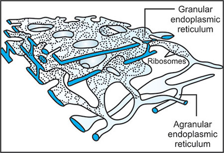
RER occurs mainly in the form of cisternae and concerned with protein synthesis. (ii) Smooth endoplasmic reticulum (SER) lacks ribosomes on their surface, occurs mainly in the form of tubules. SER is concerned with lipid synthesis.
ER provides skeletal framework to cells and gives mechanical support to the colloidal cytoplasm. It also plays a role in detoxifying the xenobiotics.
Golgi Complex
Golgi complex is membrane bound structure similar to ER, discovered in 1873 by Camillo Golgi. It is a stack of flattened membrane vesicles (cisternae) surrounded by net working of tubules of 300–500A° diameters. Cisternae gently curved, convex part cis side faces ER and concave part trans side locates near plasma membrane (Fig. 1.4).
Golgi complex functions in association with ER, is a center of reception, finishing, packaging and transportation of variety of materials. Proteins synthesized in ER added with sulfate, carbohydrates, lipid moieties etc. and dispatched in the form of secretory vesicles. Golgi complex also gives rise to lipoprotein of plasma membrane and lysosomes.

Lysosomes
Lysosomes are packets of hydrolases. These are spherical 1µm in diameter surrounded by tough carbohydrate rich lipoprotein membrane enclosing about 50 types hydrolases such as proteases, lipases, carbohydrases nucleases, transferases sulfatases etc.
Lysosomes provide an intracellular digestive system through which macromolecules, foreign bodies and worn out unwanted structures are got digested.
Peroxisomes
Circular membrane bound organelle having about 0.25 µm diameters contain enzymes peroxidases and catalase. Peroxiosomes detoxify various toxic substances and metabolites through peroxidative reactions catalyzed by peroxidases. Catalase degrade H2O2 resulted from break down of fatty acid and amino acids.
Mitochondria
They are spherical, oval or rod like bodies, about 0.5–1µm in diameter and up to 7 µm in length.

DNA molecules, which encode information for certain mitochondrial proteins (Fig. 1.5).
Mitochondria are considered to be the powerhouse of the cell, where energy released from oxidation of foodstuffs is trapped as chemical energy in the form of ATP. Mitochondria are respiratory center of cell where pyruvate oxidation, citric acid cycle, electron transport chain and ATP generation takes place. Beta-oxidation of fatty acid and ketone body synthesis also taking place.
Centrioles
Two cylindrical rod shaped structures of 0.3–0.7µm lengths and 0.1–0.25µm diameters, which lie right angle to one another near nucleus called Centrioles. Centriole is an array of 9-triplet microtubules equally spaced from central axis, made up of structural protein tubulin. Centrioles form mitotic poles during cell division. They also give rise to cilia and tail of sperm.
Nucleus
Cell center, prominent spherical structure where genetic material is confined. All cells in the human body contain nucleus, except matured RBCs and upper dead skin cells. Generally nucleus spherical or oval in shape and of 3–25µm in diameter. But squamous epithelial cells contain discoidal and multilobed in polymorphonuclear leucocytes. Nuclear envelope, which encircles the nucleus, consists outer and inner nuclear membranes, typical lipoprotein membranes. Outer nuclear membranes continuous with membranes of ER and found attached with ribosomes on its outer surface. Nuclear envelope contains numerous nuclear pores of 100–1000A° diameter, which regulate nucleo-cytoplasmic trafficking of ions, nucleotides, proteins, mRNA, tRNA and ribosomal subunits
Nucleoplasm consists genetic material (chromosomes), and nucleolus. Nucleolus is ribonucleoprotein structure, is the site of formation of ribosomal subunits. Nucleoplasm composed of mainly the nucleoproteins, proteins, enzymes, minerals, organic and inorganic substances.
TRANSPORT ACROSS MEMBRANE
Biological membranes are lipoprotein viscous barriers exist around all living cell and also form structural and functional component of all cell organelles. Membranes contain mainly lipids, proteins and very little amount carbohydrates. The contents of these vary according to the nature of the membrane. Lipids are mainly amphipathic phospholipids, glycolipids and cholesterol. Proteins are of two types (i) Peripheral or extrinsic proteins and (ii) Integral or intrinsic proteins.
Organization of biological membranes, the arrangement of lipids and proteins was best explained in fluid mosaic model of Singer and Nicolson (1972) (Fig. 1.6). According to this model, membrane is viscous fluid phospholipid bilayer, in which globular proteins inserted in a mosaic pattern.

Amphipathic phospholipid consists polar phosphate head, glycerol neck and nonpolar two fatty acid tails. Hydrophobic tails or fatty acids form the middle core of lipid bilayer. Hydrophilic heads line both sides. Both phospholipids and proteins are amphipathic form permeability barrier. Degree of saturation and unsaturation of fatty acids, presence of cholesterol and carbohydrates regulate the fluidity and movement of molecules. Hydrophilic heads of inner and outer surface keep constant circulation of water. But hydrophobic fatty acid core acts as selective permeable barrier saves the cells and cell organelles from osmotic shocks.
Important function of the membrane is to with hold unwanted molecules but permit entry of molecules necessary for cellular metabolism. Transport across the membrane occurs in following ways: (i) Passive transport. (ii) Active transport. (iii) Exocytosis. (iv) Endocytosis.
Passive Transport
Passive transport of molecules across the membrane is along the concentration gradient without using energy. Movement of molecules from higher concentration to lower concentration takes place without using energy. Solutes and gases enter into the cells passively. They are driven by the concentration gradient. The rate of transport is directly proportional to the concentration gradient of that solute across the membrane. Passive transport of molecules across the biomembranes is in two ways. (i) Simple diffusion. (ii) Facilitated diffusion.
- Simple diffusion: Small uncharged molecules such as H2O, O2, CO2, CH4, other gases, urea, ethanol, etc. cross lipid bilayer by simple diffusion.
- Facilitated diffusion or carrier mediated passive transport: Diffusion of molecules across the membrane along the concentration gradient through carrier proteins or permeases. It differs from simple diffusion in certain aspects. Firstly, the process is stereospecific, i.e. only one of the two possible isomers, L and D, is transported. Secondly, it shows saturation kinetics. Thirdly, a carrier is required for transport across the membranes (Fig. 1.7).
The carrier proteins or permeases are specific integral membrane proteins are highly specific for molecules, which they transport. Carrier proteins specific for individual sugars, amino acids, phosphate, etc. Whenever there is concentration gradient of a solute across the membrane, solute molecules from hypertonic side bind to specific permease of the membrane. This binding triggers some conformational change producing a pore or tunnel in carrier protein through which ions, glucose etc., may cross. After the transportation permease regains its original structure.
Uniport, Symport and Antiport
Carrier proteins, which simply transport a single solute from one side of the membrane to the other, are called uniports.

Transport of one solute depends on the simultaneous transfer of a second solute, either in the same direction (symport) or in the opposite direction (antiport). Both symport and antiport are collectively called co-transport (Fig. 1.8).
Symport: Example–Glucose-Na+ symport protein in intestinal epithelial cell.
Antiport: Example–Na+-K+ -ATPase pump, Cl− HCO3− anion exchange permease in erythrocytes.
Active Transport
Active transport of molecules across the membrane against the concentration gradient using energy. Molecules are transported from lower concentration (hypotonic) to higher concentration (hypertonic) with the use of energy (Fig. 1.9). In all cells a significant portion of energy goes in maintaining concentration gradient of ions across plasma membrane and intracellular membranes. In human RBC 50% of (cellular metabolism) energy is used for the above purpose. In active transport there are mainly two types
- ATP driven active transport or primary active transport: Transmembrane proteins or carrier proteins form channels to bring the transport of molecules and ions across biological membranes using energy from ATP.The most important active transport in cells is Na+/k+-ATPase pump. All cells maintain high internal concentration of k+ and low concentration of Na+. This Na+/k+ gradient across the membrane is maintained using energy from hydrolysis of ATP. ATPase is large carrier protein, hydrolysis of ATP brings the binding of 3Na+ to ATPase, which bring some conformational changes in ATPase so that 3Na+ pumped outside and in exchange of 2K+ pumped in opposite direction.
- Ion driven active transport or secondary active transport: Secondary active transport takes place in the presence of ionic gradient maintained across the membrane by primary active transport. Example: Glucose absorption in intestinal epithelial cells. Concentration gradient maintained by Na+/k+-ATPase pump across the cell brings the symport of Na+ and glucose molecules into the cell.


Exocytosis
Secretions of cell such as proteins, lipids and carbohydrates are released out of the cell through exocytosis.

These secretions are packed in the form of secretary vesicles. As per necessary stimulation these vesicles move towards and fuse with plasma membrane. In this way materials inside the vesicles are externalized. Examples: Release of acetylcholine from synaptic vesicles in presynaptic cholinergic nerves; release of trypsinogen by pancreatic cells; release of insulin by β cells of Langerhans etc.
Endocytosis
Endocytosis is the mechanism by which cells uptake macromolecules in the form endocytic vesicles. Plasma membrane invaginates and encloses the materials which results into vesicles. There are two types (Fig. 1.10).
- Phagocytosis: Ingestion of large particles such as bacteria, cell debris etc., and plasma membrane invaginates in the form pseudopodia and encloses particles in the form of phagosome. Materials of phagosomes will be digested by lysosomes. Examples: Engulfment of bacteria by macrophages and granulocytes.
- Pinocytosis: Uptake of nonspecific or specific extracellular molecules in the form of endocytic vesicles. Later is termed as receptor mediated endocytosis. Plasma membranes internalize these receptor-attached molecules in the form of vesicles. Examples: Uptake of chylomicrons by liver cells; internalization of LDL through LDL receptors of plasma membrane.
SELF-ASSESSMENT QUESTIONS
- Briefly discuss the ultra-structure of a typical cell.
- Add a note on the structural aspects of mitochondria and mention the metabolism which takes place in mitochondria.
- Explain the fluid mosaic model of plasma membrane.
- Write the features and importance of active transport mechanism.
- How do you explain the ATP driven active transport and ion driven active transport?
- Mention few significances of endocytosis and exocytosis.