

DEVELOPMENTAL DISTURBANCES OF THE JAWS
Agnathia
It is also called as ‘hypognathous’. It is a extremely rare congenital defect characterized by absence of maxilla or mandible. There may be absent ears, absent or hypoplastic tongue, cleft palate, dysplastic ears, hypertelorism, microstomia and narrow auditory canal.
Micrognathia
It means small jaws and either maxilla or mandible may be affected. It can be apparent (It is due to abnormal positioning or abnormal relation of one jaw to another, which produces illusion of Micrognathia) or true (it is due to small jaw). It can be congenital or acquired in origin. In many instances it is associated with other congenital abnormalities, particularly congenital heart disease and Pierre-Robin syndrome (cleft palate, micrognathia, glossoptosis). Acquired type is post-natal type and result from disturbances in the area of temporomadibular joint (Ankylosis) (Fig. 1.1).2


Clinically it is the one of the cause of abnormal alignment of teeth. This can be seen by observing the occlusion of teeth. In true micrognathia, the jaw is small enough to interfere with feeding of the infant and may require special nipples in order to feed adequately.
It is managed by surgical and orthodontic treatment.
Macrognathia
It refers to the condition of abnormally large jaws. It is also called as ‘megagnathia’. It is hereditary in nature. Some other causes are pituitary gigantism, Paget's disease of bone, acromegaly and leontiasis ossea.
There is much “show” when the patient smiles, so that there is a so-called “gummy” smile. This is due to the upper jaw being too long. There is prominent chin button.
It is managed by osteotomy.
Facial Hemihypertrophy
It is also called as ‘Friedreich's disease’. It is caused by hormonal imbalance, incomplete twinning and chromosomal abnormalities.
It may occur alone or in generalized hemihypertrophy. Enlargement of one half of the head present since birth. Enlarged side grows at a rate proportional to uninvolved side (Fig. 1.2). Pigmentation and hemangioma may occur on skin. Syndromes associated with facial hemihypertrophy are Proteus syndrome and Klippel-Trenaunay-Weber syndrome.


Fig. 1.3: Facial hemihypertrophy seen on left side(Courtesy: Dr Datarkar, Asso. Professor, Oral and Maxillofacial Surgery, SPDC, Wardha, India)
Rate of development of permanent teeth on the affected side is more rapid and erupt before their counter-parts on the uninvolved side.
Radiologically enlargement of bone on affected area. The malar bone, zygomatic process and temporal bone may be enlarged in all diameter (Fig. 1.3). The alveolar process is enlarged in some cases.
It is managed by cosmetic repair.
Facial Hemiatrophy
It is also called as ‘Parry-Romberg syndrome’. It is a rare disorder characterized by slowly progressive wasting (atrophy) of the soft tissues of half of the face (hemifacial atrophy).5
It is caused by atrophic malfunction of cervical sympathetic nervous system, trauma, infection and hereditary factors.
Onset noted as a white line furrow or mark on one side of face or brow near midline. In rare cases, the disorder is apparent at birth. Severe headache, visual abnormalities, nausea and vomiting occurs. There is graying (blanching) of hair. Many individuals also experience atrophy of half of the upper lip and tongue. There is reduced growth of jaws and eruption of teeth is retarded. There is also malocclusion on the affected side.
Radiologically reduction in size of bone on affected side. Reduction in size of condyle, coronoid process or overall dimension of body and ramus of mandible. The affected side of the face is smaller in all dimensions than the opposite side (Fig. 1.4).

It is managed by orthodontic treatment, plastic surgery and hearing aids.
Cleidocranial Dysplasia
It is also called as ‘cleidocranial dysostosis’, ‘Marie and sainton disease’, ‘craniocleido-dysostosis’.
It is hereditary and when inherited, it appears as a true dominant Mendelian characteristic.
It primarily affects skull, clavicle and dentition. There may be complete absence of clavicle and patients have unusual mobility being able to bring their shoulders forward until they meet in midline (Fig. 1.5). The head is brachy-cephalic (reduced anterior-posterior dimension but increased skull width) or wide and short. Nasal bridge is depressed with a broad base.

Maxilla and paranasal sinus are underdeveloped, resulting in maxillary micrognathia. Prolonged retention of primary dentition and delayed eruption of permanent dentition. Numerous unerupted teeth are found which are most prevalent in the mandibular premolar and incisor area (Fig. 1.6). There is presence of supernumerary teeth usually in anterior region.
Radiologically skull film reveals open sutures, presence of wormian bones, widened cranium, delayed ossification of fontanelles, frontal and occipital bossing and basilar invagination. Examination of chest reveals malformation and absence of clavicles (Fig. 1.8). Jaw examination reveals prolonged retention of primary dentition, multiple super-numerary teeth and small under-developed jaw (Fig. 1.7).

Fig. 1.6: Enamel hypoplasia and delayed eruption of teeth seen in patient of cleidocranial dysplasia


Craniofacial Dysostosis
It is also called as ‘Crouzon's disease or syndrome’. In some instances, Crouzon syndrome is inherited as an autosomal dominant trait. In other cases, affected individuals have no family history of disease. The disorder is characterized by distinctive malformations of the skull and facial (craniofacial) region.
Facial abnormalities typically include unusual bulging or protrusion of the eyeballs (proptosis) due to shallow eye cavities. Outward deviation of one of the eyes (divergent strabismus or exotropia); widely spaced eyes (ocular hypertelorism). There is protuberant frontal region with an anterior-posterior ridge overhanging the frontal eminence and often passing to the root of nose (triangular frontal defect).
Maxillary hypoplasia with shortened antero-posterior dimension of maxillary arch. Dental arch width is reduced and this gives an appearance of highly arch palate (Fig. 1.10). In some cases, facial angle is exaggerated and the patient nose is prominent and pointed, resembling ‘parrot beak’. Unilateral or bilateral cross-bite is evident (Fig. 1.9).
Radiologically digital marking in skull as a result of increased intracranial pressure from early synostosis of cranial sutures. The cranial walls are thin with multiple radiolucencies appearing as depressions or scalloped appearance of ‘beaten silver’ (Fig. 1.11).

Fig. 1.9: Patient is having Crouzon's syndrome. Note parrot beak and bulging of frontal bone in midline


Mandibulofacial Dysostosis
It is also called as ‘Treacher-Collin syndrome’ and ‘Franceschetti syndrome’.
Underdevelopment of zygomatic bone, results in midfacial deformities. Craniofacial malformations includes underdeveloped (hypoplastic) or absent cheek (malar) bone. Downward inclination of palpebral fissure. There is deficiency of eyelashes. There is varying degree of visual impairment in some cases. Affected infants may also have underdeveloped (Fig. 1.12) (hypoplastic) and/or malformed (dysplastic) ears (pinnae) with blind ending or absent external ear canals (microtia), resulting in hearing impairment (conductive hearing loss). Absence of external auditory canal resulting in partial or complete deafness.12

Underdevelopment of mandible with steep mandi-bular angle. Facial appearance sometimes resembles fish or bird (Fig. 1.13). High arch palate with cleft palate.
Radiologically there is reduction in size of zygomatic bone. Maxillary sinus is underdeveloped or completely absent (Fig. 1.14). Articular eminence is either shallow or absent. Hypoplasia of mandible and maxilla showing accentuation of antegonial notch and steep mandibular angle, which gives impression that the mandible is bending in an inferior and posterior direction (Fig. 1.15).


Fig. 1.15: Hypoplasia of mandible showing impression that mandible is bending in inferior and posterior direction
Phlebectasia
This term is first used by Gerwig in 1928. It is an isolated, abnormal, fusiform or saccular dilatation of veins. It is also known as venous congenital cyst, venous aneurysm, venous ectasia or essential venous dilatation.
It is caused by mechanical compression of left innominate vein by a high tortuous aorta in hypertension or of venous structures between the sternum and the left innomi-nate artery in pectus excavatum.
Usually appears as isolated swelling. Swelling is not visible before the compression (Fig. 1.16) but as soon as patient clinch the jaws swelling is visible (Fig. 1.17).15


Fig. 1.17: Note the swelling seen on left side of face below the ear after the compression(Courtesy: Dr Abhishek Soni, Lecturer, Periodontia, VSPM Dental College and Research Center, Nagpur, India)

Fig. 1.18: Decompression image on sonography of phlebectasia(Courtesy: Dr Abhishek Soni, Lecturer, Periodontia, VSPM Dental College and Research Center, Nagpur, India)
Ultrasonography—dilated blood vessels are usually seen on ultrasonography (Fig. 1.18).
Venography—dilated venous channels seen on veno-graphy (Fig. 1.19).
It is managed by surgical, embolisation or injection of sclerosing agents.
Gardner Syndrome

Fig. 1.19: Venography picture of jugular vein, venous dilatation (green arrow) and left jugular vein (red arrow)(Courtesy: Dr Abhishek Soni, Lecturer, periodontia, VSPM Dental College and Research Center, Nagpur, India)
Osteomas (Fig. 1.20) are most common in frontal bone, mandible, maxilla and sphenoid bone. There is presence of unerupted supernumerary teeth (Fig. 1.21) in the jaws. There may be cutaneous sebaceous cyst, subcutaneous fibroma, and multiple polyp or small and large intestine.
DEVELOPMENTAL DISTURBANCES OF SIZE OF TEETH
Microdontia
It refers to teeth that are smaller than normal.
Peg shaped lateral is the one of the common form of localized microdontia in which the mesial and distal sides converges or taper incisally, forming peg shaped or cone shaped crown (Fig. 1.22).18

Fig. 1.20: Osteoma seen on back of patient(Courtesy: Dr Datarkar, Asso. Professor, Department of Oral Surgery, SPDC, Wardha, India)

Fig. 1.21: Multiple supernumerary teeth seen in case of Gardner syndrome(Courtesy: Dr Datarkar, Asso. Professor, Department of Oral Surgery, SPDC, Wardha, India)

Macrodontia
It is also called as ‘megadontia’. These are the teeth which are larger than normal (Fig. 1.23).
It is occasionally seen in facial hemi-hypertrophy, in which half of the teeth in unilateral distribution are affected. Angioma of face, pituitary gigantism and genetic component.
There is crowding, which may result in malocclusion. As space is less, there is impaction of teeth. It should not be confused with fusion.20

SHAPE OF TEETH
Gemination
It refers to the process whereby, single tooth germ invaginates resulting in incomplete formation of two teeth that may appear as bifid crown on single root. It occurs during the proliferation stage of the growth cycle of tooth.
It appears clinically as bifid crown on single root.
Fusion
It is also called as ‘synodontia’. It represents the embryonic union of normally separated tooth germs. It represents junction at the level of dentin between juxtaposed normal tooth germs. It can be complete or incomplete (Fig. 1.24).21

It is transmitted as autosomal dominant trait with reduced penetration. Physical force or pressure generated during development causes contact of tooth germs.
It may occur between a normal tooth and a super-numerary tooth such as ‘mesiodens’ or ‘distomolar’. Tooth is almost twice in size than normal, with or without bifid crown. Tooth may have separate or fused root canals (Fig. 1.25). Dental caries is common in fused teeth.
Endodontic treatment should be carried out.
Concrescence
It is a form of fusion that occurs after the root and other major parts involved in teeth are formed or when the roots of two or more teeth are united by cementum, below the cementoenamel junction. It is also called as ‘false gemination’. It can be congenital or acquired.22

Fig. 1.25: Fusion of teeth with confluent dentin(Courtesy: Dr Amit Parate, Lecturer, Oral Medicine and Radiology, GDCH, Nagpur, India)
It is caused by traumatic injury, overcrowding of the teeth with resorption and interdental bone loss.
It usually involved are only two teeth, roots are fused by cementum (Fig. 1.26). Teeth may fail to erupt or incompletely erupt. There may be malocclusion or the teeth may be impacted.
Diagnosis is made by radiographs. Dentist must be careful while doing extraction.
Talon's Cusp
It projects lingually from cingulum area of maxillary and mandibular teeth or it is an anomalous hyperplasia of cingulum on the lingual of maxillary and mandibular incisors, resulting in the formation of supernumerary cusp.23

Fig. 1.26: Fusion tooth by roots(Courtesy: Dr Mody, Professor and Head, Oral Medicine and Radiology, GDCH, Nagpur, India)
It resembles like an ‘Eagle's talon’. It blends smoothly with the erupted tooth, (Fig. 1.27) except that there is deep a developmental groove where the cusp blends with sloping lingual tooth surface. Cusp may or may not contain pulp horn and is usually ‘T’ shaped. It is associated with Rubinstein-Taybi syndrome.
Radiologically outline is smooth and a layer of normal appearing enamel is distinguishable.

Dilaceration
It refers to angulations or sharp bends or curve in the roots and crowns of the teeth (Fig. 1.28). It is cause by mechanical trauma, developmental defect.
There is curve or bending occurs anywhere along the length of tooth, sometimes at cervical portion or midway along the root or even just at the apex of root. Sometimes, angles are so acute that a tooth does not erupt.
Radiologically it will show angular distortion of unusual relationship between coronal and radicular portion of the tooth, on either side of defect (Fig. 1.28).25

Fig. 1.28: Dilaceration of root in a very unusual way(Courtesy: Dr Mody, Professor and Head, Oral Medicine and Radiology, GDCH, Nagpur, India GDCH, Nagpur, India)
Dens in Dente
It is also called as ‘dens invaginatus’ or ‘dilated composite odontome’ or ‘gestant odontome’. Infolding of the outer surface of the tooth into its interior surface occurs (Fig. 1.29). It is a developmental variation which is thought to arise as a result of an invagination in the surface of crown before calcification.
It can be coronal dens invaginatus or radicular dens invaginatus.
In coronal type there is a deep pit in cingulum (mild), pocket of enamel is formed within tooth (moderate), it may exhibit an invagination extending nearly to the apex of the root (severe).26

In some cases there appears to be a grossly magnified cingulum rising to the level of the incisive edge of the tooth, but lacking the normal contour of a cingulum.
In radicular type crown is small, short and conical with small orifice at the extreme summit of the convexity. Lingual marginal ridge is prominent.
Radiologically infolding is recognized by its greater radiodensity (Fig. 1.29). In radicular type it appears as a poorly defined, slightly radiolucent structure running longitudinally within the root. The tooth resembles ‘inverted open umbrella’, the handle of which is short, being represented by the conical, nipple-shaped crown.
Dens Evaginatus
It is also called as ‘Leong's premolar’, ‘evaginated odontome’ or ‘occlusal enamel pearl’. Dens evaginatus is a developmental condition that appears clinically as an accessory cusp or globules of enamel on occlusal surface, between buccal and lingual cusps of premolar.
Radiologically dentin core is covered with opaque enamel. Fine pulp horns may be apparent.
If tubercle is a cause of occlusal interference, it should be removed under aseptic conditions.
Taurodontism
It is described in 1913 by Sir Arthur Keith. In this, body of tooth is enlarged at the expense of root. It is characterized by clinical and anatomical crown of normal shape and size, an elongated body and short roots with longitudinally enlarged pulp chambers.
It is cause by failure of Hertwig's epithelial root sheath to invaginate at proper horizontal level.
It can be hypotaurodont, mesotaurodont and hyper-taurodont.
Involved teeth tend to be of rectangular shape rather than the normal tapering towards root (Fig. 1.30). In hypertaurodont, the bifurcation or trifurcation occurs near the apex of root. It is associated with Klinefelter syndrome and Trichodentoosseous syndrome.
Radiologically pulp chamber is extremely large with much greater apicoocclusal height than normal (Fig. 1.30). Extensions of rectangular pulp chamber into elongated body of the tooth.28

Supernumerary Roots
Teeth that are normally single rooted exhibit two roots. They develop as slender outgrowths at the center of furcation area of molar teeth (Fig. 1.31).
Radiologically if the two apices are on the labial and lingual side they may get superimposed on each other appearing as a bulbous root, which may mimic hyper-cementosis.
NUMBER OF TEETH
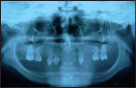
Anodontia
It is congenital absence of teeth. It is caused by hereditary ectodermal dysplasia, cleidocranial dysplasia, craniofacial dysostosis and cleft lip and palate, genetic factors, evolutionary trend towards few teeth and X-ray radiation (Fig. 1.32).29

It can be total (oligodontia) or partial (hypodontia). In partial anodontia there is absence of one or more teeth. Commonly missing are 3rd molar, maxillary lateral incisor, maxillary or mandibular 2nd premolar.
Ectodermal Dysplasia
It is also called as ‘hereditary hypohidrotic (anhidrotic) ectodermal dysplasia’. It is a X-linked, recessive mendelian character.
It is characterized by hypotrichosis, hypohidrosis and anhidrosis with saddle nose appearance. The hair of scalp and eyebrows tend to be fine, scanty and blond (Fig. 1.33).30


Supraorbital and frontal bosses are pronounced. Skin is often dry, soft, smooth and scaly with partial or complete absence of sweat glands.
Patient with this abnormality invariably manifest oligodontia (Fig. 1.34) or partial absence of teeth, with frequent malformation of any present tooth in deciduous and permanent dentition. There is reduction of the normal vertical dimension of alveolar ridge (Fig. 1.35) resulting in protuberant lips.
In dental point of view partial and complete dentures should be constructed for both functional and cosmetic purpose.
Supernumerary Teeth
It is also called as ‘hyperdontia’. It is associated with cleft palate, cleidocranial dysplasia, orofacial digital syndrome and Gardner's syndrome.32

It can be mesiodense (in the midline in the incisal (Fig. 1.36) region of maxilla between central incisors) distomolar (found in molar region frequently located distal to 3rd molar) Para molar (situated buccally or lingually to one of the maxillary molars or inter-proximally between 1st, 2nd and 3rd maxillary molars) and peridens (Fig. 1.37) (erupt ectopically, either buccally or lingually to the normal arch are referred as peridens).33

Occlusal radiograph will aid in determining the location and number of unerupted teeth.
Natal Teeth
Premature eruption of teeth or teeth like structures that are present at birth. They are hyper mobile because of their limited root development. Teeth may be conical or may be normal in size and shape and opaque yellow-brownish in color (Fig. 1.38). Teeth appear to be attached to a small mass of soft tissue.34

Fig. 1.38: Natal teeth seen in lower anterior region. Also note the ulcer seen ventral surface of tongue
Some teeth are so much mobile that there is danger of displacement and possible aspiration and in this case, removal is indicated.
STRUCTURE OF TEETH
Amelogenesis Imperfecta
It is also called as ‘hereditary enamel dysplasia’, ‘hereditary brown enamel’ and ‘hereditary brown opalescent teeth’.
It can be Hypoplastic type (there is defective formation of enamel matrix) Hypocalcification type (there is defective mineralization of formed matrix) hypomaturation type (in this enamel crystal lattice remains immature).35
Hypoplastic type (Fig. 1.39)—it appears as thin enamel on teeth that do not contact with each other mesiodistally. Horizontal rows of depressions or one large hypoplastic area with hypocalcification adjacent to and below the hypoplastic area is found.
Hypocalcified type—the enamel is so soft that it may be lost soon after eruption, leaving crown composed of only dentin. Enamel has cheesy consistency and can be scraped from dentin with an instrument or penetrated easily by dental explorer.
Hypomaturation type—the enamel can be pierced by an explorer point under firm pressure and can be lost by chipping away from the underlying, normal appearing dentin.


Snow capped teeth—in this condition varying amount of enamel on incisal or occlusal aspect of crown is present and has opaque white appearances. Opacity may be solid or flecked and may involve enamel surface. Pattern of defect on teeth anterior to the posterior teeth resemble that which would be obtained when ‘dipped into white paints’.
Radiologically squarish type of crown being devoid of the normal mesial and distal contours (Fig. 1.40). The normal enamel cap is missing and in its place, there is thin and opaque layer of enamel.
Cosmetic improvement should be done.
Enamel Hypoplasia
It is an incomplete or defective formation of organic enamel matrix. Local and systemic factors that interfere with the normal matrix formation can cause enamel surface defects and irregularities. It can occur due to nutritional deficiency, exanthematous disease, congenital syphilis, hypocalcemia, 37birth injury, local infection, trauma, fluoride, tetracycline and chronic lead poisoning.
Drinking water that contains in excess of 1 PPM (part per million) fluoride can affect the ameloblasts during the tooth formation stage and can cause the clinical entity called as ‘mottled enamel’ (Fig. 1.41). It is due to disturbance in tooth formation caused by excessive intake of fluoride, during the formative period of dentition. It frequently becomes stained as unsightly yellow to brown color, (Fig. 1.42) which is caused by coloring agents from food, medicine and by disintegration of the increase protein contain in the hypomineralized parts of the enamel.

Fig. 1.41: Hypoplasia due to fluoride(Courtesy: Dr Abhishek Soni, Lecturer, Periodontia, VSPM Dental College and Research Center, Nagpur, India)

Radiologically extensive lesion appears as a series of rounded dark shadows crossing the tooth in straight lines (Fig. 1.43).
It is managed by bleaching with 30 percent H2O2 (hydrogen peroxide)—this technique is enhanced by micro-abrasion or grinding of the surface layer.
Dentinogenesis Imperfecta
There are various names for dentinogenesis imperfecta like ‘hereditary opalescent dentin’ and ‘odontogenesis imperfecta’. It can be Shield type I (dentinogenesis imperfecta always occurs with osteogenesis imperfecta), Shield type II (it does not occur in association with osteogenesis imperfecta) and shield type III (it has got shell teeth appearances and multiple pulp exposure).39

Fig. 1.43: Enamel hypoplasia showing absence of enamel on incisors with periapical pathology(Courtesy: Dr Amit Parate, Lecturer, Oral Medicine and Radiology, GDCH Nagpur, India)
Color of teeth may vary from brownish violet to yellowish brown (Fig. 1.44). Amber translucency of both primary and permanent dentition may be seen. Enamel may be lost and dentin undergoes rapid attrition. Usual scalloping of dentinoenamel junction is absent. The neck of teeth suddenly narrows down. The appearance of crowns may be described as ‘dumpy’.
Radiologically constriction of cervical portion of tooth that imparts bullous appearance (Fig. 1.45). Partial or complete obliteration of pulp chamber. Pulp obliteration may take place before or after eruption of teeth.40


Root canals may be absent or thread like or may be blunted. In Brandywine type, enamel of tooth appears normal, while the dentin is extremely thin and the pulp chambers are enormous. Teeth appear as ‘shell teeth’. In some cases the radicular portion of pulp cavities is very narrow, while the pulp chambers have a bulbous expansion terminating in a point deep to the occlusal aspect which resembles ‘flame’.
Cast metal crown should be given.
Dentin Dysplasia
It is a rare disturbance of dentin formation, characterized by normal but atypical dentin formation, with abnormal pulp morphology. It can be radicular or coronal types. It is transmitted as autosomal dominant trait.
Affected teeth are occasionally slightly amber and translucent. Malalignment and malpositioning due to extreme mobility. Primary teeth with yellow, brown, bluish, grey-amber translucent appearances.
Radiologically tooth has short roots with sharp conical and apical constriction (Fig. 1.46). Pulp chamber and canals are obliterated before eruption and appears as half moon shaped. Obliteration of pulp chamber produce ‘crescent shaped’ pulp remnants (Fig. 1.46). In coronal type permanent teeth exhibit large chamber in coronal portion which is described as ‘thistle tube’ in shape which is due to radiating extension of pulp chamber.

GROWTH OF TEETH
Embedded and Impacted Teeth
Embedded teeth are those which are unerupted, usually because of lack of eruptive force. Impacted teeth are those prevented from erupting by some physical barrier in eruption path (Fig. 1.47).
It is caused by lack of space, rotation of tooth bud, and systemic disease like Osteopetrosis, ectodermal dysplasia, cleidocranial dysostosis, rickets and cretinism can be associated with impactions.
Teeth may be impacted distally, mesially, horizontally, etc (Figs 1.48 and 1.49). Periodontal pocket formation and subsequent infections may occur. Because of location, impacted tooth may cause resorption of roots of adjacent teeth.43


Transposition
Tooth may be found occupying an unusual position in relation to other teeth, in the dental arch, i.e. two teeth apparently exchanging their position (Fig. 1.50).
Teeth often exchange their positions. Permanent canine is most often involved, with its position interchanged with lateral incisor. Second premolar is infrequently found between first and second molar. Transposition of central and lateral incisor is rare. Transposition does not occur in primary dentition.
It can be recognized on radiograph by the unusual sequence of teeth in dental arch (Fig. 1.51).
