

- Neck nodes are important predictors of outcome in patients with head and neck cancers. The survival rates drop by 50 percent in cases of squamous cell cancers with palpable nodes.
- The primary sites most commonly involved in the spread of this carcinoma are the mucosal areas of the upper aerodigestive tract, particularly the larynx, oropharynx, hypopharynx, and oral cavity.
- The survival rate is less than 5 percent in patients who previously underwent surgery and have a recurrent metastasis in the neck. Therefore, the control of the neck is one of the most important aspects in the successful management of head and neck cancers.
- To plan the optimum management and to prognosti-cate the disease it is therefore necessary to assess the number of lymph nodes involved, their size, fixity and extracapsular spread. These features are however best understood after the final histological examination of the lymph nodes has been done making neck dissections an essential part of management of head and neck cancers.
- The assessment involves a careful attention to both sides of the neck, clinically as well as with imaging modalities in order to stage the disease and predict the outcome and plan the optimum management in these cancers.
- The term “neck dissection” refers to a surgical procedure in which the fibrofatty contents of the neck are removed for the treatment of cervical lymphatic metastases. This operation falls in to the domain of any surgeon with special interest in the region and one with a thorough understanding of the anatomy of this tightly packed area. While operating in the neck for sure, “eyes will not see what the mind does not know.” (Figs 1.1 to 1.3B).
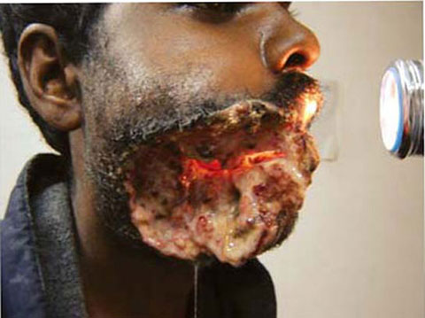
- The radical neck dissection (RND) described by Crile in 1906 and popularized by Martin and colleagues during 1950s, was followed with almost religious consistency by most head and neck surgeons until the late twentieth century, when the principles of ‘functional’ neck dissection, developed by Suárez and popularized by Bocca, Gavilán, Ballantyne, Byers and others (1960s), led to the acceptance of modified radical neck dissection as treatment for lymph node disease in various stages The main goal of RND was to remove, en bloc, the entire ipsilateral lymphatic structures from the mandible superiorly to the clavicle inferiorly and from the infrahyoid muscles to the anterior border of the trapezius.Fig. 1.1: The commonest Indian cancer is oral cancer and patients continue to present late with locally advanced cancers. This cancer is linked to the tobacco chewing habits of rural Indians i.e. of keeping the quid in the lateral gingivo-buccal sulcus. The typical “Indian oral cancer” is a lateral sulcus cancer. The large fungating and ulcero-proliferative growth is seen here with a persistent and discharging oro-cutaneous fistulaThe resection also included the spinal accessory nerve, the internal jugular vein, the sternocleidomastoid muscle, and the submandibular gland. The anatomic structures that were preserved included the carotid arteries, vagus nerve, hypoglossal nerve, brachial plexus, and phrenic nerve.
- Classic radical neck dissection is still the criterion standard for surgical control of neck metastases. It is a well-designed operation that is relatively easy for the trained head and neck surgeon to learn and perform (Figs 1.4 and 1.5).
- In the last three decades, progressive advances have occurred in the understanding of cervical fascial planes, lymphatic drainage patterns, preoperative staging, and extracapsular spread. A concern to maximize control and to minimize morbidity has prompted modifications to the classic neck dissection. One such modification (modified radical neck dissection) is the preservation of one or more non-lymphatic structures (i.e. spinal accessory nerve, internal jugular vein, sternocleidomastoid muscle) (Figs 1.6 to 1.8B).Fig. 1.6: Bilateral neck dissections (left RND and right MRND-I) performed for an extensive thyroid malignancy that had infiltrated in to the larynx. Wide local resection including total thyroidectomy with laryngectomy including anterior wall of pharynx (the nasogastric tube may be seen) has been performed for the primary tumor
- More recently, selective neck dissections (SND), involving removal of nodes confined to the levels at greatest risk of metastasis from primary tumours at various sites, have become an accepted practice for elective and, in some instances, therapeutic treatment of the neck (Fig. 1.8C).
- Sentinel node biopsy (SNB) may replace SND for patients with N0 disease in future and endoscopic neck dissections to replace the existing procedures are still under evaluation.
- It is vital to reinforce the fact that although the approach in cancer management now has shifted more and more 12towards preservation of form and function, the gold standard operation for positive necks in the hands of relatively less experienced surgeons practicing at low volume centers continues to be radical neck dissection (RND), being simple to learn, practice and teach.
- In order to avoid confusion amongst surgeons as a result of the multiple modifications to the radical neck operation and evolution of new terms to describe such changes, varying from author to author, standardization was necessary. Therefore in 1991, the American Academy of Otolaryngology-Head and Neck Surgery published an official report that standardized the terminology for the different types of neck dissections.
- In 2001, the report was updated with only a few changes, which dealt with the application of various types of selective neck dissection procedures for oral cavity cancer, pharyngeal and laryngeal cancer, thyroid cancer, and cutaneous malignancies. In addition, two new neck sublevels, Va and Vb, were added, for a total of six neck levels and six neck sub-levels. (The 1991 version of the report listed only 4 neck sublevels). With the exception of the two added neck sublevels, the terminology in the updated report is the same as that of the 1991 version. This nomenclature is widely used today.


“It is more difficult to preserve than to destroy”.
- Modified radical neck dissections (MRND) are more demanding and author would like to recommend that “whenever in doubt take it out” is the dictum i.e. RND if there is any doubt regarding oncological safety.
- A diligent effort should be made to preserve spinal accessory nerve in most of the cases, as the morbidity of drooping shoulder is generally unacceptable (Fig. 1.9).Ideally internal jugular vein must be preserved at least on one side (if bilateral neck dissection is contemplated) to prevent severe facial and cerebral edema
- The overall functional advantage of preserving the IJV has been debated extensively as nearly one fourth of the preserved veins eventually get blocked after sometime. This has been observed particularly in patients where the myocutaneous flaps are used for reconstruction of the facial defects (apparently due to pressure of the flap pedicle). The author and his group has conducted a study (Chintamani, Tushar, Bhatnagar et al. submitted for publication) to assess the incidence of thrombosis of Internal jugular vein after modified neck dissections. The results indicate a very low or negligible incidence, if a meticulous technique is followed, thus recommending MRND. Author does radical neck dissections in very selected cases with more and more modified and/or selected neck dissections being performed more often.
- The minimum acceptable surgery in any cancer is R0 resection (i.e. microscopic freedom from the disease) therefore no effort should be made to preserve at the cost of optimum clearance. In the event of lymph nodes involving or being too close for comfort to the three vital structures, preservation should take the back seat as the residual disease may compromise actual outcome.

Modifications to the Radical Neck Dissection
- Type I: The spinal accessory nerve is preserved.
- Type II: The spinal accessory nerve and the internal jugular vein are preserved.
- Type III: The spinal accessory nerve, the internal jugular vein, and the sterno-cleidomastoid muscle are preserved.
- Selected neck dissections: where only the involved or the likely to be involved lymph nodes are removed thus preserving the form and function but requiring the expertise. These are now described as SND—the levels removed, e.g. supra-omohyoid neck dissection may be described as SND-II, III, IV. The terms like anterolateral or posterolateral neck dissections were found to be a little confusing, mentioning the levels removed does help in standardizing the nomenclature.
- Extended radical neck dissection: The lymph node groups and/or additional structures not included in the classic neck dissection are resected.
The cervical node metastases may be treated with combination of surgery and radiotherapy and sometimes with 16radiotherapy alone. The controversy regarding the role of elective neck dissections in N0 necks still exists. Only two studies have shown no significant survival difference between elective and delayed neck dissections in N0 necks. These studies were however criticized on account of small number of patients. Recent studies (Yuen et al, Kligerman et al.) in patients of carcinoma tongue with N0 necks have shown distinct survival advantage with elective neck dissections.3,4
Factors that Contribute to the Risk of Neck Metastases
- Anterior portions of the oral cavity are associated with smaller risk of neck metastasis than posterior portions.
- Young patients with oral carcinoma have a higher risk of developing nodal metastasis than older patients.
- Risk of neck involvement by metastasis increases with an increase in tumor size.
- Perineural and perivascular invasion are associated with a high risk of nodal metastasis. The extracapsular spread of the nodes also carries a high probability for lymphatic spread.
- Poorly differentiated tumors are associated with a higher risk of neck metastasis than well-differentiated tumors.
Incidence and Frequency of Neck Metastases
- The rate of ipsilateral metastatic disease in patients with stage T3-T4 squamous cell carcinoma of the oral cavity, oropharynx, hypopharynx, or supraglottis is approximately 50 percent.
- The rate of bilateral or contralateral metastatic disease in these patients varies from 2–35 percent.
- Nasopharyngeal carcinoma appears as a neck mass in approximately 50 percent of patients. Metastatic neck disease in thyroid gland tumors occurs as follows: papillary, 55 percent; medullary, 50 percent; and follicular, 25 percent.
- Tumors localized in the oral cavity, oral mucosa, oropharynx, hypopharynx, and supraglottis have a higher incidence of metastasis than tumors of the superior gingiva, hard palate, and glottis.
Shah et al. reviewed 1119 radical neck dissection specimens done at the Memorial Sloan Kettering Hospital between 1965 and 198610 and following observations were made:
- The oral cavity most commonly involved nodal levels I, II, and III, with level IV (20%) and level V (4%) affected less often.
- Most oropharyngeal primaries metastasized to levels II, III, and IV with level- I involvement in 17 percent and level V disease present in 11 percent.
- The oral cavity most commonly involved nodal levels I, II, and III, with level IV (20%) and level V (4%) affected less often.
- Most oro-pharyngeal primaries metastasized to levels II, III, and IV with level I involvement in 17 percent and level V disease present in 11 percent of cases.
- The hypopharyngeal cancers involved levels II, III, and IV in the majority of cases, while levels I and V were positive for metastasis in 10 and 11 percent respectively.
- Primaries of the larynx metastasized to levels II, III, and IV most commonly, and to level I in 8% and level V in 5 percent of cases.
Further evaluation of drainage pathways can be reviewed by referring to Lindberg's 1972 paper. The stage and site of the primary tumors (T- stage) may be utilized to predict the probability of neck metastases in patients with N0 necks.
REFERENCES
- Crile G. Excision of cancer of the head and neck with special reference to the plan of dissection on one hundred and thirty-two operations. JAMA 1906;47:1780–86.
- Rigual NR, Wiseman SM. Neck dissection: current concepts and future directions. Surg Oncol Clin N Am 2004; 13(1):151–166
- Alfio Ferlito, Alessandra Rinaldo. Neck dissection: past, present and future? Journal of Laryngology and Otology 2006.
- Fakih AR, Rao RS, Patel AR, et al. Prophylactic neck dissection in squamous cell carcinoma of oral tongue: a prospective randonized study. Stud Surg Oncol 1989.
- Vandenbrouck C, Sancho-Garnier H, Chassagen D. et al. Elective versus therapeutic radical neck dissection in epidermoid carcinoma of the oral cavity. Cancer 1980; 46:386–90.
- Yuen APW, Wei WI, Wong YM, et al. Elective neck dissection versus observation in the treatment of early tongue carcinoma. Head Neck 1997; 19583–88.
- Kligerman J, Lima RA, Soares JR, et al. Supraomohyoid neck dissection in the T1/T2 squamous cell carcinoma of the oral cavity. Am J Surg 1999;168:391–94.
- Ballantyne AJ. Significance of retropharyngeal nodes in cancer of the head and neck. Am J Surg 1964;108:500–504.
- Bocca E, Pignataro O. A conservation technique in radical neck dissection. Ann Otol Rhinol Laryngol 1967;76:975–87.
- Byers RM. Modified neck dissection. A study of 967 cases from 1970 to 1980. Am J Surg 1985;150:414–21.
- Byers RM, Clayman GL, Guillamondequi OM, Peters LJ, Goepfert H. Resection of advanced cervical metastasis prior to definitive radiotherapy for primary squamous carcinomas of the upper aerodigestive tract. Head Neck 1992;14:133–38.
- Candela FC, Kothari K, Shah JP. Patterns of cervical node metastases from squamous carcinoma of the oropharynx and hypopharynx. Head Neck 1990;12:197–203.
- Carter RL, Barr LC, O’Brien CJ, Soo K, Shaw HJ. Transcapsular spread of metastatic squamous cell carcinoma from cervical lymph nodes. Am J Surg 1985;150:495–99.
- Clarke RW, Jones AS. Neck dissection for non-squamous malignancy. Clin Otolaryngol 1992;17:540–44.
- Coster JR, Foote RL, Olsen KD, Jack SM, Schaid DJ, DeSanto LW. Cervical nodal metastasis of squamous cell carcinoma of unknown origin: indications for withholding radiation therapy. Int J Radiat Oncol Biol Phys 1992;23:743–49.
- Crumley RL, Smith JD. Postoperative chylous fistula prevention and management. Laryngoscope 1975;86:804–12.
- de Langen ZJ, Vermey A. Posterolateral neck dissection. Head Neck Surg 1988;10:252–56.
- DeSanto LW, Beahrs OH. Modified and complete neck dissection in the treatment of squamous cell carcinoma of the head and neck. Surg Gynecol Obstet 1988;167:259–69.