

TATTOO MARKS
Tattooing is common among Indians in the lower social strata of society. Various pigments like indigo, carbon, vermilion, Indian ink and Prussian blue are introduced into the dermis using pointed needles. Designs and patterns are usually found tattooed over the inner aspect of forearm and the dorsum of hand. Tattoo marks, are important identification marks both of the living as well as of the dead. They especially aid the verification of identity of decomposed bodies where the facial features are grossly altered. This is because the tattoo marks remain intact even after the epidermal layer of skin has been peeled off or destroyed due to putrefactive changes. These marks establish the identity of the individual from their designs, patterns, situations and extent. They may indicate the religion of a person and are particularly useful in the identification of an exhumed body (Figs 1.1 to 1.5).


Fig. 1.4: A tattoo mark (Anjaneya Swamy) (MLI: It indicates religion and God of Worship of the deceased also)

Fig. 1.5: Tattoo mark of scorpion on forearm dermis in a body recovered from water (Cuticle of forearm is peeled off. Palmar skin and of fingers shows “washer-woman's hand” appearance)
SCARS
Scars form important marks for identification. The shape, size and site of a scar may indicate the purpose and the nature of the weapon or agent that caused it.
Scar of Surgical Incision
The scar so produced is linear and permanent as the dermis is incised during surgery. It is composed of fibrous tissue covered by epithelium. A scar produced in childhood grows in size with age. The age of a scar can be approximately determined as follows: A 5-6 days old scar appears reddish and becomes pale, soft and sensitive by two weeks. It turns brownish in two to six months and thereafter becomes pigmented, wrinkled and insensitive (Fig. 1.6).

Fig. 1.6: A linear, oblique surgical scar over the right iliac region (appendicectomy) in a female helps in identification
RADIOLOGICAL EXAMINATION FOR AGE DETERMINATION
X-ray studies of closure of skull sutures, teeth eruption and appearance of ossification centres and their fusion with their shafts are useful for estimating age and for identification.
Closure of Skull Sutures
Before 30 years of age there is absence of skull suture closure and the fusion is first seen in the inner surface of the skull than the outer surface (Figs 1.7 and 1.8).
Age of Closure of Different Skull Sutures
- Sagittal: 30 to 50 years
- Coronal: 40 to 60 years
- Lambdoid: 50 to 70 yearsFig. 1.7: X-ray of the skull and lower jaw (lateral view). The sagittal, coronal and lambdoid sutures are not closed. Age: Below 60 years
- Parieto-mastoid and squamous suture: 55 to 60 years
- Spheno-parietal: About 70 years The anterior fontanelle is closed at 1½ years of age
- Metopic suture (i.e. interfrontal): 2 to 6 years It may persist in 10% of cases
- Palate suture: 45 to 50 years.


X-ray Studies of Teeth Eruption
Lower Jaw for Third Molar Tooth
- Complete eruption of the third molar tooth (wisdom tooth) occurs at the age of 17 to 25 years.
- The third molar tooth may be impacted and unerupted.
In congenital absence of the third molar tooth space in the lower jaw behind the second molar tooth is not formed (Fig. 1.11). Eruption of the third molar tooth is more variable and uncertain.

Fig. 1.9: The crown of lower first deciduous molar tooth showing two buccal cusps and two lingual cusps separated by a fissure. The roots are resorbed prior to shedding for the eruption premolar (permanent tooth) in its place in the lower jaw at the age of the nine years

Fig. 1.10: X-ray of lower jaw for third molar tooth. The third molar tooth shows only crown formation. Age: About 12 years

Important Medicolegal Points
- When all the epiphyses of long bones of the skeleton are united, it is possible to estimate the age of the individual as above 25 years.
- All the epiphyses of wrist joint are completely fused at 18 to 20 years of age.
- The fusion of epiphyses in bones takes place two years earlier in the female than in the male.
- Within the range of two years from the appearance and fusion of ossification centres, the age of an individual can be determined.
X-ray Studies of Appearance of Ossification Centres and their Fusion
Ossification Centres of the Shoulder Joint
- Humerus (Fig. 1.12)a. Head: Appearance: 1 yearFusion to shaft: 14 to16 yearsb. Greater tubercle: Fusion of head of humerus: 4 to 5 yearsFusion to lesser tubercle: 5 to 7 years
- Scapulaa. Coracoid process: Tip appears: 10 to 11 yearsb. Acromion: Appearance: 14 yearsFusion: 18 to 20 years
- Collar boneInner end/sternal end: Appearance: 14 yearsFusion: 20 to 22 years
- Sternum
- Four middle pieces of sternum fuse with one another from below upwards between 14 and 25 years of age.
- Xiphoid process of sternum unites with the body at around 40 years of age.
- Manubrium sternii unites with the body in old age at around 60 years.

Ossification Centres of the Elbow Joint
- Distal end of humerus (Fig. 1.13)a. Capitulum: 1 yearb. Trochlea: Appearance: 10 to 11 yearsFusion: 10 to 12 yearsc. Medial epicondyle: Appearance: 6 yearsFusion: 14 to 16 yearsd. Lateral epicondyle: Appearance: 10 to 12 years
- Proximal end of radius and ulnaa. Head of radius: AppearanceFusion: 6 to 8 years: 14 to 16 yearsb. Olecranon: AppearanceFusion shaft: 9 to 10 years: 14 to 16 years
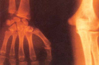
Ossification Centres of the Wrist Joint
- Epiphysis of distal end of radius: AppearanceFusion: 1 year: 16 to 18 years
- Epiphysis of distal end of ulna: AppearanceFusion: 8 to 10 years: 16 to 18 years
- Ossification of carpal bonesCapitate: 6 monthsHamate: 8 to 14 monthsTriquetrum: 2 to 3 yearsLunate: 5 yearsTrapezium: 5 to 6 yearsTrapezoid: 7 to 8 yearsScaphoid: 6 to 7 yearsPisiform: 10 to 12 years
- Ossification of metacarpal bonesEpiphysis of the base of first metacarpal bone appears at 2 to 3 years it along with and epiphysis of the head of the other four metacarpal bones unite with their shafts at 15 to 16 years of age.
Ossification Centres of Pelvis
- Epiphysis of crest of ilium: AppearanceFusion: 14 years: 18 to 20 years
- Tri-radiate cartilage in acetabulum with bone formation: Disappears/fusion: 13 to 15 years
- Epiphysis of ischial tuberosity: AppearanceFusion: 16 years: 21 to 22 years
- Ramii of pubis and ischium unites with bony union: 7 to 8 years
- Sacral vertebrae are separated by cartilages and unite with bony union one another from below upwards in 23 to 25 years.
Ossification Centres of Hip Joint
1. Head of femur | : Appearance Fusion | : 1 year : 14 to 16 years |
2. Greater trochanter | : Appearance Fusion | : 4 to 5 years : 14 to 16 years |
3. Lesser trochanter | : Appearance Fusion | : 12 to 14 years : 16 to 17 years |

Fig. 1.14: X-ray of the hip joint and pelvis. Epiphyses of the proximal end of femur are not fused completely. Inferior-ramus of the pubis is united with the ischial ramus

Fig. 1.15: X-ray of the hip joint and pelvis. Y-shaped cartilage is not obliterated and epiphyses of the proximal end of femur are not fused

Fig. 1.16: X-ray of the hip joint and pelvis. Y-shaped cartilage in the acetabulum is fused. Epiphyses of the proximal end of femur are fused
Ossification Centres of Knee Joint
1. Epiphysis of distal end of femur | : Appearance | : Before birth |
(Nearly 5 mm in diameter) | Fusion | : 14 to 16 years |
2. Epiphysis of proximal end of tibia | : Appearance Fusion | : Before birth : 14 to 16 years |
3. Patella | : | 3 to 5 years |
Ossification Centres of Ankle Joint and Foot
1. Epiphysis of distal end of tibia | : Appearance Fusion | : 1 year : 16 to 18 years |
2. Calcaneum | : Appearance | : 6 months IUL |
3. Talus | : Appearance | : 7 months IUL |
4. Cuboid | : Appearance | : 8 months IUL |
(Centres of ossification are present in calcaneum, talus and cuboid before birth)