

CELL MEMBRANE
Cell forms the building block of life. They are highly specialized in various organs to carry out specific functions, but the structures present inside, namely the organelles are identical in all types of cells (Fig. 1.1). With the advent of recent techniques in the cell biology, it is now possible to separate organelles by ultracentrifugation.
Each cell has a boundary called plasma membrane, which has a diameter of 7.5 nm or 75 A°. The membrane is made up of phospholipids and proteins. The Lipid is arranged as bimolecular layer, with the hydrophilic polar groups facing the aqueous medium on both sides of the membrane, while the nonpolar hydrophobic groups extend into the interior of the membrane. The phosphate in the lipids is hydrophilic and attached to the head, whereas, the lipid is the nonpolar and attached to the tail. Two types of proteins are present in the cell membrane namely, peripheral and structural proteins. The proteins which are attached to the polar groups of the cell membrane are globular type and they are the peripheral proteins. Peripheral proteins are present on both sides of the membrane and more on the exterior. Proteins also extend through the cell membrane forming structural proteins or integral proteins (Fig. 1.2).


Attached to the peripheral proteins on the exterior, mucopolysaccharides are present. The important constituent of this is sialic acid and its presence on the cell membrane gives negative electrical charge to the cell surface. These glycoproteins are also responsible for the antigenicity of the cell membrane.
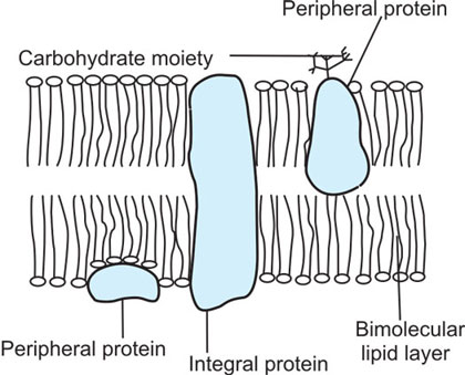
Fig. 1.2: Fluid mosaic model of cell membrane
The carbohydrate group is attached to the peripheral proteins on the outside of the membrane and form glycoproteins. They function as receptors and antigens. They also form glycocalyx which helps in intercellular connections
Fluid mosaic model
The lipids and proteins on the cell membrane are in a dynamic state, in the sense, that they are in a fluid mobile state and can change their shape and position. In addition, there is also turnover of membrane lipids and proteins.
Functions of cell membrane proteins
- The proteins serve as structural support to the membrane
- Act as antigen on the surface of the cell
- As channels or pores for the movement of ions and water
- Act as carrier proteins for the transport of solutes across the membrane
- Proteins on the surface of the cell membrane act as enzymes catalyzing chemical reactions
- roteins also act as receptors for the hormones and neurotransmitters.
CELL ORGANELLES
They are the structures present inside the cell. It includes nucleus, endoplasmic reticulum, Golgi apparatus, mitochondria, lysosomes, microsomes, microtubules and microfilaments. The detailed descriptions of these structures are given below.
Nucleus
Nucleus is surrounded by a double layered membrane with pores 70 nm size. These pores serve as transport channels for RNA to migrate to cytoplasm. Nucleus contains chromatin and one or more nucleoli. The nucleoli contain RNA, while, the chromatin has the DNA, which carries the genetic information. The structure of DNA shows two strands, which are folded into a double helical structure called chromosomes. The number of chromosomes present in the nucleus of human being is 46, which includes 22 pairs of autosomes and one pair of sex chromosomes.
DNA is about 10 to 20 nm in size and chemically made up of four nucleotides namely, adenosine, thymidine, guanosine and cystidine. Each nucleotide consists of a pentose sugar deoxy ribose, phosphoric acid residue and a side chain consisting of bases namely, adenine, thymine, guanine, and cytosine.
The sequence with which the bases are arranged forms the genetic code.
RNA is a single strand structure, which contains ribose sugar instead of deoxyribose and uracil instead of thymine base.
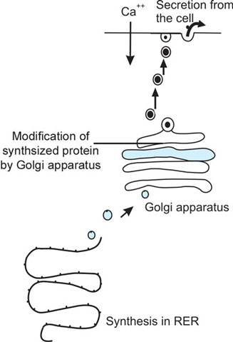
Protein synthesis depends on the genetic code present in DNA. The transfer of genetic information from DNA to the ribosomes occurs through mRNA and is called transcription. The mRNA binds to polyribosomes in the cytoplasm and assemble the amino acids with the help of tRNA. This process is called translation. The rough endoplasmic reticulum contains ribosomes on its surface and protein synthesis takes place on it. The synthesized protein undergoes post-translational modification, with cleavage of 3bonds and side chain changes. It moves to Golgi apparatus for packaging and forms secretory vesicle.
Endoplasmic reticulum
It is one of the constituents of microsomes. It is of two types namely, smooth endoplasmic reticulum (SER) and rough endoplasmic reticulum (RER). The rough endoplasmic reticulum consists of flat vesicles which are interconnected to form a network of channels throughout the cell. The surface of the RER contains ribosomes on which the proteins are synthesized.
The SER has no ribosomes on the surface and is mainly concerned with the synthesis of lipids and steroids.
Golgi apparatus
It consists of stacks of flat vesicles and is closely associated with RER. Golgi apparatus receives the synthesized protein and modifies it by concentration, processing and packaging. The concentration of the synthesized molecule occurs by the removal of water, while processing involves cleavage of the precursor molecule and addition of carbohydrate moiety. The packaging of the synthesized molecule forms the secretory vesicle. The secretory vesicle is transported to the surface of the cell guided by the microtubules and released from the cell by exocytosis.
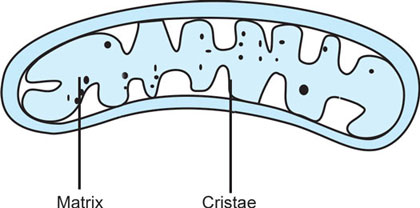
Mitochondria
It is oval shaped and has double layered cell membrane. The inner membrane has been thrown into folds called cristae which increase the surface area. The cristae includes the matrix (Fig. 1.3). The respiratory enzymes are present in the cristae while the matrix contains the nuclear material DNA and RNA. The RNA is involved in the synthesis of enzymes of oxidative phosphorylation, while the DNA facilitates mitochondrial replication. The mitochondria is called the power house of the cell, as the energy rich compound ATP is synthesized from them. The number of mitochondria in a cell varies in relation to its metabolic activity. The cells that are concerned with the secretion and those that are metabolically active, such as liver cells, will have more number of mitochondria.
Lysosomes
They are the vesicles containing hydrolytic enzymes. They are formed from endoplasmic reticulum and Golgi apparatus. The main function of lysosomes is in the digestion of the engulfed material. The substances that are engulfed into the cell by pinocytosis or phagocytosis, fuse with the lysosomes and form the digestive vacuole. The lysosomes which are not attached to the phagocytic vacuole are known as primary lysosomes and those which are attached are called secondary lysosomes. The acid hydrolases in lysosomes digest the engulfed material and the indigestible ones are in the residual body. It is released from the cell by exocytosis. The lysosomes also can digest the breakdown products of cell's own organelles. In certain pathological conditions such as gout, rheumatoid arthritis, the release of lysosomal enzymes is considered to be the causative factor in the pathogenesis of the disease.
Lysosomal storage diseases such as, Gaucher's disease, Fabry disease and Tay- Sachs disease are due to congenital absence of lysosomal enzymes. The absence of β-galactocerbrosidase causes Gaucher disease while lack of α-galactocerebrosidase results in Fabry's disease.4
Peroxisomes
It is one of the constituents of microsomes and can be obtained by ultracentrifugation of the cell organelles. The peroxisomes are more in number in liver and kidney. They contain an enzyme called catalase which causes breakdown (detoxification) of hydrogen peroxide. They are also involved in theβ oxidation of lipids and thereby regulate their level in the plasma.
Cytoskeleton
The cell contains fibers which include microtubules, intermediate filaments and microfilaments. These are known as cytoskeletal structures. They give structural stability to the cell and produce cell movement.
Microtubules
The microtubules give shape to the cell. They facilitate the transport of secretory vesicles and organelles to the cell surface. The microtubules are in a dynamic state as they can assemble and disintegrate in various conditions.
Microtubules are considered as cytoskeletal structures and they are made of 2 globular proteins, α and β tubulin. During cell division, thecentrosomes produce a third subunit γ tubulin, which helps in the movement of chromosomes. These subunits of tubulin combine with proteins like dynein, kinesin and dynamin to give various functions. The movements of chromosomes during cell division and anterograde transport in the axon involve the tubulin interaction with dynein. The retrograde transport in the axon is due to the tubulin interacting with kinesin, while the sliding of microtubules with one another is by the interaction of dynamin with tubulin.
Within the cell, the proteins and cell organelles move with the help of molecular motors, which include kinesin, dyneins and myosins.
Microfilaments
Microfilaments are made of actin and their interaction with myosin I cause cell movement. Most of the cells contain actin at the cell surface. The interaction of actin with myosin II gives muscle contraction.
Cilia
Cilia and flagella are the cell processes surrounded by the extensions of plasma membrane. The intercellular structure consists of microtubules. There is an outer 9 units of microtubules and an inner pair present in the center. The movement of cilia or flagella is caused by the sliding of microtubules.
Cell adhesion molecules
Cell adhesion molecules(CAMs) are proteins, which facilitate attachment of the cell to basal lamina and between cells. Some of these molecules also project into the cell and are attached to the cytoskeleton. CAMs are broadly divided into integrins, IgG superfamily of immunoglobulins, cadherins and selectins.
These protein molecules assist intercellular connections, cell movement and transfer information between cells. The integrin molecules help in the attachment of cell to the extracellular matrix and in the absence of this molecule the cells tend to show death (apoptosis) earlier. CAMs are also involved in the patho-physiology of inflammation, wound healing and in the metastasis of tumors.
INTERCELLULAR CONNECTIONS
The cells are interconnected mainly in three ways namely tight junction, gap junction and desmosome (Fig. 1.4).
Gap Junction
It provides gaps or channels from one cell to another and allows the passage of small molecules and ions.5
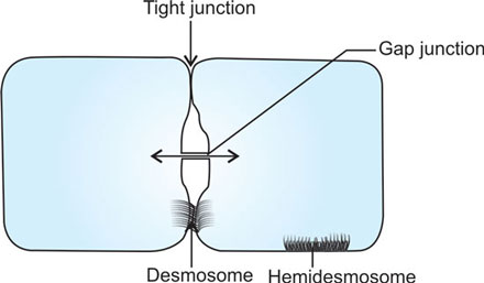
In smooth muscles and cardiac muscle, the electrical excitation spreads to adjacent cells by the movement of ions through the gap junctions.
Tight junction
It is a connection between two adjacent cells at the apical surface. The fusion of the lateral borders at the apical surface forms a barrier against the entry of substances. This kind of intercellular connection is seen in the intestinal epithelial cells, renal tubular cells and choroid plexus.
Desmosome
Two adjacent cells are held together by the electron dense fibrous substance in the extracellular space. There are intermediate filaments projecting from each cell, at the point of cell adhesion. The usual width of this connection is 20 nm. This type of connection between two cells helps to overcome mechanical stretching that occurs in the tissues like muscle and skin.
TRANSPORT MECHANISMS
The plasma membrane forms the boundary between the cell interior and exterior. The membrane shows transport of substances across the membrane in both ways.
Substances and ions move into the cell for metabolism, to give excitability of tissues and finally to maintain cell volume. There is also movement of substances from the cell into the exterior, for the sole purpose of maintaining homeostasis (constancy of internal environment).
Endocytosis
The uptake of molecules into the cell occurs by a process called endocytosis. The molecule at first fuses with the cell membrane and invaginates. The invagination is separated from the cell membrane and becomes a vesicle.
If fluid with the dissolved substances are engulfed into the cell, it is pinocytosis and the vesicle inside the cell is called pinocytic vesicle.
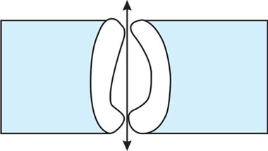
If particulate matter and bacteria are engulfed into the cell, it is known as phagocytosis (Fig. 1.5). The vesicle inside the cell forms the phagosome.
In pinocytosis, the substances that are taken in to the cell include insulin, viruses, growth factors and low density lipoproteins. The process is mediated through receptors present on the cell surface.

They are also other proteins namely clathrin, actin and myocin present beneath the receptors, which help the pinocytic vesicle to pinch off from the cell membrane. The process of endocytosis requires the supply of ATP as it is an active process.
During endocytosis process a part of the cell membrane is removed which is compensated by the addition of membrane to the cell surface during exocytosis when the secretory vesicle fuses with the cell membrane. This explains how the surface area of the cell membrane remains constant when these processes occur.
Exocytosis
Exocytosis refers to the release of substances from the cell. It could be the proteins synthesized by the RER or the cell organelles that are disintegrated. The synthesized proteins form the secretory vesicle and are guided by microtubules to the cell surface. The membrane of the vesicle and plasma membrane fuse and the contents of the vesicle are extruded out (Fig. 1.6). This process requires Ca++ entry into the cell and supply of energy.
Transcytosis
Transcytosis also can be observed in the transport across the membrane. It refers to the movement of macromolecules (protein hormones) from one side of the cell to the other side unchanged. This kind of transport is seen in endothelia.
Passive transport
When there is a concentration gradient across the membrane, diffusion of water and solutes occur. The diffusion will be from a region of higher concentration to a region of lower concentration (Fig. 1.7).
The process of diffusion depends on:
- Membrane permeability for the diffusing substance
- Concentration gradient
- Size of the molecule in relation to the pores
- Cross section of the diffusing area
- Temperature
- Distance involved during diffusion.
Diffusion of solute
Diffusion of solutes across the membrane follows Fick's law. It states that the rate of 7diffusion (J) is directly related to the cross sectional area (A) through which diffusion takes place and concentration gradient (C/X)

D = diffusion coefficient and– sign denotes the direction.
Diffusion of solvent
Osmosis
Water molecules move down along the concentration gradient through a semipermeable membrane and this process is called osmosis (Fig. 1.8). It is the movement of solvent into a region of greater concentration of solute to which the membrane is not permeable. Examples of water diffusion by osmosis are seen in renal tubules, intestinal mucosa, between cells and exterior, between capillaries and interstitial fluid. The process of osmosis creates hydrostatic pressure in the region to which the solvent diffuses. The movement of solvent can be prevented by applying pressure over the region of higher solute concentration. This pressure is known as osmotic pressure. It depends on the number of solute particles and in an ideal solution, the osmotic pressure (p) is related to:

n = number of solute particles
R = gas constant
T = absolute temperature
V = Volume
In our body such an ideal solution does not exist. Hence the osmotic pressure in the body fluids depends on the number of solute particles per unit volume of fluid.
Passive diffusion of water occurs whenever there is active solute movement. The concentration of solute in a fluid gives the tonicity. If the solute concentration is more, the tonicity of the fluid is referred as hypertonic and conversely, decreased solute concentration in a fluid gives rise to less tonicity and it is known as hypotonic. If fluids from two different compartments show the same tonicity, it is said to be isotonic. In isotonic state, there is no movement of water on either side of the cell membrane due to the osmotic equilibrium.
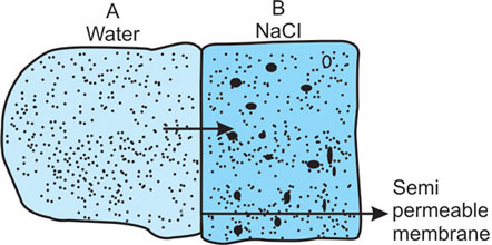
Fig. 1.8: Osmosis
Movement of water from A to B occurs through the semipermeable membrane as the solute (NaCl) concentration in B is greater
In mammals, solutions of 0.9% NaCl are isotonic to plasma. The osmotic concentration of solutes in plasma is 290 mEq/L. This is equal to the freezing point of plasma −0.54° C. The osmotic concentration of solutes in plasma is known as osmolality and is due to the presence of osmotically active solutes like Na+, C1−, HCO−3 urea and other solutes. Plasma proteins contribute very little to plasma osmolality, inspite of being present in greater amounts.
Plasma osmolality is altered when there is electrolyte and fluid disturbance in the body. In severe dehydration, hyperosmolality results. In renal failure, the retention of urea in plasma leads to hyperosmolar state, which results in coma.
Solvent drag
Whenever solvents diffuse, they tend to drag some solute with it. This is called solvent drag, and can be observed in capillaries and renal tubules.
Carrier mediated transport
A substance that cannot diffuse through the pores or leaky channels in the membrane can be transported by a carrier across the membrane.8

Fig. 1.9: Facilitated diffusion of glucose
Facilitated diffusion of solutes like glucose occurs by the conformational change of carrier protein in the cell membrane
Lipid soluble substances and respiratory gases can easily diffuse through the membrane. There are also substances of low molecular size, which can pass through the leaky channels. The solutes that cannot pass through these pores depend on the carrier proteins to transport them. If the transport occurs along the concentration gradient, it becomes a facilitated diffusion. It is a downhill transport, because, the transport does not require energy.
Facilitated diffusion is determined by:
- Concentration gradient
- Saturation of carrier protein.
The carrier protein shows:
- Specificity
- Competition
- Inhibition.
Facilitated diffusion can be seen in the transport of glucose and amino acids from the lumen of the renal tubule to the PCT cell and also from the lumen of intestine to the mucosal cell (Fig. 1.9). The carrier protein which transports glucose is called glucose transporter.
Cotransport
There are instances where two solutes are transported together by the carrier and such transport is called symport or cotransport. Examples of this transport are glucose transporter (SGLUTI) which transports both glucose and Na+ from the lumen of intestine to the mucosal cell (Fig. 1.10). Amino acids are also transported as symport with Na+ in the intestine. Similar cotransport also exists in the renal tubules.
In cotransport, the movement of solutes occurs by facilitated diffusion.
Active transport
Active transport of solutes occurs from a region of lower concentration to a region of higher concentration. This requires a carrier protein and the supply of energy, as it is an uphill transport (Fig. 1.11). Examples of active transport include Na+ pump, secretion of H+ from the parietal cells of gastric glands, iodide transport in the thyroid gland acinar cells, etc.

Fig. 1.10: Cotransport (symport)
In intestinal mucosa and renal tubules glucose enters the cell by cotransport with Na+. The carrier protein is common to both. The attachment of both the solutes to the carrier molecule brings about conformational change, which causes the release of glucose and Na+ into the cell interior

Fig. 1.11: Active transport mechanism by Na+- K+ ATPase. The unit of the carrier protein has five binding sites. 1. Na+ binding site; 2. K+ binding site; 3. Ouabain binding site; 4. Phosphorylation site; 5. ATP binding site.
Sodium is actively transported from the interior of the cell to the exterior by the enzyme Na+-K+ ATPase. The attachment of sodium and potassium occurs to the α subunit of the carrier molecule which is a larger globular protein spanning through the cell membrane. On the inner side of the α subunit, 3 binding sites for Na+ and on the outer side 2 binding sites for K+ are present. The α subunit also functions as ATPase on the inside of the protein, close to the sodium binding site. The function of the other smaller globular protein β is not clear.
At the intracellular surface, the carrier protein is phosphorylated causing ATP to hydrolyse into ADP and Pi. This causes attachment of 3 molecules of sodium and the conformational changes bring sodium molecules to the exterior of the cell membrane. Dephosphorylation of the carrier protein causes attachment of 2 molecules of K+ and the conformational change of the carrier protein brings the K+ molecules to the cell interior. Sodium pump is also known as electrogenic pump, as it causes more positivity on the outside of the membrane. Cardiac glycoside ouabain inhibits sodium pump by acting on the carrier molecule on the outside of the membrane. Sodium pump is also inhibited by metabolic poisons like dinitrophenol.
Secondary active transport
Active transport of Na+ by the Na+-K+ ATPase carrier across the basolateral border of the intestinal mucosal cells and renal tubules, results in entry of glucose into the cell. As said above, glucose and Na+ are cotransported from the lumen into the cell. The active transport of Na+ in the basolateral surface provides the chemical gradient for further movement of Na+ and glucose into the cell. Glucose transport into the cell forms the secondary active transport, as it depends on the active transport of Na+ at the basolateral surface (Fig. 1.12).

Fig. 1.12: Secondary active transport
The entry of glucose into the cell depends on the primary active transport of Na+ at the basolateral border
Counter transport (Antiport)
Antiport
The solutes, when exchanged in the opposite direction by the carrier protein, form the counter transport or antiport (Fig. 1.13). Examples are:
- Na+– H+ exchange in the renal tubes
- Na+– K+ exchange (Na+ pump)
- HCO3−– C1− exchange (chloride shift)
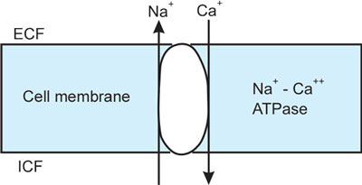
- Na+– Ca++ exchange (heart and brain)
Gibbs Donnan's equilibrium
Donnan and Gibbs have shown that the sum of anions and cations on either side of the cell membrane are equal, eventhough their distribution across the membrane varies due to the selective permeability of the membrane. It is known that unequal distribution of ions gives electrical potential. The cations and anions on either side of the membrane are distributed in such a way that in each compartment their total number will be equal. This is called Donnan's electrochemical equilibrium.

Examples of Donnan's ionic equilibrium can be observed in chloride shift which occur in the transport of CO2. The exit of anion HCO−3 from RBC disturbs the electrochemical equilibrium. But, it is set right by the movement of another anion Cl− from plasma into the RBC.
The entry of Na+ into the cell through leaky channels of the membrane will also disturb Donnan's ionic equilibrium. But, the Na+ pump mechanism does not allow this to happen. In addition to maintaining the electrochemical equilibrium, the Na+ pump maintains the cell osmolality and cell shape.
Membrane potential
The unequal distribution of ions across the cell membrane, selective permeability of the cell membrane for K+ and the electrogenic Na+– K+ pump creates potential difference between inside and outside of the cell membrane. In excitable tissues namely nerve and muscle this electrical potential is significant for excitation of the cell. In these tissues the electrical potential does not alter the Donnan's ionic equilibrium as only a small number of ions are involved in the genesis of membrane potential.
CHEMICAL MESSENGERS
Communication from one cell to another usually takes place by the chemical messengers. These substances can be proteins, amino acids or lipids. Chemical messengers enter the cell through gap junctions, paracrine, endocrine and neural communications. There is also cell's own secretion that can act on itself which forms the autocrine secretion.
Chemical messengers produce their effects by acting on specific receptors situated on the surface of the cell. They form the first messengers or ligands. The intracellular effects are mediated through the release of other chemical substances known as second messengers and third messengers.
The examples of second messengers are:
- Cyclic AMP
- Inositol tri phosphate (IP3)
- Diacyl glycerol (DAG)
- Ca++
How are second messengers formed?
Second messengers are formed through a nucleotide regulatory protein called G protein. When a first messenger (ligand) binds to the receptor on the cell membrane, the ligand receptor (Gs) complex converts GTP to GDP and activates adenylyl cyclase, if cyclic AMP is formed as second messenger. The binding of the ligand with the Gi inhibits the adenylyl cyclase activity and prevents the formation of cyclic AMP. Cyclic AMP activates protein kinase A, which phosphorylates proteins, leading to the physiological effects (Fig. 1.14). The cyclic AMP is inactivated by the enzyme phosphodiesterase, which converts 3’ 5’ AMP to 5’ AMP and it is inactive. If this enzyme is inhibited, the activity of cyclic AMP can be prolonged. Substances like caffeine, theophylline show such effect when administered.
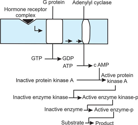
Fig. 1.14: Formation of cAMP and its action as second messenger
Activation of protein kinase A by cAMP causes phosphorylation of enzymes leading to its activation. The effect of the hormone occurs through the metabolic regulation of these enzymes or the activation of protein kinase A can lead to phosphorylation of protein, leading to protein synthesis
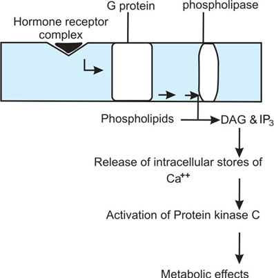
Fig. 1.15: Mechanism of action of DAG and IP3 as second messenger
The membrane phospholipids release Diacylglycerol and Inositol tri phosphate by the action of phospholipase. The DAG and IP3 that are formed release Ca++ from the intracellular stores such as mitochondria and endoplasmic reticulum. The rise in intracellular Ca++ and DAG activates Protein kinase C enzyme, which phosphorylates enzymes and regulates metabolic effects
Cyclic GMP
Similar to cyclic AMP, there is another nucleotide known as guanosine mono phosphate (cyclic GMP), which functions as the second messenger. Atrial natriuretic peptide (ANP) and nitric oxide (NO) mediate their effects through cyclic GMP. In the retina, during excitation of rods, GMP is converted to 5 GMP which is responsible for the photochemical changes.
IP3 and DAG
In the formation of IP3 and DAG as second messengers, the ligand receptor (Gs) complex converts GTP to GDP and activates phospholipase enzyme. This facilitates the formation of IP3 and DAG from the phosphorylation of phosphatidyl inositol released from cell membrane.
The second messengers such as IP3 and DAG cause rise in the intracellular Ca++ by promoting its release from the Ca++ stores such as endoplasmic reticulum, mitochondria and also by facilitating its entry into the cell from ECF, through the opening of Ca++ channels (Fig. 1.15). The intracellular Ca++ binds to the calcium binding protein called calmodulin, which activates various kinases such as myosin light chain kinase, phosphorylase kinase, etc. These kinases produce the physiological effects. The rise of Ca++ within the cell mediates such effects and hence, Ca++ forms the third chemical messenger. The rise in intracellular Ca++ also activates protein kinase C, which leads to the activation of many enzymes in the cell. Through this, the actions of the ligand can also occur.
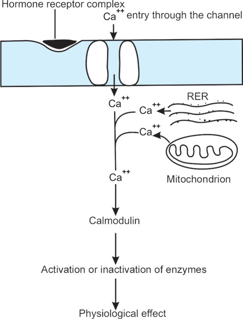
Ca++ as second messenger
The binding of ligand with the receptor on the cell surface, opens up the Ca++ channels, facilitating its entry into the cell. Inside the cell, the calcium binds to a protein calmodulin and this complex causes activation or inactivation of enzymes. Through these changes, the physiological effects of the ligand can be observed (Fig. 1.16).
Ligands mediating their effects through Cytoplasmic and nuclear receptors
Hormones such as T3 (tri iodo thyronine) has receptors in the nucleus. The hormone after binding to the receptor in the nucleus, stimulates transcription of DNA in the chromatin. This leads to mRNA formation, which leaves nucleus and enters the endoplasmic reticulum. Here, new proteins are synthesized by translation. The synthesized protein can be an enzyme, secretory protein, structural protein, or receptor protein. The biological effects of the ligand are mediated through these new proteins (Fig. 1.17).
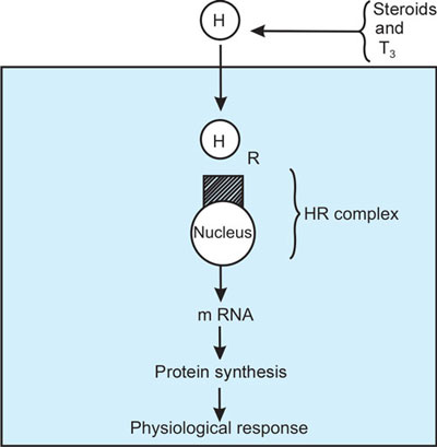
Fig. 1.17: Ligands mediating their effects through intracellular and nuclear receptors
Steroids have their receptors situated in the nucleus. The receptors are attached to a protein called Heat shock protein (HSP). This prevents the receptors getting attached to DNA and promote transcription, when the hormone is not present. When the hormone is attached to the receptor, the hormone receptor complex releases the heat shock protein. The release of HSP increases the affinity of the receptor for DNA and they are called HRE. (Hormone Response Elements). Thyroid hormones have their receptors in the nucleus. There is no HSP in the thyroid homones but the HRE are formed after the hormone binds to the receptor. This causes transcription and then translation leading to synthesis of proteins. Steroids have generally receptors in the cytosol or nucleus. The hormone receptor complex move to the DNA and produce changes as described for T3
Receptors and its Regulation
As described earlier, the cell membrane has structural proteins, which project on the outer and inner surface of the membrane. It is the peripheral proteins present on the outer surface of the membrane which function as receptors for ligands and neurotransmitters. These receptors are not static structures, as they are dynamic and mobile on the cell membrane. Their number is also not constant, as there is addition and removal to their numbers during the process of regulation.
The regulation of receptors number is done by the ligand concentration in the ECF. If more amount of ligand is present, the number of receptors decreases which is called down regulation. On the other hand if the ligand concentration is less in the ECF, the receptor number increases on the cell membrane and it is known as up regulation.
Whenever a ligand binds to a receptor, ligand receptor complex is formed, which moves laterally on the membrane and form coated pits. These coated pits are pinched off from the membrane and taken inside the cell by endocytosis. This process is called internalization of receptors. 14Lysosomes fuse with the endocytosed vesicle and release carbohydrate, amino acids and lipids into the cytoplasm, which are utilized for cell metabolism.
ION CHANNELS IN THE CELL MEMBRANE
Ion channels are formed in the cell membrane by the integral proteins. These channels are guarded by gates, which open and close, regulating the movement of ions. The gates are regulated either by voltage or chemical ligands. Accordingly, the channels are called voltage gated and ligand gated channels respectively.
Study of channels
Ion channels can be studied by patch clamp technique (Fig. 1.18). A microelectrode is kept on the surface of the membrane and a high resistance seal is applied around the tip of the microelectrode. By a suction force, a patch of cell membrane containing a few ion channels is taken inside the seal. The activity of the ion channels can be studied in situ or the small part of the membrane is removed and kept in solutions of known ion concentration to study the channel activity.
Sodium channel
It is a tetramer with four units. Each unit crosses the membrane six times. The pores with 0.5 nm diameter is present surrounding the subunits. The channel is guarded by gates, which are present in both inner and outer sides of the membrane. On the ECF side, there is activation gate. On the inner side of the membrane, there is an inactivation gate. For sodium to enter inside, both the gates should be in the open state (Fig. 1.19). The sodium channels are blocked by poisons like tetrodotoxin and saxitoxin.
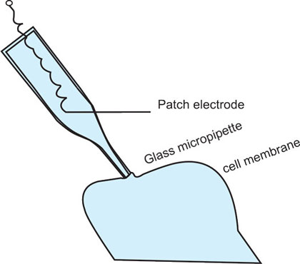
Fig. 1.18: Diagram to show patch clamp of membrane channels
A glass micropipette is kept on the cell membrane and a piece of membrane containing a few channels is sucked into the micropipette and the edges of the pipette form the seal from the rest of the membrane. The electrode measures the current flow through the ion channels present in the patch of the membrane sucked into the glass micropipette
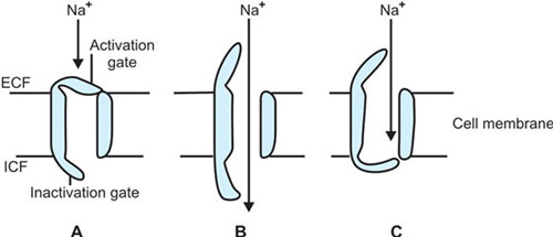
Figs 1.19A to C: Voltage gated Na+ channel A. Resting (− 90 mv) B. Activated (− 90 mv to + 40 mv) C. Inactivated (+ 40 mv to − 90 mv)

Potassium channels
The structure of the potassium channel is similar to the sodium channel and they are the voltage gated channels. There is another type of K+ channel which has only two subunits and crosses the membrane twice. These are called inward rectifier potassium channels which allow K+ entry and not its exit. Unlike Na+ channel, K+ channel (voltaged gated channel) has only one gate and it is present on the inside of the membrane. There is no inactivation gate for the potassium channel. The gate should open for K+ movement (Fig. 1.20). The voltage gated potassium channels can be blocked by chemicals like TEA (tetra ethyl ammonium) and 4 - amino pyridine. Ligand gated potassium channels also exist and activated by:
- Acetylcholine
- Ca++
- Arachidonic acid
- ATP
Calcium channels
Calcium and chloride channels are similar to sodium channel in their structure. The ligand gated Ca++ channel is activated by hormones and neurotransmitters. The voltage gated Ca++ channel are of three types namely:
- Long lasting (L)
- Transient (T)
- Neuronal (N)
The long lasting voltage gated Ca++ channel is significant in the cardiac muscle and it is blocked by verapamil, nifedipine and diltiazem drugs. These drugs are extensively used in the treatment of cardiovascular diseases.16
Self-study Questions
Multiple Choice Questions
Choose the single best answer
- The cell membrane proteins function as all of the following except:
- Transporter
- Ligand
- Receptor
- Channels
- The carbohydrate attachment to the peripheral proteins on the outer surface of the plasma membrane shows all of the following except:
- Antigenicity
- Surface electrical charge
- Intercellular connections
- Carrier
- Endoplasmic reticulum in a cell perform all of the following functions except:
- Granular endoplasmic reticulum shows protein synthesis
- In the skeletal and cardiac muscle granular endoplasmic reticulum forms sarcoplasmic reticulum
- Lipid and steroid synthesis occur in the smooth endoplasmic reticulum
- In the granular type endoplasmic reticulum translation of mRNA occurs on the ribosomes.
- Peroxisomes functions include:
- Transport
- Packaging
- Detoxification
- Lipid synthesis
- Molecular motors include all of the following except:
- Dynein
- Centrosome
- Actin
- Myosin
- The protein which is attached to the basal lamina and between cells include:
- Microfilaments
- Cell adhesion molecules
- Microtubules
- Molecular motors
- The process of pinocytosis does not include the entry of which of the following into the cell?
- Viruses
- Growth factors
- Bacteria
- Insulin
- Which of the following does not require a channel protein for its transport?
- Oxygen
- Sodium
- Water
- Chloride
- Which of the following would decrease the rate of diffusion of substance across cell membrane?
- Increase in temperature
- Increase in membrane permeability
- Decrease in molecular size
- Decrease in concentration gradient
- The function of tRNA in the cell include:
- Transcription
- Translation
- Replication
- Transduction
- In cotransport, the solutes are transported by:
- Diffusion
- Facilitated diffusion
- Active transport
- Counter transport
- Which one of the following is an example of transport against electrochemical gradient?
- Diffusion
- Facilitated diffusion
- Active transport
- Secondary active transport
- Channels in the cell membrane are formed from:
- Peripheral protein
- Integral protein
- Glycoprotein
- Bilipid layer
- The example of secondary active transport includes:
- Sodium
- Water
- Chloride
- Glucose
- The plasma oncotic pressure is due to the presence of:
- Sodium chloride
- Glucose
- Albumin
- Fibrinogen
- The plasma osmolality is due to all of the following except:
- Urea
- Glucose
- NaCl
- Albumin
- Which one of the following is not an example of counter transport?
- Na+- Glucose
- Na+ - Ca++
- HCO3− - Cl−
- Na+- K+
- The hormone which mediates its effects through cGMP is:
- Vasopressin
- Oxytocin
- Atrial natriuretic peptide
- Angiotensin II
- Voltage gated sodium channels can be blocked by:
- Tetrodotoxin
- α bungarotoxin
- Tetraethyl ammonium
- δ-tubocurare
- The increase in the intracellular Ca++ level can occur due to release of Ca++ from all of the following except:
- Sarcoplasmic reticulum
- Mitochondria
- Calmodulin
- Endoplasmic reticulum
ANSWER KEYS
1. (B) | 2. (D) | 3. (B) | 4. (C) | 5. (B) | 6. (B) | 7. (C) | 8. (A) | 9. (D) | 10. (B) |
11. (B) | 12. (C) | 13. (B) | 14. (D) | 15. (C) | 16. (D) | 17. (A) | 18. (C) | 19. (A) | 20. (C) |
Short Answer Questions
- List the functions of cell membrane proteins.
- Describe the process of protein synthesis and its secretion from the cell.
- Describe the function of lysosomes and explain its pathophysiology.
- List the cytoskeletal proteins and their functions.
- Describe the functions of cell adhesion molecules.
- Describe the process of pinocytosis and phagocytosis with examples.
- List the factors that influence diffusion.
- List the differences between facilitated diffusion and active transport.
- Describe carrier mediated transports with examples.
- Define uniport, symport, and antiport processes in the cell.
- What is the normal osmotic concentration of plasma?.
- Describe Donnan's electrochemical equilibrium.
- What are second messengers ? Describe how are they produced.
- Name the hormones that mediate their effects through c AMP and cGMP.
- Name the ligands that mediate their effects through IP3 and DAG.
- Explain the process of down regulation and up regulation of receptors.
- Name the agents that block the voltage gated channels of sodium, potassium and calcium