


DEFINITION
Gingiva is the part of oral mucosa that covers the alveolar processes of the jaws and surrounds the necks of the teeth.
MACROSCOPIC FEATURES
Anatomically, the gingiva is classified into three distinct domains: Free/marginal gingiva, attached gingiva and interdental gingiva (Fig. 1.1).
- Free gingiva
- Interdental gingiva
- Attached gingiva
- Marginal gingiva is the terminal edge of the gingiva surrounding the teeth in a collar – like fashion. The marginal gingiva is called free as it is not attached to the underlying periosteum of alveolar bone. The gingival margin is demarcated from attached gingiva by an indentation called as free gingival groove which is positioned at a level corresponding to the level of the cementoenamel junction (CEJ). Free gingival groove is only present in about 30–40% of adults. Functionally, the marginal gingiva forms the soft tissue wall of the V - shaped gingival sulcus. Gingival sulcus is a shallow groove between the tooth and normal gingiva that extends from the free surface of the junctional epithelium coronally to the level of free gingival margin.
- Interdental gingiva is the part of the gingiva which is present in the interdental space beneath the area of tooth contact. In the presence of diastema the interdental papilla is absent (Fig. 1.2).Col is a valley like depression which connects the facial and lingual papillae and conforms to the shape of the interproximal contact areas (Fig. 1.3). Col epithelium is identical to junctional epithelium having the same origin (from dental epithelium), non-keratinized and gradually replaced by continuing cell division.
- Attached gingiva is firm, resilient and tightly bound to the underlying periosteum of alveolar bone and cementum by connective tissue fibers. The attached gingiva is thus, firmly entrenched between two movable structures – the marginal gingiva coronally and the alveolar mucosa apically.


Width of attached gingiva: It is the distance between mucogingival junction and the projection on the external surface of the bottom of gingival sulcus/periodontal pocket. The dimensions of the attached gingiva vary from the anterior to the posterior teeth. Width of attached gingiva on facial aspect of – maxillary incisor region is 3.5 to 4.5 mm, mandibular incisor region is 3.3 to 3.9 mm, maxillary first premolar is 1.9 mm and mandibular first premolar is 1.8 mm approx. Width of attached gingiva increases with age and in supraerupted teeth. This increase in dimension occurs as a result of an increase in the height of the alveolar process which in turn is the result of passive eruption.
Significance of attached gingiva:
- It gives support to the marginal gingiva.
- It provides attachment or a solid base for the movable alveolar mucosa for the action of lips, cheeks and tongue.
- It can withstand frictional and functional stresses of mastication and toothbrushing. When the marginal tissue is alveolar mucosa, it does not resist the functional stresses of toothbrush trauma imposed on it. Frequently, the result is apical shifting of the marginal tissue and additional recession. Attached gingiva has more densely organized connective tissue and is more firmly bound to the underlying periosteum and bone. Consequently, it is more resistant to the functional stresses placed upon it. Alveolar mucosa is thin, delicate tissue, poorly attached to bone and cementum and is not capable of withstanding these same functional stresses.
- Attached gingiva acts as a barrier for passage of inflammation. A tooth having alveolar mucosa at its margin seems to show clinical signs of inflammation in the presence of microbial flora more readily than does a corresponding tooth that has a sufficient band of attached gingiva. Such marginal tissue appears to be more susceptible to the products of inflammation that may result in pocket formation or apical migration of both attachment apparatus and marginal tissues.
- It provides resistance to tensional stresses. Attached gingiva serves as a buffer between the mobile free gingival margin and mobile alveolar mucosa. There are skeletal muscle fibers within the alveolar mucosa that exert a force in an apical direction on the attached gingiva. This force is dissipated by bound down keratinized tissue.
Width of attached gingiva can be measured:
- Anatomically: Stretch the lip/cheek to demarcate the mucogingival line while pocket is being probed. Measure the total width of gingiva (gingival margin to mucogingival line) and subtract the sulcus/pocket depth from it to determine width of attached gingiva.
- Functionally:
- Tension test: Stretch the lip or cheek outward and forward to mark mucogingival line. Measure the total width of gingiva (gingival margin to mucogingival line) and subtract the sulcus/pocket depth from it to determine width of attached gingiva.
- Roll test: Push the adjacent mucosa coronally with a dull instrument to mark mucogingival line. Measure the total width of gingiva (gingival margin to mucogingival line) and subtract the sulcus/pocket depth from it to determine width of attached gingiva. A more reliable method of identifying the mucogingival junction would be to take the side of a periodontal probe or similar blunt instrument and jiggle the alveolar mucosa in an apicoronal direction. Since the alveolar mucosa is mobile, it will roll up ahead of the blunt instrument.
- Histochemically: Iodine staining test: Paint the gingiva and oral mucosa with Schiller's or Lugol's solution (iodine and potassium iodide solution). The alveolar mucosa takes on a brown color owing to its glycogen content, while the glycogen free, attached gingiva remains unstained. Measure the total width of the unstained gingiva and subtract the sulcus/pocket depth from it to determine width of attached gingiva.The other dimension that may play a significant role in the maintenance of the periodontal health is the thickness of the gingiva. Gingival phenotype or biotype has been classified by Eger and Muller into thick and thin or Class I, IIA and IIB. Thick gingival phenotype seems to be more conducive to periodontal health. A thin phenotype predisposes to gingival recession and increased tendency to gingival inflammation (Fig. 1.4).
Mucogingival junction is the interface between the apically located alveolar mucosa and the coronally located attached gingiva which remains stationary throughout life. Mucogingival junction is present on the three gingival surfaces namely facial gingiva of the maxilla, facial and lingual gingiva of the mandible. The palatal gingiva of the maxilla is continuous with the tissue of the palate, which is bound down to the palatal bones. Because the palate is devoid of freely movable alveolar mucosa, there is no mucogingival junction.

MICROSCOPIC FEATURES
Histologically, gingiva is composed of gingival epithelium, epithelium-connective tissue interface and underlying connective tissue.
Gingival Epithelium
The gingival epithelium is comprised of oral epithelium, sulcular epithelium and junctional epithelium (Fig. 1.5 and Table 1.2).
- Oral epithelium/outer epithelium: It covers the crest and outer surface of marginal gingiva and surface of the attached gingiva. It is a keratinized stratified squamous epithelium.
TABLE 1.2 Differences between oral, sulcular and junctional epithelium OralSulcularJunctional1. KeratinizationKeratinizedNonkeratinizedNonkeratinized2. Rete pegsPresentAbsentAbsent3. Strata granuloma and corneumPresentLackingLacking4. Merkel cellsPresentAbsentAbsent5. Langerhans cellsPresentFewAbsent6. Type IV collagen in basal laminaPresentAbsentAbsent7. Tight junctionsMoreFewFew8. Acid phosphatase activityPresentLackingLacking9. Glycolytic enzyme activityHighLowerLower10. Intercellular spaceNarrowerNarrowerWiderFollowing are the layers of oral epithelium (Fig. 1.6):- Stratum basale: The cells are either cylindric or cuboid. The basal cells are found immediately adjacent to the connective tissue and are separated from connective tissue by a basement membrane. It is the germinative layer, having the ability to divide. When two daughter cells have been formed by cell division, an adjacent older basal cell is pushed into the spinous cell layer and starts, as a keratinocyte, to traverse the epithelium. It takes approximately 1 month for a keratinocyte to reach the outer epithelial surface, where it is shed from the stratum corneum.
- Stratum spinosum: It is a prickle cell layer in which large polyhedral cells with short cytoplasmic processes are present. The uppermost cells of this layer contain granules called as keratinosomes or Odland bodies, which are modified lysosomes. They contain a large amount of acid phosphatase, an enzyme which is involved in the destruction of organelle membranes.
- Stratum granulosum: Cells of this layer are flattened in a plane parallel to the gingival surface. Keratohyaline granules which are associated with keratin formation are (1 µm in diameter) round in shape and appear in the cytoplasm of the cell.
- Stratum corneum: It consists of closely packed, flattened cells that have lost nuclei and most other organelles as they become keratinized. The cells are densely packed with tonofilaments. Clear, rounded bodies probably representing lipid droplets appear within the cytoplasm of the cell.
- Sulcular epithelium: It lines the gingival sulcus. It is a non-keratinized, stratified squamous epithelium which extends from the coronal end of the junctional epithelium to the crest of the gingival margin.
- Junctional epithelium (JE): Junctional epithelium consists of collar like band of stratified squamous nonkeratinized epithelium. The normal length of junctional epithelium is 0.25–1.35 mm.


Development/Origin of Junctional Epithelium
Before the tooth begins its eruptive movements, the crown of the tooth is covered by a double layer of epithelial cells. The inner layer of cells called ameloblasts which have completed their formative function, develops hemi-desmosomes and becomes firmly attached to the enamel surface. The outer layer consists of more flattened cells, the remnants of all the remaining layers of the dental organ. Together these two layers are called as reduced enamel epithelium. Connective tissue present between this reduced enamel epithelium and the overlying oral epithelium breaks down, and degenerates when the tooth eruption begins in the oral cavity. The cells of the outer layer of reduced enamel epithelium and the basal cells of the oral epithelium proliferate and migrate into the degenerative connective tissue and thus eventually fuse to establish a mass of epithelial cells over the erupting tooth. Cell death in the middle of this epithelial plug leads to the formation of an epithelium-lined canal through which the tooth erupts without hemorrhage. From this mass of epithelium, together with the remaining reduced dental epithelium, the epithelial component of dentogingival junction is established. The reduced ameloblasts, which have lost and do not regain the ability to divide, change their morphology and are transformed into squamous epithelial cells that retain their attachment to the enamel surface. The cells of the outer layer of reduced enamel epithelium which retain their ability to divide, become and function as basal cells of a forming junctional epithelium.
It was first named epithelial attachment (Epithe-lansatz) by Gottlieb, but later it was examined electron microscopically and was renamed as junctional, or attachment epithelium by Stern. This epithelium synthesizes the material that attaches it to the tooth. This material, its morphology, mode and mechanism of function, is what is now called the epithelial attachment. Thus, the cellular structure is referred to as junctional or attachment epithelium and its extracellular tooth attaching substance is referred to as the epithelial attachment.
Junctional epithelium is divided into three zones: the apical, middle and coronal zone. The middle zone is the zone with the maximum adhesiveness, and the coronal zone is the most permeable of the three zones.
Junctional epithelium has three surfaces: internal surface which faces the tooth surface, external surface which faces the gingival connective tissue and coronal surface of the junctional epithelium forms the base of the sulcus. Junctional epithelium is attached to the tooth surface by means of internal basal lamina and to gingival connective tissue by an external basal lamina. The attachment of junctional epithelium to the tooth is mediated through an ultramicroscopic mechanism defined as the epithelial attachment apparatus. It consists of hemidesosomes at the plasma membrane of the cells directly attached to the tooth (DAT cells) and a basal lamina like extracellular matrix, termed the internal basal lamina, on the tooth surface (Fig. 1.7).
Junctional epithelium is easily penetrated because of the following factors:
- Along the junctional epithelium, subepithelial vessels are parallel to the surface and are made up mostly of venules rather than capillaries. These venules have a greater disposition towards increased permeability than do capillaries and arterioles and they are more susceptible to hemorrhage and thrombosis.
- Few intercellular tight junctions
- Minimal cytoplasmic filaments
- Higher number of intercellular spaces
- Lower number of desmosomes.

Functions: Junctional epithelium serves many roles in regulating tissue health:
- Provides attachment to the tooth.
- Acts as barrier attached to the tooth and thus forms an epithelial barrier against the plaque bacteria. External basement membrane laterally forms an effective barrier against invading microbes.
- Rapid cell division and funneling of junctional epithelial cells towards the sulcus hinder bacterial colonization and repair of damaged tissue occurs rapidly.
- Allow GCF flow—Junctional epithelium allows the access of GCF, inflammatory cells and components of the immunological host defense to the gingival margin. Junctional epithelium allows two - way movement of variety of substances: a). From connective tissue into crevice – Gingival fluid exudates, PMNs, Ig, complement and various cells of immune system; b. From crevice to connective tissue – Foreign material such as carbon particles, trypan blue.
- Active antimicrobial substances are produced by junctional epithelial cells. These include defensins, lysosomal enzymes, calprotectin and cathelicidin.
- Epithelial cells activated by microbial substances secrete chemokines, e.g. IL-1, IL-6, IL-8 and TNF-α that attract and activate professional defense cells such as lymphocytes and PMNs.
Cells present in the gingival epithelium are namely keratinocytes and non- keratinocytes:
- Keratinocytes: These make up 90% of the total gingival cell population. They originate from the ectodermal germ layer. Structurally, keratinocytes are like any other cells having cell organelles like nucleus, cytosol, ribosomes, Golgi apparatus. Keratinocytes have melanosomes, which are the pigment bearing granules present in these cells only and not in the other cells of periodontium. The main function of the gingival epithelium, i.e. protection and barrier against the oral environment is achieved by the proliferation and differentiation of the keratinocytes. Keratinocytes have to move from basal to superficial layers of the epithelium as the process of differentiation occurs in a basocoronal direction culminating in the formation of a keratin barrier. The microfilaments present in the keratinocytes help in cell motility and maintenance of the polarity.Keratinocyte motility requires the following steps:
- Development of lamellopodia, i.e. extensions on the leading edge of the cell towards the direction of movement.
- Attachment of this portion of the cell to the substratum.
- Movement of the cytosolic material towards the leading edge of the cell.
- Detachment of the rear end.
- Non-keratinocytes/Clear cells: The various non-keratinocytes are langerhans cells, merkel cells and melanocytes.
Langerhans cells (LCs) are modified monocytes belonging to reticuloendothelial system which reside chiefly in suprabasal layers. They are responsible for communication with immune system by acting as antigen – presenting cells for lymphocytes. These cells containg - specific elongated granules called as Birbecks granules and have marked adenosine triphosphatase activity. Paul Langerhans used gold impregnation technique 100 years ago to visualize LCs. they are the only epidermal cells which express receptors for C3 and Fc portion of IgG. Langerhans cells can move in and out of the epithelium unlike melanocytes.
Merkel cells are located in deeper layers of epithelium. These are not dendritic cells as melanocytes and langerhans cells. These cells possess keratin tonofilaments and occasional desmosomes which link them to adjacent cells. Merkel cells are sensory in nature and respond to touch.
Melanocytes originate from neural crest cells found in the stratum basale of the gingival oral epithelium. Oral mucosal melanocytes were identified in gingiva by Laidlaw and Cahn in 1932. These cells have long dendritic processes that are found interspersed between the keratinocytes of the epithelium. They lack tonofi-laments and desmosomal connection to adjacent keratinocytes. Melanocytes are the cells which are responsible for the barrier to UV damage and synthesize melanin which is responsible for providing color to gingiva. Melanin is synthesized in organelle called premelanosomes/melanosomes in melanocytes cells. Melanosomes are transported along microtubules and actin filaments to the cell periphery. Melanocytes bind to the plasma membrane and transfer the melanosomes to adjacent keratinocytes (Fig. 1.8). The precise mechanism is unknown, but has been described as cytocrine secretion.

Sometimes, in the connective tissue macrophages take up the melanosomes produced by melanocytes in the epithelium and are called as melanophages/melanophores. Melanocytes may be classified as active or inactive, depending on presence or absence of mature melanosomes. The ratio of melanocytes to keratinocytes producing epithelial cells is approx. 1:36 cells.
Epithelium—Connective Tissue Interface
Ultrastructurally, epithelial – connective tissue interface is composed of 4 elements namely basal cell plasma membrane with its specialized attachment devices (hemidesmosomes), lamina lucida an electrolucent zone of 25 to 45 nm wide and lamina densa an electrodense zone of 40 to 60 nm thickness where type IV collagen is present and last is reticular layer. From the lamina densa so called anchoring fibrils project in a fan-shaped fashion into the connective tissue (Fig. 1.9).
The various junctional complexes present in gingiva are:
- Tight/occluding junctions are formed by the fusion of external leaflets of adjacent cell membranes at a series of points.
- Adhesive junctions:Cell to cell
- Zonula adherens
- Desmosomes: It is the most common type of junction which consists of two adjacent attach-ment plaques one from each cell that are separated by an interval of approx. 30 nm.
Cell to matrix- Focal adhesions
- Hemidesmosomes
- Communicating (gap) junctions: They have intercellular pipes/channels that apparently bridge both the adjacent membranes and intercellular space. The intercellular space in gap junction is approx. 3 nm and is the major pathway for direct intercellular communication.

Gingival Connective Tissue / Lamina Propria
The gingival connective tissue consists of gingival fibers, various cells and ground substance.
Gingival Fibers
Fibers in human gingiva are made up of collagen, reticulin and elastin. Collagen fibers make up more than 50% of the volume of human gingiva. Types I, III, IV, V, VI of collagen are present in gingiva. Type I collagen predominates. The structural formula for type I collagen is [α1 (I)]2 α2. Type III collagen is fetal collagen which is important in the early phases of wound healing and remains in an unmineralized form. Type III collagen in the gingiva is partly responsible for the maintenance of space in the healing matrix. Type IV collagen is present in the lamina densa layer of the basement membrane of the epithelium. Type VI collagen is distributed with the elastin fibers along the blood vessels. The type VI collagen fibers impart rigidity required to maintain the elastic blood vessel wall from undergoing permanent deformation. Type VII collagen acts as anchoring fibrils that help to reinforce epithelial attachment to the underlying connective tissue.

The functions of these fibers are:
- To stabilize the attached gingiva to the alveolar process.
- To stabilize the attached gingiva to the tooth.
- Helps to maintain the epithelial seal to the tooth.
- To provide stability to the tooth.
- To brace marginal/free gingiva firmly against the tooth and adjacent attached gingiva.
- To provide rigidity to withstand forces of mastication without being deflected away from the tooth surface.
Gingival fibers are arranged into following groups (Fig. 1.10):
- Dentogingival group: These fibers extend from the cementum apical to junctional epithelium and course laterally and coronally into lamina propria of the gingiva. These provide gingival support.
- Alveologingival group: These fibers arise from the alveolar crest and insert coronally into lamina propria of the gingiva. Attaches attached gingiva to alveolar bone.
- Circular group: This group of fibers encircle the teeth in a cuff or ring like fashion. Maintain contour and position of free marginal gingiva.
- Transseptal fibers: These are the group of prominent horizontal fibers located interproximally that extend from cementum of one tooth to the cementum of the neighboring tooth. Maintain relationship of adjacent teeth, protect interproximal bone. The transseptal fibers collectively form an interdental ligament connecting all the teeth of the arch. This ligament, although belonging to the supraalveolar fiber apparatus, appears to be uniquely important in maintaining the integrity of the dental arch. It is rapidly reformed after excision.Residual portions of transseptal fibers are seen, even in advanced stages of resting periodontal disease.
- Dentoperiosteal group: On the oral and vestibular surfaces of jaws, dentoperiosteal group of fibers extends from the tooth, passing over the alveolar crest to blend with fibers of the periosteum of the alveolar bone. Anchors tooth to bone, protect periodontal ligament.
- Semicircular group: Group of fibers which attach at the proximal surface of a tooth, immediately below the cementoenamel junction, go around the facial or lingual marginal gingiva of the tooth and attach on the other proximal surface of the same tooth.
- Transgingival group: Fibers that attach in the proximal surface of one tooth, transverse the interdental space diagonally, go around the facial or lingual surface of the adjacent tooth, again traverse diagonally the interdental space and attach in the proximal surface of the next tooth. Secure alignment of teeth in the arch.
- Intergingival group: These fibers run parallel to dentition on vestibular and oral surfaces. They provide contour and support for the attached gingiva.
- Interpapillary group: They are seen in the interdental gingiva extending in a faciolingual direction. Provide support for interdental gingiva.

Dentogingival, dentoperiosteal and alveologingival fibers group provide the attachment of gingiva to the tooth and to the bony structure. Fibers of circular, semicircular, transgingival, intergingival and transseptal bundles connect teeth to one another.
Cells
- Fibroblasts are derived from the undifferentiated progenitor mesenchymal cells that are present in the follicle. These are elongated or spindle shaped cells having prominent rough endoplasmic reticulum and golgi apparatus. Their cytoplasm is usually rich in mitochondria, vacuoles and vesicles. They play important role in the development, maintenance and repair of the gingival connective tissue. These cells have the ability to not only respond to paracrine as well as autocrine signals but also synthesize and secrete a number of growth factors, cytokines and metabolic products.
- Mast cells are located perivascularly and are identified by their unique cytoplasmic granules which produce heparin and histamine.
- Other cells are eosinophils, macrophages, adipose and inflammatory cells (neutrophils, plasma cells and lymphocytes).
Ground Substance
The cells, fibers, nerves and vessels of the gingiva are embedded in a viscous, gel-like ground substance. The ground substance is composed of proteoglycans and glycoproteins, which facilitates cell movement and diffusion of various biologically active substances. A number of proteoglycans have been identified in the gingival tissues including decorin, biglycan, versican and syndecan. Glycoproteins identified in gingival connective tissue are fibronectin, tenascin, osteonectin and laminin.
BLOOD SUPPLY
Arterial supply: Blood vessels are easily evidenced in tissue sections by means of immunohistochemical reactions. Earlier techniques like histoenzymatic reactions and perfusion with India ink into experimental animals techniques were used. There are three sources of blood supply to gingiva namely supraperiosteal arterioles, vessels of periodontal ligament and arterioles emerging from the crest of the interdental septa (Fig. 1.11). Supraperiosteal arterioles mainly supply free gingiva and gingival sulcus. These arterioles are the terminal branches of sublingual artery, mental artery, buccal artery, facial artery, greater palatine artery, infraorbital artery and posterior superior dental artery. Vessels of periodontal ligament mainly supply col area.

Arterioles emerging from the crest of the interdental septa mainly supply attached gingiva.
Dentogingival plexus are plexus of blood vessels beneath junctional epithelium. The blood vessels in this plexus have a thickness of approximately 40 µm, which means that these are mainly venules. No capillary loops occur in it, in healthy gingiva. Subepithelial plexus are plexus of blood vessels beneath oral epithelium of free and attached gingiva, yield thin capillary loops of 7 µm to each connective tissue papilla.
The venous and lymphatic vessels follow a course closely paralleling that of arterial supply. Lymphatic drainage starts in the connective tissue papillae and drains into regional lymph nodes. Buccal gingiva of maxilla, buccal and lingual gingiva of mandibular premolar and molar region drains into submandibular lymph nodes. Mandibular incisor region drains into submental lymph nodes whereas third molars region drains into jugulodigastric lymph nodes. Their main function is to return fluids and filterable plasma components to the blood via the thoracic duct.
NERVE SUPPLY
The various regions of gingiva are innervated by end branches of trigeminal nerve. The gingiva on the labial aspect of maxillary incisors, canines, premolars is innervated by the superior labial branches from infraorbital nerve. Buccal gingiva in maxillary molar region is innervated by branches from posterior superior dental nerve. Palatal gingiva is innervated by greater palatal nerve except incisors area which is innervated by sphenopalatine nerve. Lingual gingiva in mandible is innervated by sublingual nerve, a branch of lingual nerve. Gingiva on the labial aspect of mandibular incisiors and canines is innervated by mental nerve. Buccal aspect of molars is innervated by buccal nerve. Innervations of mandibular premolars is by both mental and buccal nerve. In the attached gingiva, most nerves terminate within the lamina propria, and only a few endings occur between epithelial cells. Meissner type tactile corpuscles, krause type end bulbs and encapsulated spindles are the types of neural terminals.
CLINICAL CRITERIA OF NORMAL GINGIVA
Color
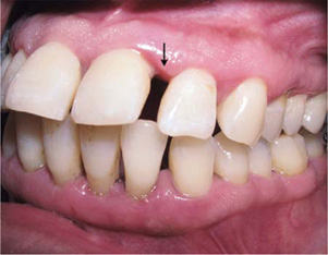
Color of the gingiva is described as coral pink which depends upon vascular supply, thickness of epithelium, degree of keratinization of epithelium and presence of pigment containing cells (Fig. 1.12).

A variation in gingival pigmentation is not produced by variation in the number of pigment forming melanocytes but by genetically determined variation in their pigment producing capacity. Thus, variations in gingival pigmentation are related to complexion and race. It is lighter in blond individuals with a fair complexion than in dark complexioned individuals. In the Caucasian individuals pigmentation is minimal, in African or Asian individuals there are brown or blue- black areas of pigmentation while in Mediterranean people occasional patches of pigmentation are found.
Gingival pigmentation was classified according to modification of melanin index:
Category 0: | No pigmentation |
Category 1: | Solitary unit(s) of pigmentation in papillary gingiva without formation of continuous ribbon between solitary units. |
Category 2: | At least 1 unit of formation of continuous ribbon extending from two neighboring solitary units. |
Surface Texture
The surface texture of free gingiva is smooth whereas of attached gingiva is stippled. Pitted surface texture giving orange peel appearance is called as stippling which is more prominent on the labial than on the lingual gingival surfaces. Stippling is normally present on attached gingiva and center of interdental papilla. It is best viewed by drying the gingiva and switching off the chair light. Stippling varies with age, it appears usually in children of about 5 years and increases with age but is absent in old age.

Histologically: The bottom of the pits correspond to deep ridges or projections of epithelium into lamina propria of the connective tissue. The protruding parts correspond to thinner epithelium overridges or projections of the connective tissue. The ridge and the peg arrangements between the epithelium and connective tissue provide excellent mechanical stability between the two tissue components as well as large contact interphase for metabolic interchange. In erythematous tissue stippling may disappear, although it may be present in thick fibrotic tissue (Fig. 1.13), which is diseased. Stippling is not an absolute sign of health and the absence of it is not necessarily a sign of disease.
Contour
The marginal gingiva follows a scalloped outline normally and straight line along teeth with relatively flat surfaces. Attached gingiva has festooned appearance with intermittent prominence corresponding to contour of roots. When the teeth are placed more labially, then the normal arcuate contour is accentuated and gingiva is located farther apically. When teeth are lingually placed, the gingiva is horizontal and thickened. Thus, contour of gingiva depends upon shape and alignment of the teeth in the arch. It also depends upon the location and size of the area of proximal contacts and dimensions of the embrasures.
Shape
Shape of interdental gingiva depends upon contour of the proximal tooth surface, location and shape of the proximal contact and dimensions of the gingival embrasures. The interdental papilla is pointed and pyramidal in normal contact areas and in anterior regions. But it is flat or saddle shaped in spaced teeth and in molar regions.
Size
The size of the gingiva corresponds to the sum total of the bulk of cellular and intercellular elements and their vascular supply.
Consistency
On palpation with a blunt instrument, attached gingiva should be firm, resilient and tightly bound to the underlying hard tissues. The abundant collagen fibers and the non-collagenous protein combines to give gingiva, the firm consistency.
LANDMARK STUDIES RELATED
Ainamo A, Ainamo J. The width of attached gingiva on supraerupted teeth. Journal of Periodontal Research 1978;13:194–18.
This study comprised 28 first and second maxillary molars which in the lack of antagonists had erupted beyond the occlusal plane. The maxillary mucogingival junction was marked with short pieces of metal wires, orthopantograms were taken and the distance from the mucogingival junction to the floor of the nasal cavity and to the cementoenamel junction were measured to the nearest mm. Eleven measurable occluding contralateral teeth were used as controls. A comparison was also made between the supraerupted teeth and the previously measured normally occluding teeth. The results indicated that even during pronounced supraeruption, the teeth tend to erupt with their investing tissues while the location of the mucogingival junction remains constant. This finding is of special interest as it would make possible to treat the problem of a too narrow zone of attached gingiva by grinding the tooth out of occlusion and allowing it and its gingival margin to erupt. The anatomical width of attached gingiva, i.e. the distance from the mucogingival junction to the cementoenamel junction was found to be 3.7 mm wider in the supraerupted teeth than in the normal occluding control teeth.
Caffesse RG, Nasjleti CE, Castelli WA. The role of sulcular environment in controlling epithelial keratinization. Journal of Periodontology 1979; 50:1–6.
The influence of the sulcular environment on the keratinization of the outer surface gingival epithelium 14was tested in three young adult rhesus monkeys. A total of 40 mucoperiosteal flaps were raised and inverted so as to bring the outer surface epithelium in contact with the tooth and were sutured. The monkeys were sacrified after giving H3 thymidine one hour prior. The material was prepared for histologic and radioautographic evaluation. Results indicated that the outer surface epithelium changes its morphology to a nonkeratinized epithelium devoid of deep rete pegs when in close contact with the tooth, resulting in the anatomical characteristics normally seen in sulcular epithelium. It was concluded that the sulcular environment has the capability of controlling the keratinizing potential of the outer surface epithelium. The constant irritation of bacterial plaque and its product may be responsible for the premature desquamation of the sulcular epithelium which in turn might not allow its full differentiation.
POINTS TO PONDER
- ✓ The junctional epithelium and gingival fibers together forms a functional unit called as dentogingival unit.
- ✓ The pH of the gingiva ranges from 6.5 to 8.5.
- ✓ Junctional epithelium is the only attachment in the body between soft tissue and a calcified tissue which is exposed to the external environment.
- ✓ Gingival fiber groups enable the gingiva to form a rigid cuff around the tooth that add stability especially when a significant portion of the periodontal ligament and alveolar support is lost. This explains that the increased mobility in periodontally involved teeth immediately after surgical procedures is because these procedures disrupt or remove the gingival fiber groups.
BIBLIOGRAPHY
- Baktold PM, Walsh LJ, Narayanan AS. Molecular and cell biology of the gingiva. Periodontol 2000;24: 28–55.
- Eley BM, Manson JD. The periodontal tissues. In, Periodontics 5th ed Wright 2004; 1–20.
- Grant DA, Stern IB, Listgarten MA. Gingiva and dentogingival junction. In, Periodontics. 6th ed CV Mosby Company 1988; 25–55.
- Itoiz ME, Carranza FA. The Gingiva. In, Newman, Takei, Carranza. Clinical Periodontology. 9th ed WB Saunders 2003; 16–35.
- Lindhe J, Karring T, Araujo M. Anatomy of the Periodontium. In, Lindhe J, Karring T, Lang NP. Clinical Periodontology and Implant dentistry. 4th ed Blackwell Munksgaard 2003; 3–49.
- Ramfjord SP, Ash MM. Connective tissue. In, Periodontology and Periodontics, Modern Theory and Practice. 1st ed AITBS Publisher and distributor India, 1996; 15–20.
- Ramfjord SP, Ash MM. Epithelium. In, Periodontology and Periodontics, Modern Theory and Practice. 1st ed AITBS Publisher and distributor India, 1996; 5–14.
- Stern IB. Oral mucous membrane. In, Bhaskar SN. Orban's Oral histology and Embroylogy. 11th ed Mosby 1991; 260–336.
- Squier CA, Finkelstein. Oral mucosa. In, Tencate AR. Oral histology Development, Structure and Function. 5th ed Mosby 1998; 345–87.
MCQs
- The mucogingival junction is located between the:
- Free gingiva and attached gingiva
- Free gingiva and tooth
- Base of the sulcus and alveolar mucosa
- Attached gingiva and alveolar mucosa
- Stippling is seen in:
- Marginal gingiva
- Attached gingiva
- Interdental gingiva
- Attached gingiva and center of interdental papilla
- The area of periodontium more susceptible to tissue breakdown is:
- Free gingiva
- Gingival sulcus
- Interdental col
- Interdental papilla
- Dentogingival unit comprises:
- Gingival fibers
- Gingival fibers and junctional epithelium
- Periodontal fibers and ligament
- None of the above
- Gingiva is supplied by:
- Supraperiosteal vessels
- Vessels of periodontal liagment
- Arterioles emerging from alveolar crest
- All of the above
- Which of the following fiber group is not attached to alveolar bone:
- Odland bodies are:
- Modified mitochondria
- Modified lysosomes
- Modified ribosome
- Modified centrioles
- Which of the following cells of the gingival epithelium is not a clear cell?
- Keratinocyte
- Langerhans cell
- Merkel cells
- Melanocytes
- The length of the junctional epithelium ranges from:
- 0.25 – 0.75 mm
- 0.15 – 0.75 mm
- 0.25 – 1.35 mm
- 0.5 – 1.0 mm
- The width of attached gingiva is greatest in:
- Maxillary anterior region
- Maxillary molar region
- Maxillary premolar region
- Mandibular premolar region
- The color of attached gingiva in health, is determined by:
- The presence of melanophores
- Degree of keratinization of epithelium
- Vascular supply
- All of the above
- If a diastema is present, the interdental papilla is:
- Larger in size
- Smaller in size
- Absent in the region
- None of the above
- Which of the following enzymes increase their activity towards surface in gingival oral epithelium?
- Succinic dehydrogenase
- Nicotinamide adenine dinucleotide
- Cytochrome oxidase
- Glucose-6-phosphatase
| Answers |
1. D | 2. D | 3. C | 4. B | 5. D |
6. A | 7. B | 8. A | 9. C | 10. A |
11. D | 12. C | 13. D |