

INTRODUCTION
Conventional two-dimensional (2D) echocardiography requires mental conceptualization of a series of multiple orthogonal planar or tomographic images into an imaginary multidimensional reconstruction for better understanding of complex intracardiac structures and their spatial relation with surroundings. The emerging three-dimensional (3D) echocardiographic imaging technique is a major step ahead toward the final goal of complete visualization and comprehensive assessment of cardiac anatomy, physiology, pathomorphology and pathophysiology in real-time, which could potentially facilitate image interpretation and reduce inter-observer variability.1–3
The concept of 3D echocardiography started in the early 90s. Previously, dynamic cardiac 3D-rendered images were possible using an off-line process, by sequentially acquiring 2D images, outputting these images to disk or CD-ROM, and using a workstation to input the 2D images for Cartesian coordinate conversion. The result was a tedious, time- consuming process. Outside of a research setting, this cumbersome process proved costly in terms of productivity and the ability to provide quality patient care.
While three-dimensional imaging of the heart and its structures is already state of-the-art with other tomographic imaging technologies including radionuclear, magnetic resonance and computed tomography, progress in three-dimensional echocardiography has been rather slow. In its early stages, three-dimensional echocardiography was applied mainly in volume measurement of the left ventricle using static wire frame pictures, which may demonstrate the shape of the ventricular cavity, but does not provide tissue-depicting information. Recently, along with the rapid evolution in computer technology, three-dimensional echocardiography has grown into a well-developed technique able to display dynamic images of the heart that contain tissue information and show the depth of the structures in their realistic forms. These capabilities undoubtedly provide an improved understanding of unpredictable pathomorphology and decrease the variability both in the quality and interpretation of complex pathology among investigators. Advantages of three-dimensional echocardiography are presented in Table 1. In particular, the availability and versatility using the volumetric data set, showing cardiac anatomy and pathology in projections that have not been possible until now, offer significant advantages.
OFF-LINE 3D RECONSTRUCTION
IMAGE ACQUISITION
Currently available methods for data acquisition include:
Linear Scanning
The transducer translates perpendicularly to the imaging plane, linearly over the patient's skin, in discrete, equal steps. This is accomplished by mounting a conventional linear or curved transducer in an assembly housing a motor and drive mechanism. Parallel images are stored and digitally combined to create a third dimension. Linear scanning has been used in conjunction with intravascular ultrasound catheters to create endocardial casts, representations of the inner arterial architecture that are particularly useful in the elucidation of complex vascular lesions.2
Rotational Scanning
A motor rotates the transducer array from 0 to 180° around an axis that is perpendicular to the array. This rotation geometry allows the axis of rotation to be fixed, while the acquired images sweep out, producing a conical imaging volume.
Fan-like or Tilt Scanning
The mechanical assembly tilts the transducer about an axis parallel to the axis of transducer. The process of acquiring 2D images at fixed intervals during the tilt motion or at regular angular intervals results in the computer storage of a set of 2D image planes arranged in a fan-like geometry. This technique allows image acquisition from a small acoustic window. The array is moved from a fixed point in a sweeping motion that spans approximately −60° to +60°, producing a pyramidal image volume.
FREE-HAND SCANNING
Although the mechanical scanning approach to ultrasono graphy offers speed and accuracy at times the bulkiness and weight of the devices hinder the scan. To overcome this problem many investigators have attempted to develop various free-hand scanning techniques in which the operator can hold the transducer with a position sensor attachment and manipulate it over the anatomy in the usual manner. This technique requires the operator to scan steadily over the precordial surface at a uniform speed. A number of free-hand scanning approaches have been developed that make use of four basic position sensing techniques: acoustic, articulated 3arm, image correlation methods and magnetic field. In the last one approach, a pulsed magnetic field is used to track the array's position in 3D space. A small magnetic receiver is attached to the transducer housing and a magnetic field transmitter is located close to the patient chest. Because of the magnetic field created around the chest, freehand scanning should not be used if the patient's heart rate is pacemaker-dependent.
IMAGE PROCESSING AND ANALYSIS
Off-line 3D reconstruction requires a series of steps including:
CONVERSION
The digital data are stored in their original 2D format and must be converted to a rectangular coordinate system for reconstruction (post-processing).
INTERPOLATION
It consists of assigning pixel intensities to fill in existing gaps. After conversion and interpolation, the pixels are treated as spatially correct 3D image elements known as voxels.
SEGMENTATION
It consists of delineating the ROI that contains the information required for reconstruction. Segmentation allows tissue and blood to be separated and displayed individually.
IMAGE ENHANCEMENT
Because of fuzzy ultrasound images and noise, including artifacts, the manipulation of 3D data sets from a 2D video screen requires graphics enhancements such as histograms and filtering.
VOLUME RECONSTRUCTION
After the 2D images have been acquired and their relative position and orientation determined, the 3D data can be reconstructed. This process refers to the generation of a 3D representation of the anatomy and involves placing each acquired 2D image at its correct relative position to all the other images. The 3D reconstruction process can be implemented using two distinct methods: feature based and voxel based.
REAL-TIME 3D ECHOCARDIOGRAPHY OR LIVE 3D ECHOCARDIOGRAPHY
Live three-dimensional echocardiography is a breakthrough in the field of medical ultrasound. The idea of real-time 3D cardiac sonography is not new. Clinicians have been experimenting with the manipulation of 2D images to come up with 3D images for twenty years. In the past, dynamic cardiac 3D rendered images were possible by sequentially acquiring 2D images and then using a workstation to input 2D images for Cartesian coordinate conversion and volume rendering. Outside research settings, this time-consuming process proved cumbersome and was simply impractical. Real-time volumetric 3D echocardiography employs a matrix array that generates a pyramidal burst of ultrasound. It requires very rapid beam formation and data processing, only possible with parallel-processing system, which permits the reception of 16 signals in return for each transmitted impulse. The beam is steered in both elevational and azimuthal directions.
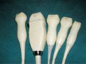
MATRIX TRANSDUCER
The first matrix array transducers for live 3D echocardiography were developed by Duke University, but the currently available probes and the system are commercially manufactured by Philips Corporation (Bothell, WA, USA). Laser is used to cut the piezoelectric crystal into many equal-sized minute square elements, forming an element matrix. The most commonly used transducer at present consists of more than 3600 (60_60) to 6400 (80_80) elements with operating frequencies ranging between 2 and 4 MHz. These elements are housed in the tip of the transducer so that they can be in close contact with the surface of the body for easy transmission and reception of ultrasound pulses.
STEERING 3D SOUND BEAM BY PHASED ARRAY
The matrix transducer generates ultrasonic beams in a phased array manner. The computer controls emissions in multiple directions and controls the timing of element firing. Each fired element produces an ultrasonic wave beam, and following Huygens principle, the wave beams combine to form a wave front. If all of the elements are fired simultaneously, 4the direction of the wave front will be perpendicular to the surface of the probe. By delaying the firing time of some elements, ultrasonic wave beams are created in different phases. Thus, the direction of the wave front can be altered by adjusting this delay time. So, although the probe is maintained in a stable orientation, the ultrasonic beam can change direction and reach any targeted area. The ultrasonic beam goes along the predetermined X-axis and produces a scanning line from near to far. The scanning line performs azimuth steering along Y-axis in the phased array manner and produces a 2D sector image. The 2D sector image then performs elevation steering along Z-axis and finally produces the pyramidal 3D image data set.
BOTTLENECK OF REAL-TIME 3D ULTRASOUND
With the matrix transducer, the theory of rapidly scanning and obtaining a pyramidal 3D dataset was established. The remaining problem was how to control the procedure to image in real time and to emit pulses with a computer. For example, suppose the size of the pyramidal 3-D image dataset is 60° _ 30° and one scanning line in every 1” of 60° 2D sector image is generated by azimuth steering along Y-axis. Then, at every 1” in the 30° elevation steering along Z-axis, there will be one frame of 2D sector image as well, so 60 Y-axis sections by 30 Z-axis sections makes 1800 scanning lines for one pyramidal 3D image dataset. According to a study by Roget in 1824, the persistence of human vision is about 1/24 of a second. Therefore, in order to achieve live 3-D images, we must obtain at least 24 pyramidal 3-D image datasets per second. To observe the stereo continuous movement of heart structures, the scanning lines generated by the live 3D system every second would be the scanning lines (1,800) of one pyramidal 3D data set multiplied by 24 (frame rate). Then, 43,200 scanning lines are needed in 1 second, the minimal pulse repetition frequency is 43.2 KHz and, subsequently, the maximal interval between the two consecutive pulses is only 23.1 _s. Given that the average propagation speed of ultrasound is about 1500 m/sec in soft tissues, these pulses could only travel a distance of 3.47 cm in that time for an imaging distance of only 1.73 cm. Such a small depth is not adequate for clinical imaging, which usually requires a depth of over 10 cm.
SOLUTION TO BOTTLENECK PROBLEM
In order to resolve this difficulty, the researchers at Duke University (Durham, NC) and Philips Corporation (Bothell, WA) developed a new microelectronic pulse transmitting-receiving technique, which is characterized by16:1 parallel processing to scan a pyramidal volume instead of the usual 1:1. This technology allowed many ultrasonic scanning beams to be emitted at the same time. As the pulse repetition frequency increases, the interval of the pulses increases 16 times with 16:1 parallel processing, which also increases the depth of ultrasound transmission through body tissues. By this approach, when the pulse repetition frequency is 43.2 KHz, the pulse interval is increased 16 times from 23.1 to 370 s, and the imaging depth from 1.73 cm to 27.7 cm without depth ambiguity.
BIPLANE MODE
Another benefit of the xMATRIX transducer technology is the possibility to display two orthogonal images in real time.
CLINICAL APPLICATIONS
Three-dimensional echocardiography has produced promising results from both experimental and clinical studies in the past two and half decades. Favorable experience has been gained in its clinical applications with both transthoracic and transesophageal image data acquisition.
VOLUME QUANTIFICATION
Echocardiography is a widely available clinical method, which consents to measure diastolic and systolic volumes and derived parameters, such as ejection fraction. Precision and accuracy of echocardiographic measurements is typically compromised by user subjectivity and geometrical assumption about left ventricular shape. The rationale behind the accuracy and reproducibility of volume measurement with three-dimensional echocardiography in comparison with any two-dimensional method is that the three-dimensional approach obviates any geometric assumptions of the shape of the measured object. Good correlations with angiography, magnetic resonance imaging and anatomical measurements (in vitro) have been reported. At present, ventricular volumes are calculated by manual endocardial tracing of sequential short-axis views derived by parallel slicing through three-dimensional data set at prescribed thickness intervals at either end-systole or end-diastole. Volume quantification is achieved by the summation of the voxels included in the traced area with 5the subsequent summing of the subvolumes of each slice with known slice thickness. Stroke volume and ejection fraction of a given chamber can be derived from its end-systolic and end-diastolic volumes.
VALVULAR HEART DISEASE
Both qualitative and quantitative evaluation of valvular heart disease can be improved by three-dimensional echo - cardiography. Any plane and paraplane analysis of the stenotic valve helps to find the smallest orifice area for accurate planimetry. Mitral valve prolapse is visualized as a bulging or protrusion on the atrial side together with its exact location and extension, allowing the surgeon to better plan the repair procedures. Three-dimensional echocardiography has been shown to be highly accurate for identifying the flail scallop. Three-dimensional echocardiography may further help in the quantitative evaluation of valvular abnormalities by improved analysis of the proximal flow convergence. Research is currently directed towards reconstruction of regurgitant jets showing the site of origin, trajectory and both the geometric distribution and morphology of the jet.
CONGENITAL HEART DISEASE
Three-dimensional echocardiography has been proven valuable in congenital heart disease for better evaluation of morphologic abnormalities and understanding of complex spatial relationships. Computer reconstruction of enface views of an atrial or ventricular septal defect, visualized from its right or left side, not only provides a surgeon's view of the defect before the heart is open but also enables accurate measurement of the dimensions of the defect and of the tissues surrounding the defect, the latter being crucial for planning closed-chest closure of the defect using a transcatheter closing device.
CORONARY ARTERY DISEASE
Coronary heart disease is one of the most commonly encountered diseases for the cardiologist. Three- dimensional echocardiography has shown its potential in accurate evaluation of volumes and function of the ventricles, in the analysis and quantitative measurements of regional wall motion abnormalities and myocardial perfusion territories using contrast agents Initial experience indicates that three-dimensional visualization of the proximal segments of the coronary arteries is possible.
CARDIOMYOPATHY
High-resolution Live 3D Echo allows a comprehensive analysis of LV wall motion before and during CRT and in contrast to conventional 2D Echo—the comparison of all LV segments. Live 3D Echo used in conjunction with a quantitative contour tracing algorithm allows comparison of asynchronism between all segments, while providing intuitive display of ventricular synchronicity. In addition it allows clinicians to obtain quantifiable data, it may provide a good prediction of CRT responders, can aid in optimizing pacemaker settings and Live 3D Echo enables rapid image acquisition.
CARDIAC MASSES
Three-dimensional echocardiography has been used in almost all aspects of cardiac disorders and various benefits have been derived. Evaluation of intracardiac or intravascular masses including vegetations, tumors, thrombi or plaques is facilitated both qualitatively (by three dimensional display of their site, size, attachment and mobility) and quantitatively (by accurate measurement of their dimensions and volumes).
AORTIC DISEASES
Aortic diseases such as dilation, aneurysm, dissection or coarctation with three-dimensional echocardiography and incremental information was obtained.
PLANNING CARDIAC SURGERY
Live 3D Echo improves spatial orientation and accesses critical views of the heart, aiding surgeons in the observation and quantification of size, shape and volume of the heart. One of the most important methods in assessing heart function is measuring left ventricular volumes during the cardiac cycle. Since accuracy is critical in assessing and quantifying the condition of the heart, Live 3D Echo is a much better option than other imaging methods as it enables clinicians to make measurements using an offline software tool without making geometric assumptions, which can prove costly. In addition to preoperative planning, Live 3D Echo is important in evaluating and monitoring surgical outcomes by performing real-time assessments of understanding will help improve patient throughput, training, patient communication and marketing.
Live 3D Echo contributes to improved clinical efficiencies with the potential to speed up diagnostic and interventional 6procedural time. The ability to obtain more complete diagnostic information decreases time spent studying clinical information and provides a higher degree of confidence in those results. The mobility of the ultrasound equipment also provides advantages. Ultrasound is simple and easily performed at the patient's bedside, providing real time results, thereby enhancing patient throughput and reducing wait times.
TRAINING AND PATIENT EDUCATION
Training is another area of operations that will realize significant improvements and cost savings. Two-dimensional cardiac ultrasound required a steep learning curve, which made training time consuming and expensive, since it required visualizing the heart three-dimensionally in 2D pieces in order to understand the results. Live 3D Echo provides the ability to quickly view the heart as it really appears, instantaneously in 3D, which sharply reduces this learning curve, reduces training costs and time spent getting clinicians up to speed.
Two-dimensional ultrasound makes little sense to patients wanting to know and understand their own healthcare. The ability to show patients easily understandable 3D images of their hearts will make it easier for clinicians to explain these images to patients. This not only speeds time clinicians spend with patients but also gives patients a higher level of comfort in what they are being told as they will better understand what their healthcare provider is showing them.
CONCLUSION
Live 3D Echo is a significant new advancement in ultrasound that is changing the way in which cardiology is practiced from the clinic all the way to the operating room. As a result, live 3D Echo is advancing the level of care provided to patients, which will ultimately have a positive impact on an institution's bottom line.
REFERENCES
- Lang RM. Badano LP. Wendy Tsang. et al. EAE/ASE Recommendations for image acquisition and display using Three-dimensional echocardiography J Am Soc Echocardiogr 2012; 25:3-46.
- Judy Hung. Lang RM. Frank Flachskampf. et al. 3D Echocardiography : A review of the current status and future directions J Am Soc Echocardiogr 2007; 20:213-33.
- Alexandra Gonçalves. José Luis Zamorano. Valve Anatomy and function with transthoracic three-dimensional echocardiography : advantages and limitations of instantaneous full volume color Doppler imaging. Ther Adv Cardiovasc Dis 2010;4:385.