Sataloff's Comprehensive Textbook of Otolaryngology: Head & Neck Surgery (Pediatric Otolaryngology) - Volume 6 Robert T Sataloff, Christopher J Hartnick
INDEX
Page numbers followed by f refer to figures, fc refer to flowcharts, b refer to boxes and t refer to tables.
A
Aberrant innominate artery 388
Abnormalities in development of the larynx and trachea 602
Abnormalities in development of the lip and palate 600
Abnormalities of the branchial clefts and pouches 601
Abnormally shaped ear 71
Acoustic and aerodynamic recording methods 995
parental discussion 996
physical examination 996
Acute otitis media (AOM) and otitis media with effusion (OME) 280
adjuvant therapies for AOM or OME 282
antibiotics for AOM or OME 281
prevention of AOM 281
ventilation tubes for AOM or OME 282
Acute otitis media 109f
Acute supraglottitis 470
Adaptation for speech 300
Additional soft tissue sarcomas 745
Adenoidectomy 376
Adenoids 605
Adenotonsillar disorders 605
Adolescent sleep 819
Adult and pediatric otologic diagnosis 107
Adult true vocal fold composition 926
body 926
cover 926
Aerodigestive tract structure and function 588
After implantation 196
Airway and respiratory management 437
Airway endoscopy versus sleep cine MRI 854
Airway foreign bodies 497
Airway hemangiomas 685
focal airway hemangiomas 685
segmental involvement 685
Airway management 426
Airway obstruction 609
Allergies 417
Alport syndrome 32, 47, 58, 153, 162
Alzheimer’s disease 18
American Academy of Pediatrics 821
American Academy of Sleep Medicine 824, 830
American Sign Language 232
Amino acid 38
Aminoglycosides 55
Amplification and classroom management 183
Amplification devices 184
Amyloidosis 482
Anastomoses 8
Anatomy/embryology 605
Anatomy of pediatric deep neck space infection 633
facial spaces 634
full length of neck 636
infrahyoid neck 636
suprahyoid neck spaces 634
Anatomy and physiology of the salivary glands 806
Anatomy of pediatric larynx 919
laryngeal anatomy and physiology 921
laryngeal development 919
cartilaginous structures 921
Anatomy of vocal fold innervation 953
Anesthesia reactions 418
Anterior cricoid split 545
Antibiotic ointment 88
Apert syndrome 44
Apert’s, Crouzon’s, Pfeiffer’s syndrome 530
Aplasia of the salivary glands 787
Apnea hypopnea index, 857
Approach to nonspecific chronic cough 582
Approach to the patient with sialorrhea 807
Ardipithecus ramidus 23
Arizona articulation proficiency scale 178
Arnold–Chiari malformation 955
Arnold’s ear-cough reflex 582
Arrhinia 351
epidemiology 351
management 352
presentation 352
workup 352
Arteriovenous malformations 697
Arytenoid fixation 1024
Arytenoid hooding 943
Aspiration 416
Associated pathology 384, 388
complete tracheal rings 388
congenital subglottic stenosis 386
laryngeal atresias and webs 385
posterior laryngeal clefts 384
Atlantoaxial subluxation 625
Atoh1 stimulation 14
Atresia of the oval window 50
Atresia surgery 88
Attention deficit hyperactivity disorder (ADHD) 610
Audiologic considerations for children 165
Auditory-steady state evoked potentials 62
Auditory brainstem implantation (ABI) 92
Auditory brainstem response 56
Auditory neuropathy spectrum disorder 190
Auditory neuropathy spectrum disorder 225
audiological assessment 227
acoustic immittance 227
auditory brainstem response 228
behavioral audiological evaluation 229
otoacoustic emissions 228
etiology 231
genetic links 231
risk factors 231
intervention 232
management 231
physiology 225
Auricular anatomy of external ear 70
nonsurgical intervention 71
normal dimensions 70
normal orientation 71
Abnormally shaped ear 71
nonsurgical intervention 71
patient evaluation 71
surgical intervention 73
Autism and attention-deficit hyperactivity disorder 418
Automated auditory brain stem response (aABR) 56
Autosomal recessive 32
Autosomal recessive disorders 45
Goldenhar syndrome/oculoauriculovertebral spectrum 46
genetics 46
presentation 47
Jervell and Lange-Nielsen syndrome 46
genetics 46
presentation 46
Pendred syndrome 45
genetics 45
presentation 45
Usher syndrome 46
genetics 46
presentation 46
B
Bacterial tracheitis 471
Bacteriology 606
Balance disorders in children 261
etiology 263
central causes of dizziness in children 264
children 266
peripheral causes of dizziness in
prevalence 263
office evaluation 261
imaging 263
physical examination 262
Balloon catheter sinuplasty 377
Barrier defense 325
Bartonella henselae 786
Behavioral observation audiometry 190
Benign and malignant vocal fold lesions 987
cysts 986
granuloma 989
juvenile recurrent respiratory 988
papillomatosis 988
malignancies 990
nodules 987
polyp 986
sulcus vocalis 987
webs 988
Benign nasal masses 353
inverted papilloma 355
epidemiology 355
management 356
pathophysiology 355
presentation 355
workup 355
juvenile nasopharyngeal angiofibroma 353
epidemiology 353
management 354
pathophysiology 353
presentation 354
workup 354
nasopharyngeal teratoma 356
epidemiology 356
management 356
pathophysiology 356
presentation 356
workup 356
Bezold’s abscesses 207
Big brain 23, 300
Bilateral cleft lip 1119
anatomy 1119
classification 1119
goals 1122
presurgical infant orthopedics 1123
surgical repair 1124
history and background 1124
Millard repair 1124
Mulliken repair 1124
Bilateral submandibular gland excision and parotid duct ligation 812
Bilateral vestibular schwannomas 251
Bilateral vocal fold paralysis 863
Biochemical evidence of true vocal fold 928
Biopsy 779
Bipedalism 23, 300
Bipolar radiofrequency ablation 859
Bone-anchored hearing aids 84
Botulinum toxin 27
Brachycephaly 49
Brain abscesses 209
Brainstem tumors 1054
Branches of the facial nerve 5f
Branchial arch anomalies 396
Branchio-oto-renal 57
Branchio-oto-renal syndrome 41, 79, 117
Brent technique 75
Bronchial tumors 488
Brunning’s otoscope sets 122
Bubbly nasal rustle 1055
instrumental evaluation of velopharyngeal function 1056
intraoral evaluation 1056
nasometry 1056
nasopharyngoscopy 1057
phonation 1056
speech aerodynamics 1057
speech sound production 1055
videofluoroscopic speech study 1057
Buccal branch 4f
Burkitt lymphoma 740
C
Caffeine therapy 873
Caldwell-Luc procedure 305
Canal cholesteatoma 88
Candidacy criteria 192
age considerations 193
auditory brainstem implants 195
bilateral cochlear implantation 195
degree of hearing loss and benefit from amplification 194
Canonical pathway 14
Capillary malformations 667
syndromes associated with port-wine stains 667
Cardiopulmonary complication 611
Carnegie stage 11 919
Cartilaginous otic capsule 9
Case management 427
airway reconstruction 428
burns 429
foreign bodies in the airway 429
juvenile onset recurrent respiratory papillomatosis 428
tracheostomy 428
trauma 429
Case studies 234
Catathrenia 902
Caudal main trunk 5
Caudal portion of the pons 3
Causes of chronic cough 579
Causes of otalgia in children 113
Caustic ingestion 494
Cell fate specification 14
Central apnea in children 871
diagnosis 871
evaluation 872
management 871
physiology 871
anatomic lesions 875
disordered control of breathing 872
genetic/syndromes 873
specific causes 872
treatment overview 872
Central dizziness with a structural brain abnormality 265
Central dizziness without a structural brain abnormality 264
Central nervous system 3
Central nervous system: effects of anesthesia 422
Cephalic muscles 5
Cerebral palsy 955
Cerebral vascular accident 1054
Cervical branch 4f
Cervicofacial division 4f
Charcot-Marie-Tooth (CMT) disease 231, 971, 173t
CHARGE syndrome 50
pathogenesis 50
presentation 50
Chiari malformation 875
Childhood sleep 818
Children’s oncology group 765
Choanal atresia 347, 148
epidemiology 347
management 348
pathophysiology 347
presentation 347
workup 347
Choanal atresia 363
Cholesteatoma 213, 720
diagnosis and tests 216
epidemiology 213
pathogenesis 214
acquired 214
congenital 214
pathophysiology 720
presentation and diagnosis 721
signs/symptoms 215
Chondrosarcoma 715, 745
outcomes 715
presentation and diagnosis 715
treatment 715
Chorda tympani 6
Chorda tympani nerve 5
Chordoma 713
histology 713
outcomes and prognosis 714
presentation and diagnosis 713
treatment 714
Chordoma and chondrosarcoma 772
Chromosome 17
Chronic adenotonsillar hypertrophy 609
Chronic cough 572
Chronic ear disease 51
Chronic infections 613
Chronic rhinosinusitis 366
operative technique 366
postoperative follow-up 366
preoperative workup 366
Chronic rhinosinusitis (CRS) 373
Chronic tracheostomy 1021
Cicatricial pemphigoid 483
Circadian phase markers 882
Circadian rhythm sleep disorders 879
circadian rhythms across child development 882
classification and diagnosis 879
advanced sleep phase disorder 880
delayed sleep phase disorder 879
free-running disorder 880
irregular sleep–wake rhythm 881
other circadian rhythm sleep disorders 881
nonpharmacologic treatment options 884
behavioral therapy 884
cognitive behavior therapy 884
timed light administration 885
pharmacologic treatment options 885
melatonin 885
prescription-only drugs 886
vitamin B12 886
physiology 887
Cleft care in outreach setting 1130
perioperative stage 1131
postoperative stage 1131
preoperative stage 1131
Cleft lip 1099
anatomy 1102
classification 1100
embryology 1099
epidemiology 1100
etiology 1101
multidisciplinary approach to cleft care 1102
Cleft palate 51
Clinical applications of embryology of facial nerve 10
Clinical considerations in the management of children with craniofacial anomalies 1143
airway issues 1143
feeding issues 114
otologic issues 1145
Clinical evaluation 821
history and physical examination 821
Coagulation abnormalities 693
Cochlear anatomy 246f
Cochlear aperture abnormalities 102
audiological findings 102
management 104
radiology 102
Cochlear aperture aplasia 103
Cochlear aplasia 92
audiological findings 92
management 93
radiology 92
Cochlear
implantation 56
implant team 189
ionic homeostasis 37
micromechanics 48
nerve deficiency 50
nuclei 92
pathophysiology 36
stem cells 16f
Cohen classification system 516
Common anomalies of craniofacial disorders 1140
ears 1142
cryptotia 1142
lop/cup ear 1142
low-set ears 1142
microtia/anotia 1142
posteriorly rotated ears 1142
protruding ears 1142
eyes 1141
epicanthus 1141
eyelid coloboma 1141
ocular hypertelorism 1141
orbital hypertelorism 1141
orbital hypotelorism 1141
telecanthus 1141
uveal coloboma 1141
facial skeleton 1143
absence and hypoplasia of the zygoma 1143
congenital asymmetry of the facial skeleton 1143
micrognathia 1143
midface retrusion and hypoplasia 1143
retrognathia 1143
tongue 1143
glossoptosis 1143
macroglossia 1143
Common cavity 93
audiological findings 94
electrode choice 94
management 94
radiology 93
Common complications after airway reconstruction 566
Common pediatric otologic problems 107
Comorbid states 327
Comparative anatomy of paranasal sinuses 303
Complete blood count 60
Complete labyrinthine aplasia (Michel deformity) 91
audiological findings 91
management 91
radiology 91
Complications of tonsillitis 608
Compressor naris 8
Compromised airway 416
Computed tomography 393
factors influencing patient dose 394
image quality 394
radiation risks 394
strategy for dose reduction 395
terminology 393
Concha 135f
Congenital and acquired laryngeal anomalies 503
Congenital and acquired tracheal anomalies 523
Congenital anterior skull base defects 371
Congenital aural atresia 79
embryology 80
management 84
amplification 84
observation 84
surgery 85
Congenital cardiac anomalies 51
Congenital cholesteatoma 109f
Congenital cystic neck masses 651
Congenital hearing loss 55
Congenital high airway obstruction syndrome 537
Congenital laryngeal atresia 517
Congenital laryngeal cysts 517
Congenital laryngeal web 516
Congenital lesions in the oral cavity 455
Congenital nasal cavity anomalies 361
nasal dorsal mass 361
operative technique 361
postoperative follow-up 362
preoperative workup 361
nasolacrimal duct cyst 362
operative technique 362
postoperative follow-up 362
preoperative workup 362
piriform aperture stenosis 362
operative technique 363
postoperative follow-up 363
preoperative workup 363
operative technique 364
postoperative follow-up 365
Congenital nasal malformations 347
Congenital nasal masses 445
Congenital pyriform aperture stenosis 348
epidemiology 348
management 349
pathophysiology 348
presentation 348
workup 349
Congenital synechiae 462
Considerations for surgical therapy 373
Contemporary workup of air-bone gap 1181
diagnostic tools 1181
hearing assessment 1182
history 1181
imaging 1183
physical examination 1181
rigid endoscopy of the ear 1183
diagnostic workup 1183
associated symptoms 1187
evaluation of a child with an air-bone gap 1184
evaluation of a child with congenital CHL 1186
speech delay 1188
Continuous positive airway pressure 868
Corrugator 5
Corrugator supercilii 9
Cough in the pediatric patient 571
Cranial ultrasounds 60
Craniofacial disorders 1135
dysmorphology 1136
embryology 1135
associations 1138
patterns 1137
sequences 1138
syndromes 1138
role of imaging 1138
computed tomography 1140
magnetic resonance imaging 1139
ultrasound 1139
Craniofacial growth 611
Craniopharyngioma 450, 715
presentation and diagnosis 716
staging 716
treatment 716
Cricotracheal resection/tracheal resection and reanastomosis/slide tracheoplasty 566
Crisis management 422
Criterion for implantable hearing aid placement 237
bone-anchored hearing aids 237
outcomes and complications 238
surgical procedure 238
Down dyndrome 49
intraoral bone conduction device 240
transcutaneous bone-anchored hearing device 239
Crouzon syndrome 48
genetics 48
pathogenesis 49
presentation 48
Cupid’s bow 1131f
Current upper respiratory tract infection 424
Cystic fibrosis-related rhinosinusitis 344
clinical findings 344
histology 344
imaging findings 344
medical treatment 344
microbiology 344
pathogenesis 344
surgical treatment 345
Cystic fibrosis 367
operative technique 367
postoperative follow-up 367
preoperative workup 367
Cytoplasmic mitochondria 31
D
Deafness gene panels 37
hearing loss expanded panel 37
otogenome panel 37
otoscope panel 37
Deafness variation database 38t
De grouchy syndrome 79
Dehiscent carotid canal 159f
De la Cruz system 83
Delivery routes to the inner ear 18
Derivatives of branchial arches and pharyngeal pouches 600
Dermoid cysts 655
branchial anomalies 655
first arch anomalies 657
fourth arch anomalies 660
second arch anomalies 658
third arch anomalies 659
thymic anomalies 661
Determining the cause of hearing loss 31
Developing intervention goals and measuring progress 178
Developmental 415
Developmental anatomy 599
Developmental anatomy of the unified airway 381
Developmental trajectory of nonsurgical management 1065
Development of sinonasal mucosa 296
Development of the primitive midface 293
Diagnosis and management of childhood obstructive sleep apnea syndrome 821
Diagnostic algorithm 779
Diagnostic testing of congenital heares loss 62
abnormalities 70
congenital 70
embryology 70
genetic testing 62
laboratory testing 62
radiographic imaging 62
testing algorithms 64
Diffuse thyroid enlargement (goiter) 33
Digastrics muscle 5
Dilated vestibular aqueducts 45
Diploic bone 160
Disk batteries 499
Dix–hallpike maneuver 116t
Dominant syndromes 41
Apert syndrome 44
genetics 44
presentation 44
branchio-oto-renal syndrome 41
genetics 41
presentation 41
stickler syndrome 43
genetics 43
presentation 43
Treacher Collins syndrome 42
genetics 43
presentation 43
Waardenburg syndrome 43
genetics 44
presentation 44
Dorsal cochlear nucleus 252f
Double aortic arch 388, 532
Downslanting palpebral fissures 51
Down syndrome 49, 874
pathogenesis 49
presentation 49
Dysontogenetic cysts 464
Dysphagia 398
clinical background 398
clinical questions 399
imaging modality of choice 399
interpretation 399
E
Early hearing detection and intervention 56
Effects on development 183
Electrocautery techniques 859
Embryology and relevant anatomy 1163
mandibular hypoplasia 1164
history of MDO 1164
limitations 1165
Embryology of the branchial arches and pouches 600
Embryology of the facial nerve 3
Embryology of the intracranial portion of the facial nerve 5
Emerging technologies of true vocal fold 929
Encephalocele 450, 721
Endocrine 417
Endolymph 19
Endolymphatic duct 45
Endoscopic posterior cricoid split 545
Endoscopic sinus surgery 376
Environmental causes of hearing loss 31
Enuresis 610
Eosinophilic esophagitis 592
Ephelis (melanosis) 463
Epibranchial placode 6
Epicanthic folds 49
Epidermolysis bullosa and cicatricial
pemphigoid 482
Epistaxis 310
Epstein–Barr virus 607
Escherichia coli 59
Esophageal foreign bodies 498
Esthesioneuroblastoma 708, 769
pathophysiology 708
presentation and diagnosis 708
staging 708
Etiology of congenital hearing loss 55fc, 57
genetic 57
nongenetic 59
cytomegalovirus 59
infectious causes 59
metabolic disorders 60
other causes 60
perinatal causes 59
nonsyndromic 58
autosomal recessive 58
X-linked 59
syndromic 57
autosomal dominant 57
autosomal recessive 57
Eustachian tube 24
Eustachian tube constriction 27
Eustachian tube dysfunction 51
Eustachian tube function 23
Euthyroid 45
Evaluation of airway obstruction 843
Evaluation of dysphonia 954
Evaluation of nystagmus 262
Evaluation of pediatric dysphonia 993, 1019
assuring child participation 994
initiation of referral 993
multidisciplinary care 993
pediatric voice history 993
specific assessments 994
maximum phonation time 995
oral motor 994
perceptual 994
pitch and volume 995
quality-of-life 995
respiratory 995
stimulability 995
Evaluation of pediatric sleep disorders 821
Evaluation of the child with rhinologic disease 309
history 309
nasal obstruction 309
rhinorrhea 309
Evidence-based medicine in pediatric otology 277
Evidence-based medicine in pediatric rhinology 373
Evolution’s impact on craniofacial anatomy 301
Evolution’s role in paranasal sinus and middle-ear drainage 304
Evolution’s role in the anatomy of the human head and neck 300
Ewing’s sarcoma 712, 771
histology 712
outcomes 713
presentation and diagnosis 712
treatment 712
Examination of the ear 119
facilities and environment 120
necessary instruments 121
room and setup 121
waiting room 120
handling of the instruments 134
precautions regarding instrumentation 135
inspection 124
otoscopy 125
diagnosis of ME effusion 130
external auditory canal 125
otoendoscopy 133
otomicroscopy 132
otoscopy with surgical head 131
pneumatic otomicroscopy 132
pneumatic otoscopy 128
tympanic membrane 126
palpation 125
Examination of the ear in a young child 108
assessment of the tympanic membrane 108
cleaning the ear canal 111
general examination 112
Existing clinical practice guidelines 374
Exposure to ototoxic medications 55
External canal collapse 56
External ear 69
congenital abnormalities 69
embryology 69
External ear anomalies 51
External ear malformations 47
External framework of the laryngeal complex 921f
muscles of the larynx 921
External petrosal nerve 9
Extracorporeal membrane oxygenation 55
Extraskeletal Ewing sarcoma 744
Extratemporal anatomy of the facial nerve 4f
Eyelid/orbital/periorbital hemangiomas 677
focal eyelid hemangiomas 680
indications for treatment 678
segmental eyelid hemangiomas 681
treatment alternatives 680
F
Facial musculature 3
Facial nerve 162
vascular anatomy 163
Facial nerve anomaly 96
Facilitation of early speech and language skills 1065
Facioacoustic primordium 5
Fallopian canal 3, 10f
Farrior technique 73
Fat saturation 396
Features of otologic conditions 112
facial palsy 113
headache 113
hearing loss 112
hyperacusis 114
otalgia 113
otorrhea 112
tinnitus 114
vertigo and imbalance 113
Female pelvic outlet 24
Fibrosarcoma 742
First contact 122
Flexible laryngoscopy 562
Flipside of evolution 13
Fluoroscopy of the airway and barium swallow 395
comments and caveats 396
Focal epithelial hyperplasia 462
Fordyce granules 462
Friedreich ataxia 173t, 231
Frontalis 5
Furstenberg’s test 449
G
Gastroenterological 416
Gastroesophageal reflux disease 587
Generalized lesions 463
Genes and gene expression 327
Gene tests 38t
Gene therapy 17, 18
Genetic 417, 535
Genetic causes of hearing impairment 32
Genetic counselors 39
Genetic heterogeneity 46
Genetic heterogeneity of hearing impairment 35
Genetic nonsyndromic hearing loss 32
Genetics of hearing loss 31
Genetic testing 37
Genetic testing registry (GTR) 38t
Geniculate ganglion 3
Genome 31
Germ cell tumors 718
presentation and diagnosis 718
treatment 719
Glomuvenous malformation 694
Glossopharyngeal ganglion 6
Glottic lesions 503
Glottis 503
Glottis closure 955
Goldenhar syndrome 46, 1186
Gradenigo’s syndrome 207
Granulomatous diseases 479
Gray’s external minithyrotomy 1024
Greater superficial pefrosal nerve 8f
Greisinger’s sign 208
Growth problems 611
Guillain–Barré syndrome 958, 971
H
H. sapiens 27
Habit cough 580
Haemophilus influenzae 59
Hair cells 16
Hairy polyp 463
Hamdan sliding V-cheiloplasty technique 1115
Harvest of stem cells 17f
Head thrust test 262
Head trauma 266
Hearing in difficult listening situations in the classroom 185
additional factors in the classroom 187
noise management in classrooms 185
role of the speech-language pathologist 187
Hearing intervention 66
Hearing loss 183, 258
effects on development 183
Hearing screening 284
Heck’s disease 462
Hemangiomas
cheek 682
forehead and scalp 682
lip 676
Hemangiopericytoma 744
Hemifacial microsomia 79
Hemorrhage 624
Hepatosplenomegaly 59
Heredity of auricular abnormalities 70
Heterotopic tissue 464
Heterozygous 45
Hidden airway lesion 567
Hirano’s layered structure of the human true vocal fold 926
Hirano’s seminal work 926
Hirschsprung disease 44
Histopathology of the temporal bone 159
History and physical examination 777
History of tonsillectomy and adenoidectomy 611
Hodgkin lymphoma 735
Holinger–Henick system 919
Homer Wright rosettes 708
Horner syndrome 771
Household bleach 496
Human evolution in rhinosinusitis 300
Human genome 31
Human papilloma virus 1031
Human tensor veli palatini muscle 27
Human true vocal fold 926
Hunter syndrome 173t
Huntington’s disease 33
Hydrocephalus 208
Hyperbilirubinemia 55
Hypernasal speech 1041
Hypersomnia 903
classification 903
behaviorally induced insufficient sleep syndrome 905
idiopathic hypersomnia 905
narcolepsy 903
recurrent hypersomnias 904
diagnostic evaluation 905
management 906
Hypoplasia 99
audiological findings 100
auditory brainstem implantation 102
management 100
cases with mixed type of hearing loss 100
cochlear implantation 100
radiology 99
Hypoxia 55
I
Identification of deafness genes 33
Imaging modalities in pediatric temporal bone 143
computed tomography 143
MDCT reformats 143
Poschl reformat 144
routine CT temporal bone technique 143
Stenver reformat 144
cone beam ct 144
magnetic resonance imaging 146
additional considerations 146
MRA 146
PET 147
plain film radiography 147
routine technique 146
safety considerations 146
ultrasound 147
radiation dose reduction techniques 144
factors influencing patient dose 145
image quality 146
radiation risks 144
strategy for dose reduction 146
Immunology of the adenoids and tonsils 606
Impact of evolution 23
eustachian tube function 23
otitis media 23
Impaired child 268
Implications for otitis media 302
Incomplete partition of cochlea 94
radiology 94
incomplete partition type I 94
Infant sleep 818
Infection 399, 417
clinical background 399
clinical questions 400
imaging modality of choice 400
interpretation 400
Infection prevention 441
Infections of the larynx and trachea 467
Inferior cerebellar peduncle 3
Inflammatory conditions 780
acute sialadenitis 780
bacterial sialadenitis 782
chronic sialadenitis 783
cystic 787
developmental disorders 787
neoplasm 788
Inflammatory lesions 478
Initial presentation and evaluation 494
Inner ear 160
Inner ear anomalies 51
Inner ear development 13
Inner ear regeneration 13
Innervation of the larynx 922
Inpatient versus outpatient tonsillectomy 616
Insomnia 895
classification 895
adjustment insomnia 896
behavioral insomnia of childhood 895
idiopathic insomnia 896
inadequate sleep hygiene 897
paradoxical insomnia 897
psychophysiological insomnia 897
diagnostic evaluation 897
management 898
behavioral therapy 898
pharmacotherapy 899
Instructions 419
Insulin-dependent diabetes mellitus 425
complex congenital heart diseases 426
poorly controlled systemic disease 426
sickle cell disease 426
Internal auditory canal 3f
Interscalar septa 98
Interstitial lung disease 582
Intracranial abscesses 209
Intraglandular stone 784
Intrinsic musculature of the larynx 922
differences between infant and adult larynges with clinical implications 923
respiration and phonation 923
vasculature and innervation 922
Investigations for pediatric ear problems 114
audiology 114
blood tests and other investigations 117
diagnostic imaging 115
cholesteatoma 116
complications of suppurative middle ear disease 117
conductive hearing loss with a normal tympanic membrane 117
sensorineural hearing loss 115
microbiology 115
Isotropic fast spin-echo 852
J
Jahrsdoerfer grading scale 83
Jaundice 59
Jeff strain 37
Jervell and Lange-Nielsen syndrome 32, 37, 58, 62, 153
Joubert syndrome 874
Juvenile nasopharyngeal angiofibroma 367, 368, 705, 772
operative technique 368
Juvenile onset recurrent respiratory papillomatosis and the voice 1031
adjuvant treatment 1034
clinical presentation 1032
epidemiology 1031
evaluation/diagnosis 1032
JRRP and the voice 1035
pathophysiology 1031
prevention 1034
reflux 1035
surgical treatment 1032
Juvenile recurrent parotitis 784, 793
obstructive sialadenitis 796
K
Kabuki syndrome 50
pathogenesis 50
presentation 50
Kadish staging 40 708
Kaposiform hemangioendothelioma 665
Kaposi sarcoma 743
Kartagener’s syndrome 334
allergic fungal rhinosinusitis 334
imaging 334
adjunctive treatments 337
antibiotics 335
intranasal and systemic steroids 336
surgical treatment 338
adenoidectomy with/without sinus irrigation and balloon dilation 338
functional endoscopic sinus surgery 339
Kasabach-Merritt syndrome 665
Kiesselbach’s plexus 310
assessing pediatric sinonasal quality of life 311
bacterial rhinosinusitis 311
clinical examination 312
head and neck examination 312
microbiologic assessment 315
neonates and infants 312
school aged children and older 314
toddlers and preschool 314
imaging 316
bacterial rhinosinusitis 316
nasal obstruction 316
medical ancillary workup 317
allergy workup 317
immune workup 317
nasal pain 310
orbital symptoms 310
Kleine-Levin syndrome 904
L
Laboratory 418
Laboratory studies 778
Labyrinthitis 267
Lactic acidosis 33
Laminae 6
cervical 6
mandibular 6
occipital 6
temporal 6
Langerhans cell histiocytosis 772
Large vestibular aqueduct 102
Laryngeal anatomy 503
Laryngeal chemodenervation 977
Laryngeal cleft 511
Laryngeal electromyography 956
Laryngeal reinnervation 977
Laryngeal tumors 483
malignant laryngeal neoplasms 487
neurogenic tumors 486
subglottic hemangioma 483
Laryngeal web and atresia 602
Laryngomalacia 504
Laryngopharyngeal reflux and gastroesophageal reflux disease 478
Laryngotracheal reconstruction 437, 566
Laryngotracheobronchitis 468
Larynx 381
postnatal development 383
adult larynx 383
infant larynx 383
prenatal development 381, 386
Laser treatment 673, 690
laser treatment of superficial hemangiomas 673
laser treatment of the superficial component of a compound hemangioma 674
quiescent or involuting hemangiomas 675
Lateral neck films, flexible fiberoptic nasal endoscopy 823
Leber’s congenital amaurosis 18
Levator anguli oris 9
Levator labii superioris 9
Levator labii superioris alaeque nasi Levator veli palatini 1041
Levator veli palatini muscle 26
Li-Fraumeni syndrome, BeckwithWeidemann syndrome, and Costello syndrome 741
Limb anomalies 51
Linkage analysis in human pedigrees 35
Listeria monocytogenes 59
Liver function tests 60
Loss of human prognathism 24
adaptation for speech 25
cooking 25
Loss of sox2 15
Lower airway 386
Lumbar puncture 208
Lyme disease 955
Lymphatic malformation 464, 669, 694
sclerotherapy 697
surgical resection 695
Lymphatic malignancy 735
Lymphoma 772
M
Macaca mulatta 26
Macroglossia 49
Macroview otoscope 125
Malar hypoplasia 51
Malignant peripheral nerve sheath tumors 743
Malignant tumors 356
esthesioneuroblastoma 357
epidemiology 357
management 358
pathophysiology 357
presentation 357
workup 357
management 759
diagnostic evaluation 759
lymph node dissection 760
radioactive iodine therapy 761
thyroid-specific surgical management 759
nasopharyngeal carcinoma 358
epidemiology 358
management 358
pathophysiology 358
presentation 358
workup 358
rhabdomyosarcoma 356
epidemiology 356
management 356
pathophysiology 356
presentation 356
workup 356
sinonasal lymphoma 358
epidemiology 358
pathophysiology 358
presentation 358
treatment 359
workup 358
Management of children with inner ear malformations 91
Management of pediatric dysphonia 997
surgery 999
voice therapy 997
direct therapy techniques 998
indirect therapy techniques 997
Managing and avoiding a CSF gusher 95
continuous lumbar drainage 96
incomplete partition type II 96
incomplete partition type III 97
electrode with a cork type stopper 95
large cochleostomy 95
small cochleostomy 95
subtotal petrosectomy 95
Mandible/maxilla/gingiva 460
alveolar cysts (Bohn’s nodules) 461
cherubism 461
eruption cysts 462
facial clefting 460
granular cell tumor (epulis) 461
micrognathia 460
midpalatal cysts (epstein’s pearls) 461
nasopalatine duct cyst 460
natal/neonatal tooth 461
odontogenic tumors 462
Mandibular distraction osteogenesis for airway 1161
Mandibular distraction procedure 1167
Mandibular platysma 9
Mandibular ramus 10
Mandibulofacial dysostosis (MFD) 42
Marginal mandibular branch 4f
Marginal mandibular nerve 8f
Marx classification system 74
Mastoid air cells 87
Maxillary antrostomy 374
Measuring outcomes in treatment 374
CT findings 375
nasal endoscopy 375
quality-of-life instruments 375
Meckel’s cartilage 1136
Medical evaluation of congenital, hearing loss 60
history 60
physical examination 61
Medical treatment 672
propranolol 672
systemic and/or injectable corticosteroids 673
vincristine 673
Medulla oblongata 3
Medullary thyroid carcinoma 761
Melatonin 882
Meniere’s disease 157
Meningiomas 722
Metabolic 417
Michel deformity 92
Microbiology 637
Microcolony staphylococcus/intracellular 325
superantigens and staphylococcus 325
Micrognathia 51
Microtia 73
Brent technique 76
stage 1 76
stage 2 76
stage 4 76
classification 74
clinical presentation and management 74
epidemiology 73
evaluation 80
genetics 74
microtia repair complications 76
Nagata technique 76
stage 1 76
stage 2 76
stage 3 76
staged surgical reconstruction 75
Middle-ear disease 23
Middle ear and mastoid 159
eustachian tube 160
mastoid 160
middle ear 159
Middle ear anomalies 51
Middle ear implants 240
Middle ear pathology 56
Middle meningeal artery 9
Midline nasal masses 350
epidemiology 350
management 350
pathophysiology 350
presentation 350
workup 350
Millard’s bilateral cleft lip technique 1124f
Mitochondrial causes of hearing loss 33
Mitochondrial encephalopathy 33
Mitochondrial forms of hearing impairment 37
Mitochondrial genome 32f
Mitochondrial mutations 33
Mitochondrial syndromes 33
Modifications following prosthetic treatment 1070
Mohler’s technique 1111
Mondini deformity 98
Mondini dysplasia 45
Mondini malformation 66
Monkey’s larger tensor veli palatini muscle 27
Motor function testing 262
Motor nucleus of the facial nerve 3
Motor root 3
Mouse models 35
Movement disorders circadian rhythm 886
periodic limb movement disorder 887
diagnosis 887
etiology and epidemiology 888
restless legs syndrome 886
diagnosis
epidemiology 889
etiology 887
treating sleep movement disorders in children 888
Mucocele/ranula 462
Mucosal wave abnormalities 955
Müller’s maneuver 822
Mulliken’s bilateral cleft lip 1124f
Multidisciplinary evaluation of pediatric cochlear implant candidate 189
Multilevel obstruction 567
Munchausen’s stridor 943
Muscular valve 1041
Musculus uvulae 1041
Myasthenia gravis 971
Myer–Cotton grading system 544
Myer–O’Connor–Cotton grading system 508
Myringosclerosis 201
ancillary testing 201
complications 207
disease prevention 201
extracranial 207
medical management 205
medical therapy 205
medical treatment 202
antibiotics for treatment failures 205
initial antibiotic therapy 203
nonantibiotic therapies 205
observation 202
surgical treatment 206
adenoidectomy 207
myringotomy/tympanocentesis 206
myringotomy with pressure equalization tube placement 206
N
Nagata technique 75
Nager syndrome 51, 1165
pathogenesis 51
presentation 51
Nasal dermoid 446
Nasal emission 1041
Nasal glands 5
Nasal glioma 449
Nasal hemangiomas 681
Nasal obstruction 400
clinical background 400
clinical questions 401
imaging modality of choice 401
interpretation 401
Nasal regurgitation 1041
Nasal tumors 353
Nasoglabellar region 447
Nasolacrimal duct cyst 445
Nasopharyngeal carcinoma 768
Nasopharyngeal stenosis 625
National health service 395
Neck mass 645
diagnostic studies 647
chest radiograph 647
computed tomography 648
fine needle aspiration 648
incisional/excisional biopsy 648
laboratory studies 647
magnetic resonance imaging 648
positron-emission tomography 648
radiologic studies 647
ultrasound 647
physical examination 646
Neisseria meningitides 59
Neonatal sleep 817
Neoplasms 566
Nervus intermedius 3, 6
Neural progenitor 15f
Neuroblastoma 748, 771
Neurocognitive development 610
Neurodevelopmental disorders 580
Neuroendocrine malignancy 748
Neurofibromatosis 723
pathophysiology 723
Neurolaryngological disorders 953
Neuromuscular and movement disorders 958
Neuromuscular blockade 440
Newborn hearing screening 56, 165
Newborn hearing screening technique 169
cross-disciplinary collaboration 170
role of the otolaryngologist 170
Newborn hearing statistics 172t
Nightmare disorder 901
Nitric oxide and primary ciliary dyskinesia 326
histology 327
Non-Hodgkin lymphoma 738
Noncleft velopharyngeal insufficiency 1061
etiology and presentation 1061
evaluation of VPI 1062
genetics 1061
iatrogenic causes 1062
treatment 1062
Noninvoluting congenital hemangiomas 664
Nonrhabdomyosarcoma soft tissue sarcomas 768
Nonsurgical management of pediatric obstructive sleep apnea 867
Nonsurgical management of velopharyngeal insufficiency 1065
Nonsyndromic autosomal recessive deafness 45
Nonsyndromic deafness 33
Nonsyndromic deafness gene 35
Nonsyndromic hearing loss 32, 33
Normal development of the larynx and trachea 601
Normal development of the lip 599
Normal development of the palate 599
Notch pathway 14
Nutrition 441, 417
O
Obstructive sleep apnea (OSA) 401, 821
clinical background 401
clinical questions 403
imaging modality of choice 403
interpretation 403
Occipital platysma 8
Olive 3
Open airway reconstruction 509
Operative planning 721
Oral-sensory therapies 808
Oral aperture and lips 457
lip pits 457
maxillary and mandibular labial frenula 457
microstomia/macrostomia 457
Oral cavity/oropharynx 462
Oral lymphoepithelial cysts 462
Orbicularis oculi 5, 9
Orbicularis oris 8
Orbital abscess 366
postoperative follow-up 366
Organ of Corti 17f, 162
Origins and operative repair of palatal clefts 1085
complications 1095
embryology of the face 1085
embryology of the maxilla and palate 1085
embryology of the primary and secondary palate 1086
environmental and nutritional risk factors 1087
epidemiology 1086
functional anatomy and classification schema 1088
genetics 1087
corticosteroid use 1087
epilepsy 1087
excessive alcohol use 1087
folic acid intake 1087
tobacco use 1087
outcomes 1095
pathophysiology 1088
procedures 1090
surgical techniques 1089
timeline for intervention and controversy 1089
Bardach two-flap palatoplasty 1095
Furlow double-opposing Z-plasty 1091
operative technique 1090
postoperative care 1095
V–Y pushback 1091
Oropharynx 463
OSA questionnaires 822
sleep tapes 823
Osmotic pumps 19
Ossicles of the middle ear 159
Ossicular chain reconstruction 222
prognosis 222
Ossicular mass 10f
Ossification 9
Osteogenesis imperfecta 48
genetics 48
presentation 48
Osteosarcoma 712, 745, 770
Other skull base lesions 369
craniopharyngioma 370
pituitary neoplasia 370
Other tracheal anomalies 537
Otic vesicle 6
Otitis media 23, 197
anatomy and pathophysiology 199
microbiology 199
complicatons 207
extracranial 207
diagnosis 200
ancillany testing 201
disease prevention 201
history 200
physical examination 200
epidemiology 197
acute otitis media 197
otitis media with effusion 197
risk factors 198
medical management 205
medical therapy 205
medical treatment 202
initial antibiotic therapy 203
nonantibiotic therapies 205
observation 202
surgical management 206
Otoacoustic emission 56
Otologic syndromes 41
Otologic system 41
Otosclerotic lesions 48
Otospongiosis 156
Oval window 10f
P
Palatal mucosa 3
Palatine mucosal glands 5
Paradoxical vocal fold motion 943, 963
anatomic findings 963
diagnosis 965
epidemiology 964
etiology 964
exertional 965
irritant-associated 965
psychogenic 964
investigations 965
management 966
symptoms/signs 965
otolaryngologic findings 965
pulmonary function tests 965
Paranasal sinus development 294
ethmoid sinus 295
frontal sinus 296
maxillary sinus 294
sphenoid sinus 296
Parasomnia 900
classification 900
disorders of arousal 900
management of disorders of arousal 901
parasomnias usually associated with REM sleep 901
management 902
diagnostic evaluation 903
other parasomnias 902
Paratubal muscular anatomy 26
Parkinson’s disease 958
Parotid hemangiomas 683
treatment of parotid hemangiomas 685
Parotid space 691
buccal fat space 691
cervical lesions 693
masseter 691
premaxilla/premandibular space 693
Parson’s laryngoscopes 1169
Passavant’s ridge 1042, 1043
Past surgical history 418
Pathogenesis of human otitis media 26
Pathogenesis of middle ear disease 27
Pathophysiology of pediatric rhinosinusitis 322
allergy 323
hygiene hypothesis 322
allergic fungal sinusitis 323
biofilms 324
biome 324
localized allergy 323
Patient selection and surgical treatment options 373
Pediatric airway patients 415
Pediatric anesthesia for airway surgery 421
Pediatric auditory brainstem implantation 251
anatomy/physiology 251
complications 259
description 253
history 252
indications 253
outcomes 258
postoperative care 257
preoperative evaluation 253
procedure 255
Pediatric cochlear implantation 243
anatomy/physiology 243
clinical considerations 245
description 244
history 244
complications 248
indications and preoperative evaluation 245
outcomes 248
postoperative care 247
procedure 246
special intraoperative circumstances 247
Pediatric deep neck space infections 633
anatomy 634
clinical presentation 637
imaging 638, 778
computed tomography 638
plain radiography 638
ultrasound 639
treatment 639
medical 639
surgical 640
Pediatric esophageal disease 587
Pediatric head and neck malignancies 731
Pediatric laryngeal electromyography 953
Pediatric larynx 503
Pediatric nasal tumors 353
Pediatric rhinosinusitis 331
classification 331
comorbidities 333
diagnosis 331
diagnostic workup and evaluation of comorbid conditions 332
management 331
acute rhinosinusitis 331
chronic rhinosinusitis 331
physical examination 333
allergic rhinitis 333
asthma 333
ciliary dyskinesia 334
cystic fibrosis 334
gastroesophageal reflux disease 333
immune deficiency 333
prevalence 331
quality of life 332
history 332
Pediatric rhinosinusitis 365
acute rhinosinusitis 365
operative technique 366
preoperative workup 365
Pediatric salivary gland disorders 777
Pediatric sialendoscopy 793
Pediatric sialorrhea 805
Pediatric sinonasal anatomy and embryology 293
Pediatric sinonasal and anterior skull base surgery 361
Pediatric sinonasal neoplasm 367
preoperative workup 367
Pediatric skull base lesions 705
Pediatric sleep disordered breathing and evidence-based practice 907
diagnosis 908
epidemiology 907
history 909
management 912
objective testing 911
physical examination 910
surgical management 912
Pediatric superior semicircular canal dehiscence 268
clinical features 269
diagnostic testing 271
embryonic development 269
treatment 273
Pediatric temporal bone imaging 143
Pediatric thyroid malignancy 757
Pediatric true vocal fold composition 926
Pediatric voice 917
Pelizaeus-Merzbacher disease 226
audiologic assessment 227
clinical presentation 226
acoustic immittance 227
additional electrophysiologic assessment 228
auditory brainstem response 228
behavioral audiologic evaluation 229
otoacoustic emissions 228
speech language evaluation 231
etiology 231
genetic links 231
Pendred syndrome 33, 37, 45, 57
Perceptual evaluation of velopharyngeal function 1054
Perchlorate discharge test 45
Perilymphatic fistula 266
Perilymph fistula test 262
Periodic limb movement index 827
Perioperative intensive care management in airway surgery 433
Perrault syndrome 32
Persistent obstructive sleep apnea 843
PFAPA syndrome 608
Phantom inner ear stem cell 15
Photodynamic therapy 688
Pierre Robin sequence 43, 863, 1041, 1161
Pillar cells 14
Pituitary adenoma 719
presentation and diagnosis 719
Plumper cells 927
Pneumatic otomicroscopy 132
Pneumatic otoscopy 129
Poiseuille’s law 503, 524
Polymerase chain reaction 60
Polysomnography 824, 843
biomarkers 827
indications 824
alternatives 827
components 824
interpretation 825
Pontocerebellar groove 3
Poor human physiologic tubal function 28
Port-wine stains 687
laser treatment 687
Post-polio syndrome 958
Posterior pharyngeal wall constriction 943
Postganglionic axons 5
Postobstructive pulmonary edema 624
Postoperative complications of cleft lip 1128
Postoperative follow-up 369
Postoperative management of cleft lip 1128
Potassium voltage-gated channel q1 37
Prader-Willi syndrome 874
Preciado’s sequential diagnostic paradigm 65fc
Preganglionic parasympathetic fibers 5
Prelingual and postlingual deafness 243
Prematurity 415
Preoperative admission 425
Preoperative assessment: evaluating ventilation and feeding 1165
feeding interventions 1166
Preoperative evaluation of the child needing airway surgery 415
Preparation and training for pediatric airway surgery 422
Primary immunodeficiency 325
Proboscis lateralis 350
epidemiology 350
pathophysiology 350
management 351
presentation 350
workup 351
Procedural sedation teams, child life, and alternatives to sedation 398
Pterygopalatine ganglion 3
Pulmonary artery sling 389
Putative extracellular matrix molecule 46
Putative transmembrane receptor 35
Pyramidal bend 3
Q
Quality-of-life data 626
R
Radiographic evaluation 823
lateral neck radiographs 823
Randall-Tennison technique 1110
Rapid-eye-movement 886
Rapidly involuting congenital hemangioma 664
Rapid shallow breathing index 469
Rathke’s cleft cyst 719
Reducing the environmental causes of hearing loss 31
Reed-Sternberg (RS) cells 735
Referrals and imaging strategies 398
Referrals and imaging strategies in pediatric temporal bone 147
congenital aural atresia and external auditory canal stenosis 147
clinical background 147
clinical questions 147
imaging modality of choice 147
interpretation 148
facial nerve paresis 151
clinical background 151
clinical questions 153
imaging modality of choice 153
interpretation 153
hearing loss 153
clinical background 153
clinical questions 153
imaging modality of choice 153
interpretation 154
infected ear 148
clinical questions 149
draining ear 149
imaging modality of choice 149
interpretation 149
nondraining ear 148
Reformats and reconstructions with MDCT 395
Regeneration and its pitfalls 16
Rehabilitation of balance and motor skills in the hearing 268
Reichert’s cartilage 9, 1136
Reinke’s space 927
adolescent true vocal fold 927
child true vocal fold 927
fetal or newborn true vocal fold 927
infant true vocal fold 927
Reissner’s membrane 161, 243
Relapsing polychondritis 481
Remediation of compensatory speech errors 1067
maximizing the benefits of surgical intervention 1069
modifications following oral maxillofacial, dental and orthodontic intervention in adolescence 1069
special populations 1068
Respiratory disturbance index 857
Restrain or not to restrain a child 137
children with cognitive limitations 140
children with very narrow canals 140
Results of implantation in inner ear malformations 104
auditory brainstem implantation 104
cochlear implantation 104
Retained airway foreign body 579
Retropharyngeal cellulitis and abscesses 472
Rett syndrome 874
Revised European-American lymphoma 736
Reye’s syndrome 971
Rhabdomyosarcoma 709, 740, 765
Rheumatoid arthritis 482
Rhinosinusitis in other species in the wild 302
Right-sided aortic arch rings 533
Ring-sling deformity 529
Robin sequence 43
Role of the otolaryngologist in velopharyngeal insufficiency evaluation 1047
assessment of velopharyngeal closure 1050
basic tenets of therapy prior to surgery 1051
diagnostic evaluation 1049
additional evaluation 1050
etiology and pathophysiology 1047
associated syndromes 1049
other causes 1049
postadenoidectomy 1049
future directions 1051
history 1049
pertinent anatomy 1047
closure patterns 1047
physical examination 1050
Role of the speech pathologist in the VPI evaluation 1053
Rosen needle 246
Rudimentary otocyst 92
management 92
radiology 92
Rye classification system 736
S
Salivary duct ligation 811
Salivary fistula 88
Salivary gland malignancy 745
Salpingopharyngeus muscle 1042
Samii grade 716
Sarcoidosis 480
Sarcomas 709
Scala media 161
Scala vestibuli 161
Sclerotherapy 691
Screening techniques in newborn 166
auditory-evoked responses 166
otoacoustic emissions 166
Secondary cleft lip deformities 1128
type i repair 1129
type ii repair 1129
type iii repair 1130
type iv repair 1130
Second branchial cleft cyst 463
Second genu 3
Sedation 397
alternatives to imaging under GA include 398
background 397
effect of anesthetics on the developing brain 397
procedural sedation teams 398
Sedation 439
Sellar lesions 715
Semicircular canal aplasia 50
Sensorineural hearing loss (SNHL) 33, 39, 55
Sensory cell fate 14
Sensory progenitor 15f
Sensory root 3
Sepsis 55
Serous labyrinthitis 208
intracranial 208
Shy-drager 958
Sialendoscopic technique 797
diagnostic endoscopy 798
endoscopic interventions 800
identification and dilation of the papilla 797
Siegel’s otoscope sets 122
Silicon ring 100
Sinonasal developmental anomalies 297
Six sex-linked loci 32
Sjögren’s syndrome 785
Sleep-disordered breathing and obstructive sleep apnea 829
classification and epidemiology 829
obstructive hypoventilation 829
obstructive sleep apnea 830
primary snoring 829
upper airway resistance syndrome 830
clinical presentation 833
consequences of SDB in children 831
cardiopulmonary 831
developmental 832
metabolic 831
morbidity with primary snoring 832
neurobehavioral 831
diagnostic criteria and indications for treatment 834
endoscopic evaluation of the airway 843
classification system for dise 847
drug-induced surgical endoscopy 844
office flexible endoscopy 843
technique 844
validity of DISE 846
risk factors and etiology 832
demographics and environment 833
obesity 832
superimposed airway inflammation 832
syndromic, craniofacial, and neuromuscular abnormalities 833
sleep cine MRI evaluation of the airway 848
common findings 848
general considerations 851
pros and cons 853
sleep cine 852
sleep cine mri technique 851
Sleep behavior disorder 886
Sleep enuresis 902
Sloughed epithelium 88
Soft tissue and skeletal malignancy 740
Soft tissue sarcomas 765
Specific cough diagnoses 572
classic infections 573
fungal 573
pertussis 574
tuberculosis 573
digestive tract-associated disease 578
dysphagia and aspiration 578
gastroesophageal reflux 579
noisy breathing associated with cough 576
asthma 576
spasmodic/recurrent croup 577
vocal cord paralysis 578
anatomic abnormalities of airway 578
recurrent bacterial infections 574
chronic rhinosinusitis 576
congenital thoracic malformations 576
cystic fibrosis 574
immunodeficiency 575
primary ciliary dyskinesia 575
rhinitis, emvironment, recurrent viral illness 572
bronchiolitis 572
environmental irritants 573
hypersensitivity pneumonitis 573
postinfectious cough 572
recurrent URI 572
upper airway cough syndrome 572
Specific craniofacial disorders 1146
22q11 deletion syndrome 1150
achondroplasia 1152
craniofacial microsomia 1153
craniosynostosis and deformation plagiocephaly 1154
brachycephaly 1155
craniosynostosis 1154
deformational plagiocephaly 1155
plagiocephaly 1155
scaphocephaly 1154
syndromic craniosynostosis 1155
trigonocephaly 1155
facial clefting 1156
robin sequence 1146
Treacher Collins syndrome 1147
stickler syndrome 1149
Spectrum of inner ear anomalies 92
Speech and language access 183
amplification 184
cochlear and brainstem implants 184
limits of sound processors 185
Speech and language evaluation process 176
Speech and language evaluations of children with hearing loss 175
Sphincter pharyngoplasty 1063
Spindle cell hemangioendothelioma 665
Spinocerebellar ataxia 958
Spiral ganglion 16f, 17
Spiral ligament 161
St. Jude’s classification system 739
Stahl ear deformity 73
Stapedius muscle 5
Stem cells 13
Stensen’s duct 783
Stevens–Johnson syndrome 482
Stickler syndrome 57
Streptococcal tonsillitis/pharyngitis 607
Streptococcus agalactiae 59
Streptococcus pneumoniae 59
Stress theory of true vocal fold 928
Stria vascularis 161
Stridor and respiratory difficulty 406
clinical background 406
clinical questions 406
imaging modality of choice 406
Stroke-like episodes 33
Stylohyoid muscle 5
Stylomastoid foramen 3
Stylopharyngeus 7f
Subglottic
cyst 556
hemangiomas 513, 557
lesions 541
stenosis 506, 541
clinical presentation 542
diagnostic evaluation 542
treatment 544
Subglottis 503
Sublingual gland excision 811
Submandibular duct ligation 811
Success involves detailed treatment guidelines 1005
associated problems 1010
documenting the efficacy of treatment 1011
indications for psychiatric referral 1007
laryngeal manipulation 1009
clinical case 3 1010
special considerations: treatment of puberphonia 1007
clinical case 1 1007
clinical case 2 1008
Superficial layer of the second-arch
mesenchyme 8
Superficial petrosal nerve 3
Superior salivatory nucleus 3
Suppurative labyrinthitis 208
Supra-arytenoid cartilages 503
Supraglottic anomalies 503
Supraglottoplasty 434, 505
Suprastomal granuloma 563
surgical procedure 565
Surgical approaches and device selection 1167
buried hardware 1172
closure 1173
common pitfalls 1174
description of procedures 1168
external hardware 1170
level of evidence 1179
maxillary distraction 1178
neonatal mandibular distraction osteogenesis 1169
outcomes 1176
postoperative care 1173
activation period 1173
consolidation phase 1174
latency period 1173
risks of surgery 1168
Surgical indications for tonsillectomy and adenoidectomy 612
Surgical management of pediatric obstructive sleep apnea 857
additional procedures 862
supraglottoplasty 862
tracheotomy 862
adenoidectomy 859
adenotonsillectomy 857
cardiovascular and inflammatory impact 858
neurobehavioral and cognitive impact 858
perioperative complications 859
polysomnography impact 857
quality-of-life impact 858
base of tongue procedures 860
nasal surgery 860
surgical technique 859
uvulopalatopharyngoplasty 860
additional base of tongue procedures: hyoid suspension, genioglossal advancement 861
lingual tonsillectomy 861
partial midline glossectomy 861
tongue suspension/radiofrequency to the base of the tongue 861
Surgical management of velopharyngeal insufficiency 1073
etiology 1074
preoperative considerations 1074
revision surgery 1080
selection of procedure 1080
surgical procedures 1075
velopharyngeal closure patterns 1074
furlow double-opposing palatoplasty 1077
posterior pharyngeal augmentation 1078
posterior pharyngeal flap 1075
sphincter pharyngoplasty 1075
tonsillectomy 1079
Surgical resection 691
Surgical treatment of benign vocal fold masses 1013
emphasis on accurate diagnosis 1013
pediatric patients 1013
unique considerations for phonotrauma 1013
Symptomatic infants 59
Symptoms and initial presentation of GERD 588
Syndromic hearing loss 32
Synovial sarcoma 743
Systematic approach to voice rehabilitation 1021
glottis 1022
subglottis/trachea 1024
supraglottis 1022
Systemic inflammatory response syndrome 18
Systemic lupus erythematosus 479
Systemic sclerosis 481
T
Target cells 19
Taste fibers 3
Temporal bone 3f
Temporal bone imaging 37
Temporal branch 4f
Temporofacial division 4f
Tensor veli palatine muscles 26
Tensor veli palatini 1041
Tensor veli palatini muscle 26
Therapeutic considerations 178
The surgical management of hemangiomas 675
The treatment of hemangiomas of the head and neck 672
Third arch derivative 51
Thyroglossal duct cyst 601, 651
Thyroglossal duct cyst carcinoma 762
TNM staging 708
Tongue 458
acupressure 623
ankyloglossia 458
complications of tonsillectomy and adenoidectomy 623
ectopic thyroid 459
geographic tongue/fissured tongue 458
glossoptosis 458
macroglossia 458
median rhomboid glossitis 459
opioids 622
postoperative antibiotics 623
steroids 623
complications 623
surgical technique 616
bipolar radiofrequency technique 617
description of surgical technique 618
electrocautery tonsillectomy 617
harmonic scalpel tonsillectomy 617
intracapsular tonsillectomy (tonsillotomy) 618
Tonsillectomy and adenoidectomy 433, 616
vascular malformations and cysts 460
Tonsils 606
Totipotent embryonic stem cells 15
Tracheal agenesis 537
Tracheal lesions 557
Tracheal stenosis 526, 557
clinical presentation 558
diagnostic evaluation 559
treatment 560
Tracheal tumors 488
Tracheobronchial branching anomalies 538
Tracheocutaneous fistula 565
Tracheoesophageal fistula 534
Tracheoesophageal fistula and esophageal atresia 38
Tracheomalacia 530
Tracheomalacia and vascular or cardiac anomalies 388
Transplantation of cells 18
Transverse nuchae muscles 6
Trapezius and sternocleidomastoid muscles 7f
Traumatic brain injury 1054
Treacher-Collins syndrome 1161, 1165, 1186
Treatment for vocal fold paresis/paralysis 957
Treatment of functional voice disorders 1003
clinician-based factors 1004
terminology and classification issues 1003
establishing the clinical bond: trust, empowerment, and responsibility 1004
use of negative practice: taking responsibility 1004
Treatment of vascular malformations 687
Treatment with growth factors and drugs 17
Tufted angioma 665
Turbinate formation 294
Turner syndrome 50, 1062
pathogenesis 50
presentation 51
Tympanic membrane 10f, 109
Tympanic plexus 8
Tympanoplasty 287
U
Ultrasound 397
UNHS program design 167
Unilateral cleft lip 1103
anatomy 1103
classification 1103
history and background 1106
Mohler technique 1110
cross disciplinary collaboration 170
multiple hearing screens 169
objectives 1105
overall design 167
role of the otolaryngologist 170
preoperative considerations 1105
indications 1105
preoperative workup 1105
presurgical infant orthopedics 1105
timing of repair 1106
surgical repair 1106
Hamdan technique for primary repair of unilateral cleft lip/nasal deformity 1111
history and background 1106
Mohlar technique 1110
rotation-advancement repair 1110
straght line repair 1109
surgical technique 1108
triangular flap repair 1109
Z-plasty technique 1111
surgical procedure 1112
cleft lip repair under local anesthesia 1117
complete unilateral cleft lip/nasal repair 1113
Upward slanting palpebral fissures 49
Usherin 46
Usher syndrome 32, 46, 53, 61
Usher syndrome proteins 37
Usher’s phenotype 63
Usher’s type I 162
V
Vaccination programs 31
Vagus nerve 503
Valve of Hasner 445
Valve of Rosenmuller 445
Van der Woude syndrome 1062
Vascular malformations 396, 464, 667
Vascular slings 533
Vascular tumors 663
Velocardiofacial syndrome (VCFS) 51
pathogenesis 51
presentation 51
Velopharyngeal dysfunction 1041, 1047
Velopharyngeal inadequacy 1047
Velopharyngeal incompetence 1047
Velopharyngeal insufficiency 51, 407, 625, 1041
anatomy 1041
anatomy and function 1041
causes 1043
clinical background 407
clinical questions 407
closure patterns 1043
embryology 1041
function 1042
functional abnormalities 1045
neuromuscular abnormalities 1045
structural abnormalities 1044
imaging modality of choice 407
interpretation 407
Velopharyngeal mislearning 1054
Velopharyngeal sphincter 1041
Velopharyngeal valve 1041, 1054
nasal emission 1055
resonance 1054
Venous malformations 669, 689
arteriovenous malformations 671
Ventral cochlear nucleus 252f
Venturi jet ventilation 523
Vestibular apparatus 243
Vestibular aqueduct 45
Vestibular aqueduct enlargement 95
Vestibular dysfunction 63
Vestibular dysfunction in conductive hearing loss (CHL) 268
Vestibular dysfunction in sensorineural
hearing loss 267
Vestibular dysfunctions 16
causes 16
aging 16
hereditary defects 16
infectious disease 16
noise damage 16
ototoxic substances 16
trauma 16
Vestibular hair 13
Vestibular implants 16
Vestibular neuritis 267
Vestibular organs 17
Vestibular stem cell 16f
Vestibular testing 157, 262
Vestibuloacoustic nerve 3
Vidian artery 367
Viiith cranial nerve 3
Vincent’s (Ludwig’s) angina 607
Viruses 607
Visualization, viewing,sharing, and documenting 140
Visual problems 266
Vitruvius’ principles 1135
Vocal fold irregularities 955
Vocal fold motion 955
Vocal fold paralysis 396
Vocal fold paralysis in children 969
diagnostic evaluation 972
epidemiology 969
etiology 970
cardiovascular anomalies 971
central nervous system 970
genetic causes 972
idiopathic causes 972
inflammatory causes 971
metabolic and toxic causes 972
neoplasia 971
peripheral nervous system 971
trauma 971
management 974
signs and symptoms 972
bilateral VFP 974
unilateral VFP 978
Vocal fold paresis/paralysis 955
Vocal fold pathology 1014
epithelial lesions 1014
optimizing results 1017
role of preoperative and postoperative voice therapy 1017
voice rest 1017
phonomicrosurgery 1014
equipment and surgical exposure 1015
preoperative considerations 1014
subepithelial lesions of the lamina propria 1014
surgical principles 1015
Vocal folds 943
Voice evaluation from the otolaryngologist’s perspective 933
assessment protocol 934
building a team 933
clinic 933
time management 934
endoscopy equipment 936
endoscopy in children 936
history 935
perceptual and objective voice assessments 935
physical examination 935
Voice rehabilitation after airway reconstruction 1019
Voice therapy approach 943
evaluation 945
treatment 946
Volumetric capnography 469
Von Recklinghausen disease 743
W
Waardenburg syndrome 32, 43, 57
Waldeyer’s ring 605, 736
Weber test 1182
Weerda classification system 74
Wegener’s granulomatosis 480
Wellch-Allyn otoscopes 125
Werdnig-Hoffman disease 971
West Nile virus 955
Wharton’s duct 784
Whitish eardrum masses 218
complications 218
surgery 219
acquired 220
congenital 220
treatment 218
WNT pathway 14
X
X-linked syndromes 47
Alport syndrome 47
genetics 47
presentation 48
Xerophthalmia 785
Z
Z-plasty 1063
Zebra fish 13
Zurich distractor 1172
Zygomatic branch 4f
Zygomatic deficiency 148
Zygomaticotemporal nerve 9
Zygomaticus major 5
Zygomaticus minor 5, 8
×
Chapter Notes

Save Clear


1Pediatric Otology2

Embryology of the Facial Nerve and Related StructuresCHAPTER 1

Robert Thayer Sataloff
 
INTRODUCTION
Facial nerve embryology is discussed in detail by Sataloff and Sataloff.1 This chapter is a brief overview of material presented in that book. This chapter is also modified in part from an article on this topic, with permission.2 In studying the embryology of the facial nerve, it is helpful to keep in mind the final structure toward which development progresses. In the adult, the motor nucleus of the facial nerve (VIIIth cranial nerve) is located deep in the reticular formation of the caudal portion of the pons. The axons leave the motor nucleus and extend dorsally and medially, cranially, and superficially. They bend around the abducens nucleus to form the first genu of the facial nerve. The fibers then course deep through the pons and exit from the central nervous system between the olive and the inferior cerebellar peduncle. At this point, the axons join to form the motor root. The sensory root (nervus intermedius) consists of central processes of neurons located in the geniculate ganglion and axons of parasympathetic neurons from the superior salivatory nucleus. The nervus intermedius enters the central nervous system at the pontocerebellar groove lateral to the motor root and synapses with neurons in the upper part of the solitary tract of the medulla oblongata. The facial nerve and nervus intermedius course with the vestibuloacoustic nerve from the brainstem and enter the internal auditory canal (Fig. 1.1). For approximately 20% of its course, the nervus intermedius is fused with the VIIIth cranial nerve.3
At the point where the facial nerve enters the middle ear, it bends a second time at the geniculate ganglion (the second genu) and courses horizontally through the middle ear. It then curves (the pyramidal bend) to course vertically through the mastoid bone and exits at the stylomastoid foramen (Fig. 1.2). The nerve is ordinarily surrounded by a bony sheath called the fallopian canal. Several branches are given off during the intrapetrosal course. The facial nerve spreads extratemporally to innervate the facial musculature (Figs. 1.3A and B).
zoom view
Fig. 1.1: The usual relationships of the 7th and 8th cranial nerves as they enter the internal auditory canal and temporal bone.
Medially to laterally, the facial nerve branches include 11 structures of note (Fig. 1.4):
  1. Communications in the internal auditory canal with the VIIIth cranial nerve
  2. The greater superficial petrosal nerve, which supplies taste fibers to the palatal mucosa, preganglionic parasympathetic axons to the pterygopalatine ganglion, and postganglionic axons to the lacrimal glands, nasal glands, and palatine mucosal glands; it also communicates with the lesser petrosal nerve3
The figures in this chapter originally appeared as part of Chapters 3 and 4: Embryology and Anomalies of the Facial Nerve by Sataloff RT and Sataloff JB. Philadelphia, Jaypee Medical Inc., 2014.4
zoom view
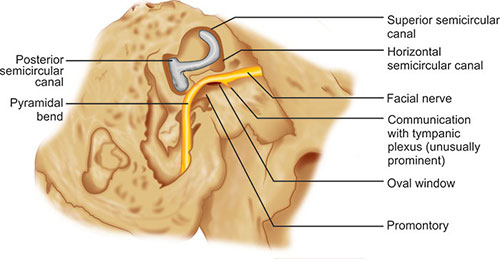
Fig. 1.2: The relationship of the facial nerve in its horizontal and vertical portions to other temporal bone landmarks.
zoom view
Figs. 1.3A and B: (A) The distribution of the facial nerve to muscles of the face after the nerve has exited from the stylomastoid foramen. The extratemporal anatomy of the facial nerve is variable. (B) Common patterns of branching of the extracranial portion of the facial nerve. Type I is seen in 20% of cases; Type II, in 37.5%; Type III in 20%; Type IV, in 15%; Type V, in 15%; and Type VI, in 2.5%, according to Coker and Fisch. (T: Temporal branch; Z: Zygomatic branch; B: Buccal branch; M: Marginal mandibular branch; C: Cervical branch).
5
zoom view
Fig. 1.4: Branches of the facial nerve.
  1. The nerve to the stapedius muscle
  2. The chorda tympani nerve, which supplies taste fibers to the anterior two-thirds of the tongue, preganglionic parasympathetic fibers to the submandibular gland, and postganglionic fibers to the submandibular and sublingual glands
  3. The posterior auricular branch, which innervates the auricularis posterior, the cranially oriented muscles of the auricle, and the occipital muscles-communicates with the greater auricular nerve, the auricular branch of the vagus nerve, and the lesser occipital nerve3
  4. The branch to the posterior belly of the digastrics muscle
  5. The branch to the stylohyoid muscle
  6. The temporal branch that supplies the lateral intrinsic muscle of the auricle, the anterior and superior auricular muscles, the frontalis, the orbicularis oculi, and the corrugator
  7. The buccal branch, which innervates the procerus, the zygomaticus major and minor, the levator labii superioris, the levator anguli oris, nasal muscles, the buccinator, and the orbicularis oris
  8. The marginal mandibular branch to the risorius muscle and the muscles of the lower lip and chin
  9. The cervical branch to the platysma; there are interconnections between the facial nerve and primarily the sensory nerves, including the trigeminal, glossopharyngeal, vagus, and cervical nerves.4
The intracranial portion of the facial nerve is supplied by the anterior inferior cerebellar artery. The intrapetrosal portion is supplied by the superficial petrosal branch of the middle meningeal artery and the stylomastoid branch of the posterior auricular artery. The extracranial portion is supplied by the stylomastoid, posterior auricular, superficial temporal, and transverse facial arteries. The anastomosis between the intratemporal branches usually occurs in the upper one-third of the vertical portion.
 
EMBRYOLOGY OF THE INTRACRANIAL PORTION OF THE FACIAL NERVE
This discussion includes information on the development of only the extracranial portions of the facial nerve. We encourage the reader to consult other literature for a brief review of the embryology of the intracranial portions,1 and for new information on differentiation and migration of the facial nerve.1 This information is very helpful for the purpose of orientation.
Fertilization through week 4: The facioacoustic primordium appears during the third week of life. It is attached to the metencephalon just rostral to the otic vesicle.6
zoom view
Fig. 1.5: A 4.2-mm embryo approximately 3-and-a-half weeks old. The region of the epibranchial placode of the second arch is labeled.
It becomes more superficial and rostral as it proceeds ventrally, and it ends adjacent to the deep surface of the epibranchial placode on the dorsal and caudal aspect of the first branchial groove. There are no branches, and the geniculate ganglion is not yet present (Fig. 1.5). During the fourth week, by the time the embryo reaches 4.8 mm in length, the facial nerve splits into two parts. The chorda tympani nerve comes off rostrally, courses ventrally to the first pharyngeal pouch, and enters the mandibular arch. The caudal main trunk terminates in mesenchyme. By the time the embryo reaches 6 mm in length, the nerve approaches the epibranchial placode, and large, dark nuclei mark the development of the geniculate ganglion.5
Weeks 5 and 6: By the time the embryo has reached 7 mm in length, near the beginning of the fifth week, the mesenchymal concentrations that form the cephalic muscles can be seen in association with their nerves (Fig. 1.6).6 The geniculate ganglion and the nervus intermedius also appear, although the latter is not always visible as a separate nerve until approximately the seventh week.5 The geniculate ganglion is lateral and rostral to the VIIIth nerve ganglion. The greater superficial petrosal nerve is present. The chorda tympani is large and enters the mandibular arch; it terminates near a branch of the mandibular nerve, which will become the lingual nerve. By the middle of the fifth week (embryo length: 10 mm), the facial nerve gives off small branches to the posterior digastric premuscle mass. The nerve terminates in mesenchyme.
In the 8-mm embryo, all the cranial nerves except the olfactory and optic nerves are recognizable. All the cranial nerves that carry sensory fibers have prominent ganglia near their points of connection with the brain. These include cranial nerves V, VII, VIII, IX, and X. The primarily efferent cranial nerves (III, IV, VI, and XII) have no external ganglia. In the 8- to 14-mm period, the posterior auricular nerve appears near the chorda tympani. Complete separation of the facial and acoustic nerves is apparent, and a discrete nervus intermedius develops.
By the time the embryo reaches 14 mm in length, the geniculate ganglion and the greater superficial petrosal nerve are well defined, and the epibranchial placode has disappeared. The greater superficial petrosal nerve courses ventrally and rostrally to the lateral aspect of the developing internal carotid artery. Here it joins the deep petrosal nerve and continues as the nerve of the pterygoid canal. It terminates in a group of cells that will become the pterygopalatine ganglion. At 16–17 mm (middle of the sixth week), a branch arises from the ventral aspect of the geniculate ganglion and courses caudally and dorsally to the glossopharyngeal ganglion. The chorda tympani and lingual nerves end near the developing submandibular ganglion. Some facial nerve fibers terminate in the mandibular arch superficially and caudally.
During this period, the superficial layer of the mesenchymal lamina of the second arch spreads to establish four laminae (Fig. 1.7):
  1. The occipital lamina (the occipitalis, auricularis posterior, and transverse nuchae muscles)
  2. The cervical lamina (the cervical part of the platysma)
  3. The mandibular lamina (the depressor labii inferioris, the mentalis, the risorius, the depressor anguli oris, the inferior part of the orbicularis oris, and perhaps the buccinator and the levator anguli oris)
  4. The temporal lamina, which spreads during the latter part of this stage (the auricularis superior).
When the embryo is between 10 and 18 mm in length, the deep layer of the second mesenchymal lamina differentiates into the posterior digastrics complex (the stapedius muscle, the posterior belly of the digastric muscle, the digastric tendon, and the stylohyoid muscle).
Weeks 7 through 9: By the beginning of the seventh week, the embryo has reached approximately 18 mm in length (Fig. 1.8). The nervus intermedius, which is smaller than the motor root of the facial nerve, passes into the brainstem between the acoustic nerve and the facial nerve motor root. The chorda tympani and lingual nerves unite proximal to the submandibular tympani nerves, and it divides into cranial and caudal branches.7
zoom view
Fig. 1.6: Diagram of a 4-and-a-half-week-old embryo illustrating the mesenchymal concentrations that will give rise to the cephalic muscles, as well as the cranial nerves associated with them.
zoom view
Fig. 1.7: A 6-week-old embryo (11 mm) showing further development of the premuscle masses of the cephalic muscles.
The caudal branches communicate with branches of cervical nerves C2 and C3. Several branches are visible in the peripheral portion of the VIIth nerve. The most caudal branches communicate with nerves from the second and third cervical ganglia in a plexus in the second arch. Another portion courses ventrally, terminating deep to the platysma myoblastic lamina. The rest of the branches course to the angle of the mouth, or caudally and superiorly into the mandibular arch. By the time the embryo is 19 mm long, some caudal branches have reached the infraorbital rim.8
zoom view
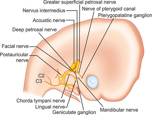
Fig. 1.8: The relationship between the facial nerve and parotid primordium at approximately 7 weeks (18 mm).
All of the peripheral branches lie close to the deep surfaces of the myoblastic laminae that will form the facial muscles. Very few fibers course dorsally. The zygomatic and temporal nerves will arise higher in the facial nerve. At 18 mm, the parotid bud is rostral and unbranched, appearing as an evagination from the lateral oral cavity area.
In the 22-mm embryo (middle of the seventh week), the posterior belly of the digastric muscle and the stapedius and stylohyoid muscles are developing (Fig. 1.9). A branch from the geniculate ganglion near the greater superficial petrosal nerve, which developed earlier, is reduced to a communication as the tympanic plexus and the lesser petrosal nerve develop from the IXth cranial nerve. The interanastomoses of the peripheral branches of the facial nerve are visible as separations of the main trunk. A small nerve branch approaches the buccal region superficial to the parotid bud.
Separations between nerve branches increase considerably in number and size by the end of the seventh week (26 mm). By then, the branch to the stapedius muscle is visible. This branch was probably present earlier, but it can be seen only after the branch separates from the main trunk.5 Peripheral branches course cranially to become the zygomatic and temporal divisions. The buccal, mandibular, and cervical divisions constitute approximately one-half of the peripheral branches. All the peripheral divisions can be identified, but the temporal branches have not yet reached the frontal region. Anastomoses are well established with the infraorbital, buccal, auriculotemporal, and mental branches of the trigeminal nerve. Previously established communications with branches of C2 and C3 have become communications with the greater auricular and transverse cervical nerves. A combined marginal mandibular-cervical branch appears between the time the embryo grows from 20 to 45 mm (seventh through the middle of the eighth week).
zoom view
Fig. 1.9: By the middle of the 7th week (22 mm), additional branching has occurred. However, the structures of the facial nerve are still anterior with respect to the external auditory meatus, which is in the process of migration from its original location low on the developing embryo.
The superficial layer of the second-arch mesenchyme differentiates into two more laminae:4
  1. Infraorbital (zygomaticus major and minor, the levator labii superioris alaeque nasi, the superior part of the orbicularis oris, and possibly the compressor naris, the depressor septi nasi, the orbicularis oculi, the frontalis, the corrugator supercilii, and the procerus)
  2. Occipital platysma.
Between 24 and 26 mm, the zygomaticus major, the depressor anguli oris, and the buccinators appear. Between 27 and 45 mm, the frontalis and zygomaticus minor appear. The branch that connects the VIIth cranial nerve with the lesser petrosal nerve (from cranial nerve IX) apparently carries small myelinated fibers that contain interspersed autonomic fibers from the auricular branch of cranial nerve X. In addition, by 26 mm the embryo develops first-order ductules of the parotid primordium, which lies next to the masseter muscle, and several branches of the facial nerve course superficial to it.7 By 27 mm, second-order ductules appear. At this time, the buccal, marginal mandibular, and cervical nerve branches approach the parotid primordium. By 32 mm (eighth week), third-order ductules are present, and the primordium has entered the parotid space.9
zoom view
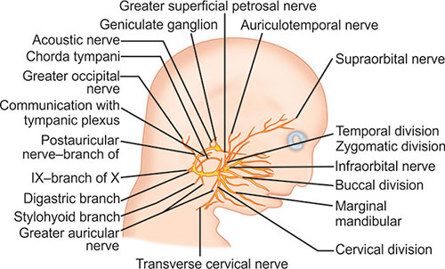
Fig. 1.10: During the 11th week (80 mm), extensive branching including communications with other cranial nerves can be seen. The vertical portion of the facial nerve is still anterior with respect to the external auditory meatus (not shown). The vertical portion and main trunk could be injured easily during surgery if the relationship of the auricle to the facial nerve were assumed to be that of an adult.
In the 37-mm embryo (8.5 weeks), fourth-order parotid ductules are present, and buccal nerve branches are superficial to the main duct. The temporal, zygomatic, and upper buccal branches are superficial on the parotid primordium. The lower buccal, mandibular, and cervical branches are deeper. The postauricular nerve goes to the occipital region, and a branch to the dorsal aspect of the auricle is present. Although there are no branches to the fused eyelids, yet a branch from the temporal division approaches the frontal region.
During the eighth week, a sulcus develops around the facial nerve, blood vessels, and stapedius muscle on the posterior aspect of the cartilaginous otic capsule. The sulcus is the beginning of the fallopian canal. The orbicularis oris, the levator anguli oris, and the orbicularis oculi also appear at approximately 37 mm.4 During the ninth week (50–60 mm), the auricularis anterior, the corrugator supercilii, the occipital and mandibular platysma, and the levator labii superioris appear. Also about the ninth week, the laterohyale fuses to the otic capsule to form part of the anterior wall of the fallopian canal and the pyramidal eminence. The segment of the anterior wall of the fallopian canal distal to the laterohyale is formed by Reichert's cartilage. The cranial nerves move closer to their adult relationships.
Weeks 10 through 15: During the 58- to 80-mm period, extensive branching of the peripheral portions of the facial nerve occurs (Fig. 1.10). Some divisions reach the midline. Extensive communication with branches of the trigeminal nerve occurs in the perioral and infraorbital regions. Communications exist between the nervus intermedius and both the VIIIth nerve and the motor root of the VIIth nerve. Despite extensive branching, the facial nerve begins its vertical course while still in the middle ear, and its relationship to the external and middle ear structures is far more anterior than it is in the adult.
During the 11th week (80 mm), the external petrosal nerve arises from the facial nerve distal to the geniculate ganglion and courses with a branch of the middle meningeal artery. Branches also arise from the facial nerve between the stapedius and the chorda tympani nerves. Together, these branches and branches of cranial nerves IX and X provide sensory innervation to the external auditory canal. Branches to the lateral aspects of the eyelids are present, and communication with the zygomaticotemporal nerve has begun to develop. Previous communications with branches of cervical nerves have now become communications with the lesser occipital and transverse cervical nerves. The horizontal portion of the facial nerve can be distinctly seen adjacent to the developing otic capsule. The nasal muscles arc also differentiated at approximately 80 mm.
The relationship between the facial nerve and the parotid gland is about the same at 12 weeks as it was at 7 weeks. However, by the time the embryo reaches 80 mm in length, complicated connections between the superficial and deep portions of the parotid primordium can be seen.7 At 14 or 15 weeks, the geniculate ganglion is fully developed, and facial nerve relationships to middle ear structures have developed more fully. During this growth period, the facial nerve has remained in association with the mesenchyme, which differentiates into the labyrinth and the mastoid.
Week 16 through birth: Between 16 and 20 weeks, the facial nerve, arterioles, venules, and the stapedius muscle lie in a sulcus on the canalicular wall. The mesenchyme in which they are surrounded is differentiating into connective tissue. Although the middle ear continues to enlarge, the facial nerve remains more superficial and anterior in relation to the auricle than it is in the adult. All definitive communications of the facial nerve are established by the 16th week (146 mm).
By 26 weeks, ossification has progressed, and growth of the outer layer of periosteal bone has resulted in a partial closure of the sulcus, forming the fallopian 10canal. The deep surface is completed first. By 35 weeks, a bony ridge has formed that separates the geniculate ganglion from the epitympanic rim. Late in fetal life, the facial canal in most cases is closed by bone except in the anterior cranial portion, where it remains open to form the facial hiatus along the floor of the middle cranial fossa. However, at least 25% of fallopian canals have this dehiscence; the most common site is adjacent to the oval window.8 The length of the dehiscence ranges from 0.5 mm to the length of the entire horizontal portion, but they are usually no larger than about 2 × 3 mm. This most common area of dehiscence is probably secondary to the failure of ossification after the stapedial artery (which passes through this area) resorbs prior to birth. The incidence of dehiscence has been reported to be as high as 55%.9
By the time of birth, the facial nerve has developed into a complex but generally consistent structure. In an interesting study of the mandibular ramus in stillborns by Sammarco et al.,10 the authors found that in 17 of 24 facial halves, some or all of the mandibular branches of the cervical facial ramus were below the angle of the mandible. Moreover, 19 of the 24 specimens had 2 or 3 mandibular branches. All mandibular branches were above the mandibular margin as they crossed the facial artery. In 16 specimens, all branches passed over the facial artery; in the other 8, they straddled the facial artery. At birth, the anatomy of the facial nerve approximates that of the adult with the exception of the nerve's exit through the superficially Iocated stylomastoid foramen. Adult anatomy occurs in this region as the mastoid tip develops postnatally.
 
CLINICAL APPLICATIONS
In patients with congenital malformations, it is usually possible to determine the fetal age at which developmental arrest occurred. This information allows the surgeon to predict the anatomy of the deformed ear and facial nerve on the basis of their usual embryologic development at the time when the arrest occurred. Moreover, if anomalies are also present in other organ systems (e.g., the kidney), they often reflect interference with development at the same time in fetal life. Hence, when a congenital anomaly of any portion of the ear can be observed visually, radiologically, or surgically, and if the clinician recognizes the fetal age at which normal development ceased in that portion, the physician should be able to predict the position of the facial nerve. If the facial nerve is anomalous, its development is most likely to have been interrupted at the same time that the development of the ear was interrupted, particularly in the case of a middle ear malformation.
zoom view
Figs. 1.11A and B: (A) Appearance of the auricle prior to otoplasty. (B) CT scan revealing absence of the external auditory canal (open arrow), an atresia plate (AP) at the level where the tympanic membrane should be, and the ossicular mass (O). The dehiscent horizontal portion of the facial nerve (curved white arrow) is seen coursing above the oval window (OW) and entering the fallopian canal (FC) at the pyramidal bend.
11
To test the validity of such predictions, surgical observations were made of 13 ears in 11 patients with congenital malformations.1,11 We describe one of these cases to illustrate the clinical application of information about facial nerve embryology. The patient was a 6-year-old boy who had been followed by the author since the age of 3 months. His auricle was malformed, and only portions of the helix, lobule, and tragus were recognizable. Computed tomography (CT) revealed the absence of the external auditory canal and the presence of a small middle ear space with an ossicular mass (Figs. 1.11A and B).
The appearance of the auricle placed the time that the defect occurred at approximately the eighth week, and the relationship of the nerve to middle ear structures had been fairly well established. However, because the middle ear was small and not fully formed, it and the vertical portion of the facial nerve appeared to the surgeon to be anteriorly and inferiorly displaced. Moreover, the vertical portion of the nerve coursed more superficially than it does in the normal adult. The nerve coursed through the region where the surgeon planned to construct the new ear canal.
After creating an ear canal and removing the atresia plate, the surgeon identified the moderately malformed ossicles. The malleus and incus were fused, but an incudomallear joint was seen. The stapes was fully developed but immobile, although it was easily manipulated. The horizontal portion of the facial nerve was in a normal position relative to the stapes, but dehiscent. As predicted, the facial nerve and middle ear were more anterior than is the case in a fully developed ear, and the facial nerve coursed anteriorly and laterally more abruptly distal to the pyramidal bend.
If adult relationships are used to gauge the position of the middle ear with respect to the position of the external meatus, the surgeon would enter the middle ear and mastoid region posterior to the vertical portion of the facial nerve and hypoplastic middle ear. Exploration from the point of entry forward would put the facial nerve at risk of injury, especially in light of its abruptly superficial course from the pyramidal bend to the stylomastoid foramen. However, with an understanding of the embryology of the facial nerve, its location can be predicted easily and accurately.
REFERENCES
  1. Sataloff RT, Sataloff JB. Embryology and anomalies of the facial nerve, 2nd edn. Jaypee Brothers Medical Publishers;  New Delhi,  India: 2013.
  1. Sataloff RT, Selber JC. Phylogeny and embryology of the facial nerve and related structures. Part II: Embryology. Ear Nose Throat J. 2003;82:764–79.
  1. Rhoton AL, Jr, Kobayashi S, Hollinshead WH. Nervus intermedius. J Neurosurg. 1968;29(6):609–18.
  1. Vidic B. The anatomy and development of the facial nerve. Ear Nose Throat J. 1978;57:236–42.
  1. Gasser RF. The development of the facial nerve in man. Ann Otol Rhinol Laryngol. 1967;76:37–56.
  1. Patten BM. Human embryology. Blakiston;  Philadelphia,  PA: 1946. pp. 112–13, 306–14, 371–5, 435.
  1. Gasser RF. The early development of the parotid gland around the facial nerve and its branches in man. Anat Rec. 1970;167:63–77.
  1. Beddard D, Saunders WH. Congenital defects in the fallopian canal. Laryngoscope. 1962;72:112–15.
  1. Baxter A. Dehiscence of the Fallopian canal. An anatomical study. J Laryngol Otol. 1971;85:587–94.
  1. Sammarco GJ, Ryan RF, Longenecker CG. Anatomy of the facial nerve in fetuses and stillborn infants. Plast Reconstr Surg. 1966;37:566–74.
  1. Sataloff RT. Embryology of the facial nerve and its clinical applications. Laryngoscope. 1990;100:969–84.