

Orthopaedics: The term was coined by Nicolas Andry. Orthos means straight and paedics means child so orthopaedics means Straight child.
Definition of orthopaedics: The branch of surgery that deals with the prevention or correction of injuries or disorders of the skeletal system and associated muscles, joints and ligaments.
ROLE OF IMAGING IN ORTHOPAEDICS
X-rays are usually the first radiological investigation done in orthopaedics and its uses involves Screening of Cortex and Marrow. X-rays are the first investigations in traumatic disorders.
Soft tissue planes (muscle and fat planes) are visualised on X-rays and often students forget this!
(Loss of soft tissue planes is earliest x-ray changes in infection/swelling in limb and it is seen after 24–48 hours of onset of disease they are more useful for infections than tumors)
Cartilage is not seen on x-rays
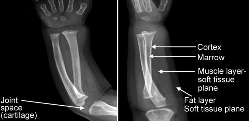

Note:
Joint space is a misnomer, Actually there is no space in areas of joints, it is the cartilage occupied area which is not visualised on x-ray. Thus whenever joint is destroyed, cartilage is destroyed hence joint space is reduced on xrays.
Types of Periosteal reaction : Indicates Bone Damage
-
No reaction: Tuberculosis of bones do not usually have a periosteal reaction.
-
Narrow zone of activity eg Solid periosteal reaction (single layer of periosteal elevation) is seen in benign lesion like.
-
In case of osteomyelitis the periosteal reaction is seen on day 7 to 10 th (or 2nd week or day 10th )
In malignant lesions there is wide area of activity e.g. Onion peel/codmans triangle and sunray appearance (all are indiative of wide area of activity) (Fig 1.5).

Onion peel or lamellated appearance- Seen in any malignant or chronic lesions (e.g. chronic osteomyelitis) but usually ewings sarcoma.

Codmans Triangle: Triangular bone growth seen at angle of lifting of periosteum it can be seen in any malignant lesion but usually osteosarcoma.

Sunray appearance/sunburst/spiculated appearance – Calcification along the vascularity of mass can be seen in any malignant lesion but usually osteosarcoma.

CT SCan: CT Scan is the investigation for cortex and calcification
Any new bone formation with 3D image – CT Scan is preferred investigation
MRI: is investigation of choice for Marrow, Soft tissues (Brain/Spinal cord/Ligaments/Tendons/nerves/vessels) and Cartilage.
Basic Images in MRI are T1 and T2.
-
T1 – 1st professional subject anatomy – so in T1 image anatomy is seen.
-
T2 – 2nd professional subject pathology – so in T2 image Pathology is seen.
‘Water is white on T2’
Water is any body fluid example, synovial fluid/C.S.F/inflammatory or traumatic edema.

Please Note:
Any occult fracture (not visualised on X-ray) e.g. Fracture neck femur ~ MRI is investigation of choice.
Any fracture in which there is marrow edema example stress fracture– MRI is investigation of choice.
Osteomyelitis starts in marrow of metaphysis– MRI is best radiological investigation.
Tumors with marrow involvement, any micrometastasis or soft tissue component – MRI can aid in diagnosis.

Bone scan: can pickup – Blastic (Osteoblastic) activity – methylene diphosphonate is taken up by osteoblasts on scanning the whole skeleton.
Bone scan show activity in areas with increased osteoblastic activity example tumors, infection or fracture. Thus in cases with bilateral stress fractures bone scan is preferred investigation.
Note: investigation of choice for unilateral stress fracture is MRI and bilateral is Bone Scan.
It can pick up tumors that go from one bone to other i.e bone to bone metastasis.
BONE:
Bone to bone/Osteosarcoma/Neuroblastoma/Ewing sarcoma (maximum incidence)

Limitation: Bone Scan cannot indentify the source of unknown primary
Note: Lesions with lytic activity do not show activity on bone scan e.g. multiple myeloma.
PET CT: Position emission tomography + CT Scan for whole body. It is a combination of 2 modalities.

18 F Deoxy glucose uptake by tumor cells (as they have anaerobic metabolism) and CT scan for all viscera so it can indentify unknown primary. (Bone Scan the uptake is by osteoblast and PET Scan by tumor cells)
Thus PET–CT is more useful than Bone Scan as it can indentify primary and is more specific for tumor cells.
Limitation: Osteoblastic lesions have limited uptake where bone scan may be more valuable.
Remember that radiological diagnosis in cases of infection and tumors is suggestive never diagnostic.
Diagnostic is always tissue diagnosis.
Tumors and infection can mimic each other (clinically and radiologically) e.g. Osteosarcoma and Ewings sarcoma are two tumors that mimic osteomyelitis. (Both have accompanying fever and increased local temperature)
Tumors and bone infections are usually metaphyseal and both need tissue diagnosis for differentiation.
-
Thus, Culture is gold standard for infection
-
Histopathology is gold standard for tumors
Osteomyelitis
-
Pyogenic Osteomyelitis on x-rays will show loss of soft tissue planes after 24-48 hours. (1st change)
-
Day 7 to 10-solid periosteal reaction is identified. (1st Bony change)
-
In tuberculosis there is no periosteal reaction.
-
Chronic osteomyelitis- sclerosed dead bone (sequestrum)is important for diagnosis and onion peel appearance is the usual periosteal reaction.
-
Mri can pick up marrow changes in metaphysis. (Best radiological investigation for Oteomyelitis and Tuberculosis)
-
Bone scan is next in preference to MRI to pick up infections by picking osteoblastic activity at the site of infection.
-
Culture and growth of organism is most definitive diagnostic modality for Oteomyelitis.
Bone Tumors
-
Xray is to localize the tumor.
-
CT scan is for extent and cortical lesion
-
MRI is for Marrow extent, micrometastasis and soft tissue involvement (Most preferred investigation for most tumors)
-
PET-CT and Bone scan for multiple lesions (PET-CT is better than Bone Scan)
-
Biopsy is definitive diagnostic modality for any tumor

IMAGING FOR ORTHOPAEDICS
-
“ORTHOPAEDICS” means:
-
Study of Bone
-
Study of Fracture
-
Straight Child
-
Study of Disease of Bone
Ans. is ‘c’ Straight Child
-
Louis Pasteur
-
Edward jenner
-
Nicholas andry
-
Kuntscher
Ans. is ‘c’ Nicholas andry
-
Investigation of choice for congenital dislocation of hip in an infant is:
-
X-ray
-
USG
-
CT Scan
-
MRI
Ans. is ‘b’ MRI
-
Sun burst appearance seen in:
-
Osteosarcoma
-
Osteopetrosis
-
Osteomyelitis
-
Osteo-radionecrosis
Ans. is ‘a’ Osteosarcoma
-
Periosteal reaction in a case of acute osteomyelitis can be seen earliest at: (March 2012)
-
5 days
-
10 days
-
15 days
-
20 days
Ans. is ‘b’ 10 days
-
Earliest sign on X-ray in TB spine is: (March 2011)
-
Paravertebral shadow
-
Narrowing of disc space
-
Gibbus
-
Straightening of spinal curves
Ans. is ‘d’ Straightening of spinal curves
-
Radiological finding of Ewings sarcoma is:
-
Soap bubble appearance
-
Sunray appearance
-
Onion peel appearance
-
Codman’s triangle
Ans. is ‘c’ Onion peel appearance
-
Mottled calcification with in the tumour is seen in X-ray is:
-
Oesteosarcoma
-
Ewing’s tumour
-
Oesteoclastoma
-
Chondrosarcoma
Ans. is ‘d’ Chondrosarcoma
-
Shepherd Crook deformity is a feature of:
-
Fibrous dysplasia
-
Post polio paralysis
-
Cerebral palsy
-
Perthe’s disease
Ans. is ‘a’ Fibrous dysplasia