

- Basic Hair Knowledge 5
- Surgery Overview35

- Assistant's Duties47

- Graft Preparation69

- Graft Placement123

This section details basic knowledge, surgery overview, assistant's duties, graft dissection, and graft placement. In the opening chapter, the basic knowledge of hair anatomy, hair growth cycles, male and female hair loss, medical management, and non-surgical options are elaborated so that an assistant can be better prepared for in-depth learning. The second and third chapter shine light on surgery overview and assistant's duties, outlining the surgery from beginning to end addressing all the tasks and duties included in assisting in hair-restoration surgery. The following two chapters cover the most important skills required for a surgical assistant. Chapter 4 describes in depth the technique of graft preparation (including slivering, graft dissection, and graft extraction), graft survival, and graft preservation. It addresses dissecting strategies and presents challenges and solutions during graft preparation and graft extraction. The chapter concludes with the critical-thinking process necessary to provide quality control. Chapter 6 delves into technique for graft preparation, elaborates on graft preservation, and the principles of proper graft placement. The chapter sets the stage for understanding graft placement by examining hair curl and hair growth directions, recipient-site type and angle, and then explains how they relate to each other. Furthermore, the chapter addresses strategies for graft placement, magnification, instruments used, and patient positioning. The chapter concludes with challenges an assistant may encounter during graft placement and offers not only solutions to those challenges but also valuable guidelines for quality control.
The true secret of happiness lies in taking a genuine interest in all the details of daily life.
William Morris
ABOUT HAIR GROWTH
Hair in General
Hair has a great social significance for human beings. It can grow on almost every area of the human body except on the palms of the hands and the soles of the feet, but hair is most noticeable in people in only a small number of areas, which are also the ones that are most commonly trimmed, plucked, or shaved. These include the face, nose, ears, head, eyebrows, legs, and armpits, as well as the pubic region. Hair is seen as an indicator of gender or aging; facial hair is one of the most visible differences between the male and female body, and while facial hair is a sign of puberty in men, white hair is an indicator of aging. Hairstyles can indicate social status, acceptance, or rejection. For example, the Manchu Qing Dynasty, beginning in the late 17th century China, ordered all Chinese citizens to adopt Manchurian hairstyles by shaving the front of their head and adopting a queue; in Islam, women would cover their hair as a symbol of modesty; while heads are shaved in prisons and concentration camps as a sign of punishment. Although in today's society a shaved head is an acceptable hairstyle for men, healthy hair indicates vitality and youth, which may explain the reason why many people experiencing baldness seek hair restoration.
Hair Anatomy
Hair has two separate structures: the hair shaft (the part we see above the skin) and the follicle (the part that is below the surface of the skin). The hair shaft is composed of strong structural protein called keratin. This is the same protein that makes up the nails and the outer layer of the skin (Fig. 1.1).
Below the surface of the skin is the hair “root” or hair follicle comprised of several structures: dermal papilla, hair bulb, hair shaft, sebaceous gland, and a tiny muscle.


Figure 1.1: Hair anatomy. Skin has three layers that are important to know: the epidermis, the dermis, and the subcutaneous adipose (fat) layer. Hair has many structures but the following are important for hair restoration: the hair shaft, the sebaceous glands, the bulge, arrector pilli muscle, and the dermal papilla (bulb)
The dermal papilla is made up of connective tissue and a capillary loop creating the place where hair production originates and where hair receives its nutrients. The hair bulb is located above and around the papilla; it contains a collection of epithelial cells interspersed with cells producing a pigment called melanin. Generally, if more melanin is present, the color of the hair is darker; if less melanin is present, the hair is lighter. This part is also called the hair matrix because it is responsible for the manufacture of the hair. The hair matrix is one of the fastest-growing cell populations in the human body, which is why some forms of chemotherapy or radiotherapy that kill dividing cells may lead to temporary hair loss.
Each strand of hair (hair shaft or hair fiber) contains three layers: medulla, cortex, and cuticle. The medulla is the innermost layer found in large and thick hair. The cortex is the middle layer, which provides strength as well as imparts color and texture to the hair. The cuticle is the outermost layer made up of tightly packed scales that form an overlapping structure similar to the roof shingles and function as a protective coat over the cortex. Most hair-conditioning products attempt to affect the cuticle by making these scales lie flat, thereby imparting a silky feel to the hair. Attached to the hair follicle is a sebaceous gland, a small sebum-producing gland found everywhere except on the palms, lips, and soles of the feet. The purpose of the sebum, an oily and waxy matter, is to lubricate the skin and hair keeping them waterproof and protected from dehydration. More sebum is produced after 7puberty. The sebum production decreases throughout life with greater reduction in women than in men. Also attached to the hair follicle is a tiny bundle of muscle fibers called the arrector pili. When this muscle contracts, it causes the hair to stand up resulting in a phenomenon commonly known as goose bumps. The bulge is located at the insertion point of the arrector pili muscle. It houses several types of stem cells, which supply the entire hair follicle with new cells and take part in healing the epidermis after a wound. This portion of the hair follicle is vital for transplanted hair to regenerate, especially if it gets damaged during the hair-transplantation process.
Hair Growth Cycles
Unlike other mammals that shed or grow hair according to the season, in humans hair growth and loss are random and not strictly but somewhat seasonal. At any given time, in humans a random number of hairs will be in various stages of growth and shedding.
The three phases of hair growth cycle are as follows: anagen, telogen, and catagen. Anagen is the active growth phase during which the cells in the matrix are dividing rapidly, adding to the hair shaft. During this phase, the hair grows approximately 1–2 cm (i.e., 1/2–1 inch) per month. The catagen stage is a short transition phase that occurs at the end of the anagen phase during which the hair follicle receives a signal to stop growing. Shortly after the hair growth stops, the dermal papilla contracts and releases the hair shaft from the follicle allowing hair to shed. The telogen phase describes the resting time for the hair follicle. During this phase, a secondary hair germ forms from a band of epithelial cells that move upward, often seen during graft dissection as a short, smaller version of the hair bulb.
Each hair follicle goes through 10–20 cycles in a lifetime, while each cycle lasts a different time: anagen lasts 3–10 years, catagen 2–3 weeks, and telogen 3–4 months. This may explain why some hairs live longer than others, as the hairs on the back of the head may be programmed to survive for twenty 10-year cycles and live for 200 years. In the human scalp, the cycles are not synchronized. Therefore, at any one time, an average of 10% of the hair is in the telogen phase (with the thought that it can range from 4% to 24%), only 1–2% is in catagen and the rest (85–90%) of the hair remain in the anagen phase. About 25–100 hairs are shed normally each day (Fig. 1.2).
Some people have difficulty growing their hair beyond a certain length because they have a short active phase of growth. However, people with very long hair have a long active phase of growth. The hairs on the arms, legs, eyelashes, and eyebrows have a very short, active growth phase (1–7 months) explaining why they are so much shorter than scalp hair.

Figure 1.2: Hair growth can be divided into three phases: anagen (active growth), catagen (active loss), and telogen (resting phase). In the scalp, 90% of the hairs remain in the anagen phase. Catagen only lasts about 2–3 weeks and is characterized by the hair-shaft separation of the dermal root, and it is recognized by a thin connective-tissue strand connecting the two. About 10% of the hairs are in telogen phase at any given moment, and this lasts about 2–3 months. Catagen phase is the shortest of the three phases and occurs prior to telogen. As the basal attachment becomes even more attenuated, the hair shaft detaches from the dermal root ultimately resulting in the hair falling out (known as exogen). Scalp hairs are asynchronously in these three cycles at any given time
ABOUT HAIR LOSS
Humans are born with approximately 100,000 hair follicles on their scalp. Scalp hair only constitutes a small fraction (100,000–150,000 follicles) of the total count for the body (approximately 5 million follicles).
Alopecia, or hair loss, is the medical term for the loss of hair from the head or body. Unlike intentional aesthetic depilation, alopecia is involuntary and often unwelcomed. However, sometimes hair loss may be caused by a psychological compulsion to pull out one's own hair (trichotillomania) or the unforeseen consequences of voluntary hairstyling routines causing mechanical hair loss (traction alopecia) from excessively tight braids often seen in African-Americans. There are many medical conditions that can cause temporary or permanent hair loss. Hair transplant is successful and possible in many cases except when the skin may be affected with a condition that would reject transplanted hair and for which 9reason a dermatologist needs to evaluate scalp before proceeding with surgery. When hair loss occurs in only one section and appears as bald patches, it is known as alopecia areata; and when complete hair loss on the body manifests including the eyebrows and eyelashes, the condition is called alopecia universalis. This condition is different from the total hair loss that follows chemotherapy. In alopecia universalis, the return of hair growth is unpredictable, while patients who lose their hair after undergoing chemotherapy are most likely to regrow their hair and do not require a hair transplant.
The most familiar type of hair loss is called baldness, and it is the state of lacking hair where it often grows, especially on the head. The most common form of baldness is a progressive hair thinning condition called androgenic alopecia that occurs in men and women. The degree and patterns of baldness can vary greatly depending on gender, age, genetics, and sometimes on one's medical condition. The most common areas of hair loss in men are frontal, temporal, and vertex, while the most commonly affected areas in women are behind the hairline and the top of the scalp and occasionally a receded hairline and/or vertex. The hair in the back of the head is rarely affected by thinning and serves as the donor area for hair transplantation. However, in some situations of female diffuse hair loss or retrograding hair loss in the male, the occipital areas could be affected, which robs those individuals of the possibility for a hair transplant (Fig. 1.3).
It was previously believed that baldness was inherited from the maternal side only. However, it is now generally accepted that both parents contribute to their offspring's likelihood of hair loss. Although baldness is not as common in women as in men, the psychological effects of hair loss, such as altering one's self-image and self-esteem, tend to be much greater in women than men.
Male-Pattern Baldness
Androgenetic alopecia (AGA) is also known as male-pattern baldness and is the most common cause of hair loss. Approximately 50% of men are affected by the age of 50, and thinning of the hair can begin as early as the age of 12 and as late as 45. Although the condition is benign, the psychosocial ramifications of AGA can be significant. A major determinant of AGA concerns testosterone (T), more specifically the enzymes that affect androgen metabolism. Testosterone is essential for health and well-being and it is a steroid hormone from the androgen group and is found in mammals, reptiles, birds, and other vertebrates. In mammals, T is secreted primarily by the testicles of males and ovaries of females, although small amounts are also secreted by the adrenal glands. In men, T plays a key role in the development of male reproductive tissues such as the testis and prostate as well as promoting secondary sexual characteristics such as increased muscle, bone mass, and the growth of body-hair.

Figure 1.3: The various regions of the hair-bearing scalp that would be rebuilt with hair transplant differently regarding angles, direction, graft sizes, etc. These regional scalp terms are referenced throughout the text and are shown in this schematic for better understanding of where they are located on the head and how they are related to one another
On average, in adult males, levels of T are about 7–8 times as great as in adult females, but, as the metabolic consumption of T in males is greater, the daily production is about 20 times greater in men.
The 5-α reductase isoenzymes (type I and II) convert T to dihydrotestosterone (DHT). The levels of both types of 5-α reductase are increased in the frontal-balding follicles compared with the nonbalding follicles found in the back of the head demonstrating that these isoenzymes contribute to AGA. Dihydrotestosterone levels are also increased in the balding scalp compared with the nonbalding scalp. Furthermore, women have 3–3.5 times less 5-α reductase than men, which probably accounts for why female AGA is less severe in most cases than with male AGA. Interestingly, individuals born without 5-α reductase type II do not develop AGA.
Aromatase is an enzyme that is part of the normal androgen metabolism that may have protective effects on AGA. Aromatase converts T into estradiol and estrone and thereby results in less conversion of T into DHT. Aromatase is found in much higher levels in female scalps with six times in the frontal scalp and four times in the occipital scalp than in men. This may explain why women who suffer from AGA may still be able to preserve their frontal hairline.
Androgen receptor proteins (ARP) are found in the outer-root sheaths and in the dermal-papilla cells of the scalp follicles. Androgen receptor proteins are found to be 30% higher in the frontal-balding scalp than in the nonbalding occipital follicles, while 40% lower in women than in men. Androgen receptor proteins are responsible 11for the signal transduction in the hair follicle that promotes the conversion of a thick, strong terminal hair into a miniaturized, fine hair. Interestingly, ARPs have the opposite effect in the beard and moustache, promoting thicker follicles in these areas at puberty.
Hair follicles produce both thick terminal hair and fine vellus hair. Vellus hairs develop on most of the human body from childhood, regardless of sex. At puberty, vellus hairs are replaced by terminal hairs in certain areas of the body, such as the axilla, pubis, and face; and this change occurs by the influence of androgenic hormones. The differentiation between vellus and terminal hair is their size and length, i.e., the hairs that grow <0.03 mm in diameter and <1 cm in length are considered vellus, and hairs that exceed the parameters mentioned above are considered terminal hairs.
One of the hallmarks of AGA is the conversion of thick terminal hairs into miniaturized, vellus-like hairs. This process of miniaturization (i.e., shrinking of hair volume and growth length) is usually an indication that one is undergoing hair loss. The main mechanism for miniaturization relates to the shortening of the anagen phase, i.e., the hair becomes “lazy” and does not grow to its full term.
Considering countless variations in hair-loss patterns, many classification schemes exist to define types and extent of baldness. The most widely accepted standard is the Norwood or Norwood-Hamilton classification for male-pattern baldness that was based on a study of a thousand adult men with male-pattern hair loss classified by the type of hair loss and age of the onset. Some of the findings were as follows:
- The patterns with least hair loss (Type I and II) are most frequently found in men aged 18–40 years. This pattern shows the recession of the hairline, frontotemporal corners, and/or temporal points.
- The Type III through V patterns are common in men of any age, and they show loss in the crown (“monk's tonsure”) and oftentimes could be combined with recession of the hairline.
- Type VI and VII, the most severe patterns of hair loss that are rare in men under age 30 and more prevalent in men older than age 60, can affect any area of the scalp, and are often characterized with an early offset and/or rapid progression of hair loss.
Typically, AGA occurs early in most cases between the age group of 15 and 25 with lifetime progression. Most oftentimes, the clinical course is a gradual one with periodic episodes of increased hair loss. Maximum pattern is usually attained by when one's age is 50, with ongoing thinning occurring throughout the remainder of one's life (Fig. 1.4).

Figure 1.4: The Norwood or Norwood-Hamilton Scale grades degrees of male-pattern baldness. Type I shows minimal to no hair loss along the frontotemporal expanse. Type II exhibits both frontotemporal recessions that do not extend farther than a line drawn through a coronal plane 2 cm anterior to the external auditory canal. Type III hair loss refers to frontotemporal recession that extends posterior to the coronal plane that lies 2 cm anterior to the external auditory canal. Type III vertex indicates hair loss that primarily affects the vertex (or crown) region with or without accompanying frontotemporal recession that does not exceed, as described in Type III. Type IV reveals greater frontotemporal loss than exhibited in Type III along with marked hair loss in the crown area but with a moderately dense swath of hair that bridges the intervening expanse between the two areas. Type V hair loss shows more extensive alopecia in both the frontotemporal and vertex areas with only a small bridge of dense hair between the two areas that remains. Type VI hair loss reveals a complete absence of any remaining hair that separates the two now confluent areas of alopecia. In addition, the hair loss is more extensive laterally and posteriorly. Type VII represents the most severe expression of male-pattern baldness with only a narrow-horseshoe configuration that remains along the posterior and lateral border of the hairline. Norwood also classified a variant of hair loss that afflicts approximately 3% of male patients with alopecia in which the frontotemporal recession marches progressively posteriorly in a uniform fashion without a central, anterior peninsula of hair. Type IIA refers to a condition in which the entire anterior hairline is receded uniformly across the forehead but does not extend any farther posteriorly than 2 cm anterior to the midcoronal line. Type IIIA reveals alopecia that extends to the midcoronal line. Type IVA signifies alopecia that has extended past the midcoronal line. Finally, Type VA indicates significant recession of the hairline into the vertex, and severer forms of this variant become indistinguishable with Types V and VI
All patterns of male-pattern hair loss tend to progress in stages and increase in prevalence with the age of the man. However, family history of male-pattern hair loss may be helpful in estimating probabilities for your future hair loss but cannot be predictive with complete accuracy.
Female-Pattern Baldness
While hair loss in men is often a genetic condition, hence a lifelong process, it is unclear what predisposes a female toward hair loss. Some females may start exhibiting hair loss as early as after puberty, while others have no signs of any unusual hair shedding until later in life when they enter menopause. In women as in men, the most likely cause of scalp hair loss is AGA: an inherited sensitivity to the effects of androgens (male hormones) on the hair follicles of the scalp. Women with hair loss due to AGA tend to have miniaturizing hairs of variable diameter over all affected areas of the scalp. While miniaturizing hairs are a feature of AGA, in women, miniaturization may also be associated with other causes. In postmenopausal women, hair may begin to miniaturize over the entire head, thereby losing its original volume and becoming difficult to style.
Although in some cases, genetics can be the causative factor, health issues such as iron deficiency or hormonal imbalance that arise from pregnancy, menopause, withdrawal of oral contraceptives, or hysterectomy can also spur hair loss in women (telogen effluvium). Pregnancy can lead to a prolonged estrogen-rich state in which hairs remain in the anagen phase until delivery. Following childbirth, the hair is shed excessively for about 4–12 weeks afterward and most likely regrows within 3–6 months. Besides these acute states of telogen effluvium, women between 30 and 60 years of age are more likely to suffer an unexplained chronic telogen effluvium marked by recurrent hair shedding that does not lead to total baldness. It is important to note that female-pattern hair loss can begin as early as the late teens to early 20s in women who have experienced early puberty. If left untreated, this hair loss associated with early puberty can progress to more advanced hair loss. In the African-American female, several types of hair loss are observed, some of which are scarring alopecias that cannot be transplanted. Consequently, hair loss in a woman (even when there is a family history of AGA) should never be assumed to be due to AGA. Examination and diagnosis by a physician, endocrinologist, dermatologist, and a hair-restoration specialist are essential before any hair transplant is undertaken. Routinely, a battery of tests is suggested to female patients exhibiting hair loss, such as a full chemistry profile, sedimentation levels to check for inflammatory diseases, serum iron levels, male/female hormones (including T and dehydroepiandrosterone sulfate [DHEAS]), and thyroid levels to check for proper function and scalp biopsy. For all of these reasons, hair loss in women can 14be more complicated than in men and should be more carefully explored for the cause so that a proper treatment plan can be instituted.
The most commonly used classification for female-pattern hair loss is the Ludwig classification (Type 1 to 3). Alternatively, for women who suffer from male-pattern baldness, the Norwood-Hamilton classification can be used. The Ludwig classification emphasizes the diffuse nature of much female-pattern hair loss with a frequently preserved hairline and thinning affecting the central top portion of the scalp, while the Norwood-Hamilton classification describes patterns of loss that are similar to male hair-loss patterns with affected hairline and/or thinning in the vertex area (Fig. 1.5).
Hair loss in women is different than in men because the areas affected can thin significantly but rarely become totally bare of hair. There are various patterns of hair loss in women, and the following are the most common types:
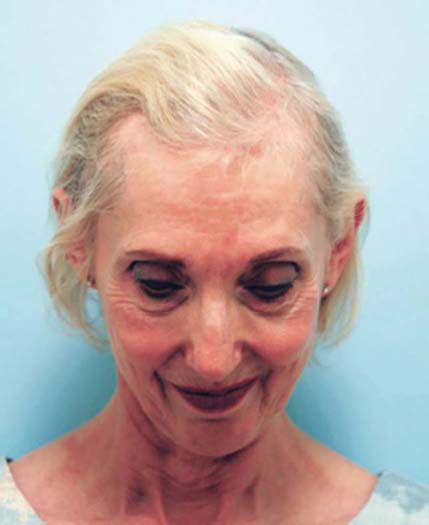
- A “Christmas tree” pattern of diffuse hair loss, with the “base” of the “tree” at the hairline and the “tip” of the “tree” at the center of the scalp. The women having this hair-loss pattern have difficulty parting their hair in the middle and often do a comb over to camouflage the thin area. This type is the most prevalent type of female hair loss and is easy to fix with hair transplantation because their donor area is oftentimes unaffected (Fig. 1.6A).
- A “Diffuse” pattern of hair loss that expands throughout the top scalp. Some studies have indicated that a diffuse thinning of hair is experienced to some degree by a majority of premenopausal women and by a large majority of postmenopausal women.There is a visible pattern of thinning that affects the top scalp and often the temporal areas as well making these women less favorable candidates for hair transplant. The most common feature for female alopecia is that the involved areas can be camouflaged with coloring or “creative” styling until it reaches a certain point of decreased density (Fig. 1.6B).
- A type of “Male-pattern baldness” with preserved central (midfrontal forelock) density. The regular female-shaped hairline is affected by the loss in both corners (i.e., frontotemporal triangles). These women often wear bangs to camouflage their bald area, which sometimes can be difficult to accomplish in cases of severe recession along the hairline. Though these women are great candidates for hair transplantation and the bald areas resemble the male-pattern hair loss of Norwood-Hamilton Types I-III, they should never be mistaken for an inflammatory scalp condition that would be unsuitable for hair transplant (Fig. 1.6C).Figure 1.6A: A woman demonstrating a Christmas tree pattern of hair loss described by Elise Olsen in which the base of the tree is noted anteriorly and the top of the tree is situated toward the crown. The Christmas tree pattern is most easily revealed with the patient looking downward with the hair parted in the midline
- A hair loss found in women (and rarely in men) called diffuse unpatterned alopecia exhibits hair thinning throughout the entire scalp, oftentimes combined with global miniaturization, depriving these individuals from ever being candidates for hair transplant (Fig. 1.6D).
- Traction alopecia is a hair loss caused by repeated pulling out of the hair from a specific area of the scalp. African-American women who wear their hair braided often exhibit hair loss in the hairline and/or in the front and above their ears. These women are good candidates for hair transplant as long as they have a good donor density (Fig. 1.6E).
- It is important to mention that there is one more “model” seen in female hair transplantation, which is not a medical condition caused by hair loss but rather an aesthetic condition of being born with a high hairline (Fig. 1.6F).


Other Types of Alopecia
There are other situations when hair restoration can be used for cosmetic or aesthetic goals, such as beard, eyelash, or eyebrow transplant. Some ethnicities, such as Asian or Hispanic, naturally lack strong facial hair and may desire to create moustache, beard, or chest hair.

Figure 1.6E: An African-American woman who had her hair in braids since an early childhood, which left her with hair loss in her temporal area

In the rare situation of someone being in an accident or enduring a scar from cleft-palate surgery, facial hair restoration could also help to camouflage the scar. Eyebrow transplant can be done in men and women. Men who are born with thin or sparse eyebrows may achieve a more masculine look by making their eyebrows appear thicker and stronger. Women may over tweeze their eyebrows or see them progressively thin out and thereby desire eyebrow restoration. 18In some cases, eyebrow loss could be an indication of hormonal unbalance or inflammatory skin condition, and these patients may not be candidates for hair transplant. Finally, eyelash transplant is a delicate procedure with unpredictable results and is often used for medical and not for aesthetic reasons.
About Hair Restoration
History of Hair Restoration
Surgical hair restoration began with the creative efforts of the pioneering Prussian surgeon and scientist, Johann Dieffenbach, in the early nineteenth century. He specialized in skin transplantation and cosmetic surgery and was very inventive and ingenious in his surgical endeavors. Significant work in hair restoration was performed by four Japanese physicians in the early to mid-1900s. Sasagawa (1930) described a hair-shaft insertion method. Okuda (1939) used round trephines (punches) to move plugs of hair from the donor scalp to the recipient areas usually into sites of scarring hair loss. Tamura (1943) used single-hair transplants in the pubic region of females, as bath houses created some embarrassment for women with sparse pubic hair; and Fujita (1953) used free grafts and subdivided grafts of 2–10 hairs in a technique that qualifies as minigrafting. These articles, unfortunately written in an older form of Kanji, were not cross-referenced to AGA and were written during times of poor communication due to the war. For these reasons, the studies were essentially lost, and for many years their work went unnoticed by the general public.
The modern era of hair transplantation in the Western world was ushered in the late 1950s when New York dermatologist Norman Orentreich began to experiment with free donor grafts to balding areas of patients who experienced hair loss. Medical grafting is a procedure similar to organ transplant, where a piece of tissue, i.e., graft, is transplanted from one area to another of the human body. It was originally believed that hair transplanted from the hair-bearing region, i.e., the donor site, into a balding region, i.e., the recipient site, would succumb to the forces of balding as did the recipient site's original hair. However, Dr Orentreich demonstrated that transplanted/new hairs grew and lasted as they did in their original home. This meant that the characteristics of the hair were preserved regardless of where they were transplanted, and this phenomenon is known as donor dominance. Otherwise, if the recipient areas were dominant, the site of transplantation would override the characteristics of the donor hair and cause transplanted hair to fall out. This was a major discovery that created the foundation of modern hair-transplant surgery and allowed its development into a mature discipline that it is today. Although his method is synonymous with unsightly “plugs”, the philosophy behind Dr Orentreich's approach is the foundation of current hair-restoration surgery.
For the next 20 years, surgeons worked on transplanting smaller grafts; and we have witnessed a tremendous evolution in hair-restoration techniques from cornrow plugs through to one-hair micrografts, now to follicular-unit grafts.
The first textbook on hair restoration, Hair Transplant Surgery, was written by Norwood in 1973. The first medical journal devoted to hair restoration, Hair Transplant Forum, was originated by Norwood in 1990 (with the name later changed to Hair Transplant Forum International). The first organization devoted solely to hair restoration, International Society of Hair Restoration Surgery (ISHRS), was founded by Drs Stough and Norwood in 1993 and is presently the world's largest organization of hair-restoration physicians. The ISHRS is a nonprofit medical association with a membership of over 1,200 members worldwide dedicated to the advancement of the art and science of hair restoration. The mission of the ISHRS is to achieve excellence in patient outcomes by promoting member education, international collegiality, research, ethics, and public awareness.
As hair-restoration surgery evolved, the number of surgical assistants participating in surgery grew and their responsibilities increased as well. Surgical assistants were involved with the ISHRS organization since its inception in 1994, and the early contributors to the field were Cheryl Pomerantz RN, Marilynn Gallespie RN, Carol Rosanelli RN JD, Helen Marzola RN, MaryAnn Parsley RN, and Joseph Greco PhD. Considering that surgical assistants are playing a significant role in patient outcome, their contribution has been recognized with an award for their achievement. Since 2003, every year one assistant is honored with the Distinguished Assistant Award recognizing his or her exemplary service and outstanding accomplishments in the field of hair-restoration surgery.
Evolution of Hair-Transplant Surgery
Initially transplanted grafts were 2- to 4-mm round plugs that were punched out from the donor area, then the bald scalp was punched out and removed from the recipient area and the “holes” on the top of the head were plugged with punched grafts from the back of the head. This procedure created a doll's head-like result and left a shotgun-riddled donor area. Although considered as a progressive and successful procedure at the time, two major downsides existed for this procedure: The donor area was rapidly depleted leaving patients “unfinished” in attempt to cover their progressively balding scalp, and the transplanted hair was extremely unnatural. Transplanted plugs were placed far apart and straight up (instead of angled to mimic the natural hair angle), which created unsightly looking doll's-hair results (Fig. 1.7A).
Dealing with the unpredictable progression of hair loss and realizing the limited availability of donor hair in the following 20 years, physicians developed several 20techniques that were revolutionary at the time but no longer now practiced. Scalp reduction was designed to make the crown area smaller by cutting out a portion of the bald scalp. The procedure was quick with immediate results but often was followed with stretchback, a phenomenon where the scalp would relax to its original size nullifying the benefits of the reduction. Sometimes, several scalp reductions were performed leaving behind an unsightly slot-deformity scar in the middle of the crown (Fig. 1.7B). Flap rotation was a procedure in which a strip of hair-bearing tissue was partially cut out from the side of the patient's head and rotated around to be sutured on the patient's head so as to recreate an immediate and full-density hairline. Rotated flaps caused many complications such as necrosis or an unnatural hairline position and appearance. Flaps rarely restored the hairline from side to side, and with the progression of the hair loss the patient would end up with an extremely thick hairline and very bald scalp behind it (Fig. 1.7C).
In the attempt to yield more natural results, surgeons worked on making hair grafts smaller, hence arriving at micrografts, tiny pieces of scalp containing only 1–2 hairs. This technique was popularized in the 1980s. Although micrografting gave more natural-looking results, the chief complaint was the lack of density and a “see-through” appearance. In 1984, Dr Headington published an article describing groupings of scalp hair. He observed that hair grows in discrete bundles of 1–4 hairs instead of single hairs as previously believed (Fig. 1.8). However, it was not until Dr Bobby Limmer's article in 1994 describing the use of a stereo microscope to divide strips of scalp into their natural hair groupings for transplantation that the hair-restoration field realized that these groupings were the units described by Headington. Thus, the term “follicular unit grafting” was given to this technique, and the shift from microtransplant to follicular unit transplant began. Mega sessions were born in which a thousand or more grafts are transplanted in a single session. The discovery of follicular units did not only allow the recreation of more naturally looking patterns but also the creation of greater density within one session of hair transplantation.
Paralleling the advancement in the recipient area, the progress in donor harvesting continued as well. There were two areas of focus for improvement: preserving donor supply and enhancing donor appearance. The progressive nature of hair loss required the physician to anticipate future needs and to plan the donor harvest accordingly. Regarding the appearance, every time a surgical procedure is performed, the donor area is left with a higher incidence of scarring. In the original punch donor harvesting after the hair was punched out, the open spaces were left to heal by themselves. Since this technique left significant donor scars behind, the next steps in evolution were in order: first to stitch the open “holes” then to cut out and connect the “holes” and suture them in one straight line.

Figure 1.7A: A photograph showing a patient who received large plugsPhoto Courtesy: M Beehner. Saratago Springs, NY, USA

Figure 1.7B: A patient who received several scalp redactions that left him with unnatural shape and a scar in the middle of his vertexPhoto Courtesy: D Didocha. Troy, MI, USA

Figure 1.7C: A patient who received Elliott flap, which restored his hairline but did not address the bald area behind the hairlinePhoto Courtesy: M Beehner. Saratago Springs, NY, USA

Figure 1.8: Hairs on the scalp do not grow singly but in groupings that have come to be known as “follicular units”. A follicular unit comprises groups of 1–4 hairs that grow in clusters spaced a short distance away from a neighboring follicular unit cluster
The improvement in the appearance of the donor area with the suturing technique was remarkable. Combining micrografting with the improvement in donor harvesting, physicians then transitioned to linear strip harvesting. This technique is still widely used in hair transplantation and is featured in this book as our preferred method (Figs. 1.9A and B)
As the progressive nature of hair loss creates the need for repeated surgeries, sometimes the donor area is left with many unsightly scars. The drawback of multiple donor harvesting over time is in the increased chances of creating wide scars with ever increasing tension on the wound edges, which becomes greater with each subsequent harvest. Thus, further refinement of the technique required better donor management. Consequently, two distinct approaches were pioneered. First, the follicular unit extraction technique was developed in order to avoid strip harvest and to circumvent a linear scar. Second, surgeons who preferred strip harvesting adopted a new suturing technique (the trichophytic closure) that allowed hair to grow through the scar and thereby camouflage its linear appearance (Figs. 1.9C and D).
In spite of all efforts to preserve the patient's hair and to maximize donor-hair capacity, some patients run out of donor hair before they achieve the desired results through hair restoration.

Figure 1.9A: A patient who received punch grafting that left his donor area filled with round scars.Photo Courtesy: M Beehner. Saratago Springs, NY, USA


Figure 1.9C: An image of a donor area 10 days after follicular unit extraction procedure. His hair above the ear is slightly thinner indicating where the grafts were harvested but the scars from the procedure are unnoticeable

Figure 1.9D: A photograph showing donor area healed with almost invisible scar using trichophytic closure
In the late 1990s, Dr Woods from Australia introduced the concept of body-hair transplant (taking hair from a patient's chest and/or beard), which gained popularity. As of the writing of this book, body-hair transplant is used primarily to cover scars or bald areas when the scalp donor hair is unavailable.
NONSURGICAL OPTIONS
Medical Treatment
Today, there are a bewildering number of medical options for hair loss. Many dietary supplements that are designed for healthy hair and that contain biotin can improve hair quality, help with telogen effluvium, but cannot prevent progression of the genetically programmed female-pattern baldness. Unfortunately, many of the herbal and vitamin hair-loss therapies lack substantiated clinical evidence of benefit. With the advent of the internet, numerous sites claim to have the product that can achieve instant hair growth and an enduring fix for baldness. Nevertheless, the Food and Drug Administration (FDA) has approved only two medical therapies for hair loss that have proven to be beneficial in selected patients, oral finasteride (marketed as Propecia by Merck) and topical minoxidil (marketed as Rogaine by Pfizer, now owned by Johnson and Johnson). Finasteride and minoxidil are approved for male hair loss, whereas only minoxidil is approved for female hair loss. Although these products may not be suitable for every person, they do provide a method to reduce or delay further hair loss and in some persons restore some hair fullness. In addition, these medications can also help to optimize the results of surgical hair restoration.
The effects of minoxidil on hair growth were discovered on the sidelines of another treatment. Originally used as a medication to treat severe hypertension, individuals who received minoxidil observed hair growth not only on their scalp but also on their body. Further investigation was conducted aiming to localize the hair-growing effects of minoxidil to the scalp only, leading to the development of the topical solution. Although the initial early reports of oral minoxidil for hypertension linked minoxidil to increasing the risk in heart-disease patients, localized topical minoxidil for hair loss is safe and is an over-the-counter treatment in the United States.
Minoxidil is currently manufactured as a 5% and 2% concentration topical solution intended for direct application to the scalp (not the hair). The 5% concentration is designed for male patients, whereas the 2% concentration is designated for women. However, women who desire a more vigorous treatment can take the 5% concentration, as it has also been recently approved for use in women as a once daily regimen. Interestingly, although the 5% concentration in women can show an increased rate of hair growth early on, there is no statistically significant difference in results between the 5% and 2% concentration after 1 year of usage.
The mechanism of action for topical minoxidil is unclear. As a potassium-channel agonist, the cellular effects on hair growth are only speculative. However, 25it is known that minoxidil can cycle hairs out of telogen (the resting phase of the hair cycle) and push them into the anagen growth phase and also lead to a more sustained anagen period. Consequently, many individuals will experience increased hair shedding early on in the treatment for several weeks and should be advised that this phenomenon actually indicates a positive effect of the treatment, as hairs move from telogen to anagen. Considering the length of each hair growth cycle, it takes approximately 6 months for new hair to regrow long enough in order for the person to realize the effectiveness of the treatment. Also, because of this effect that minoxidil has on migrating hairs from telogen to anagen, minoxidil is a recommended treatment for individuals who suffer from acute or chronic telogen effluvium.
The main side effect of minoxidil is an allergic contact dermatitis, which causes a flaky and itchy scalp and accounts for one of the two major objections for use of minoxidil. Another side effect can be heart palpitations and/or increased growth of facial hair (seen mostly in women). The most common brand name for minoxidil is Rogaine. Minoxidil is available in generic form and can be purchased in most pharmacies throughout the world.
Besides side effects, another major reason for the lack of compliance to the product is the complaint about minoxidil making one's hair look oily. Not only does oily hair lack neatness but also it is difficult to style and stick together, further exposing thin areas. Interestingly, some individuals will complain to the contrary, wherein the product renders their hair looking and feeling dry and in some situations leaves behind an unattractive white film. These complaints were mostly resolved with the release of minoxidil foam.
The original minoxidil formula found in the lotion contains the alcohol-based ingredient propylene glycol, the culprit for scalp irritation and oily-looking hair, which when removed from minoxidil foam made the product both water-soluble and more tolerable. The correct application of the product is twice a day. However, once-a-day application may be beneficial considering that the product has been thought to last for 22 hours in the scalp when applied topically as compared with a 4-hour half-life in the bloodstream when used as an intravenous preparation for hypertension.
The hair-growing effects of finasteride were also discovered accidentally when the finasteride 5-mg pill (marketed as Proscar by Merck) was given to patients to manage an enlarged prostate. Subsequent studies found that a 1-mg dosage, which is marketed as Propecia, was adequate to combat alopecia. Finasteride is a prescription-only medication and it works as a type II 5-α reductase inhibitor, where 5-α reductase is the enzyme responsible for converting T to DHT. The presence of circulating DHT impacts hair follicles susceptible to hair loss, and accordingly a 26lower level of serum DHT can slow progression of hair loss and reconvert vellus hairs back into terminal hairs. The type II 5-α reductase is found predominantly in the hair follicles (and the prostate); and, therefore, it does not promote hair growth anywhere else on the body.
Side effects occur in <2% of the patients and include decreased libido, erectile dysfunction, decreased ejaculate volume, and breast engorgement and tenderness. Side effects should fade away within 6 months after cessation and are found to be resolved in 58% of individuals who continue the treatment. Finasteride is known to reduce serum prostate-specific antigen (PSA) levels by 30–50%. Therefore, individuals over 40 years of age who are taking finasteride should be informed about their altered PSA value and are advised to make certain that their primary-care physician is consulted. Finasteride has been shown to cause potential anatomical abnormalities (hypospadias) in a male fetus in women of childbearing age who take it but not in men who father children and who are on the medication. Therefore, finasteride is absolutely contraindicated in premenopausal women and has shown only equivocal benefit in those who are postmenopausal. In several controlled studies in postmenopausal women, finasteride was shown to have no benefit,1,2 whereas one more recent uncontrolled study indicated that there might be some gain in postmenopausal women who take finasteride.3 Recently, however, a study from South Korea showed that 70 of 86 (81.4%) normoandrogenic women had global photographic improvement with a 5-mg dose of finasteride daily.4 Further, the medical literature may support the treatment with 2.5–5 mg daily dosage of finasteride for postmenopausal women who are obviously not at risk of passing on the potential fetal teratogenic effects.5,6 Although finasteride does not cause abnormalities in the fetus when men ingest it, decreased sperm count and semen volume, which may rarely occur, can diminish their fertility. Therefore, men who have difficulties conceiving may consider stopping finasteride during attempts at conception. Trials conducted in 2005 in the field of prostate-cancer prevention initially concluded that the use of finasteride increased the prevalence of prostate cancer. Additional trials conducted in 2008 rectified the original finding: since finasteride shrinks the prostate, it does not cause prostate cancer but facilitates earlier detection of cancer. Finasteride was once banned in sports because of its potential as a steroid-masking agent but recently has been approved for use in the Olympics, the Fédération Internationale de Football Association (FIFA), and many other sports.
Another medication used to treat enlarged prostate is dutasteride (Avodart by Glaxo Smith Kline), which is a potent inhibitor of both Type I and Type II isoenzymes of 5-α reductase. This medication is not FDA-approved for hair loss, and its use is considered experiential and off-label. Of note, a Phase III trial was undertaken in Korea in which 150 men with male-pattern baldness were evaluated over 6-month 27time and found statistically significant improvement compared with placebo with similar adverse events for all treatment groups.7 More recently, a randomized, double-blind, placebo, parallel 6-month study of 917 subjects involving 39 centers in 9 countries showed dutasteride at 0.5 mg daily had significantly increased hair count in an observed 2.54 cm diameter at week 24 compared with finasteride and placebo. Like the Korean study, adverse events were similar across all treatment arms.8
Initial FDA trials in the late 1990s focused almost entirely on the benefits that finasteride and minoxidil have on the vertex, also referred to as the crown, region.9 However, subsequent studies have shown both medications to be proven beneficial in the frontal, temporal, and midscalp hair. Therefore, individuals who claim that finasteride and minoxidil are only intended for restoration of the crown region are referencing outdated information.i Unfortunately, all the benefits from using these products would fade away with cessation of the medication. As it takes approximately 6 months to develop a visible effect from the medication, it usually requires about the same time for hair to reverse to its starting point once the individual stops medical treatment. Taking both medications has shown to have a synergistic benefit for the individual. When an individual decides to stop one medication, the benefits gained from the single product will disappear, but the improvement attained from the other product will be maintained for as long as the individual continues on with that product.
Although finasteride and minoxidil can provide wonderful results, they are not a replacement for hair transplantation. As standalone treatments, finasteride and minoxidil can convert many wispy vellus hairs back into thicker terminal hairs (but not universally or uniformly so). However, for those individuals who have lost all of their hairs including vellus hairs (so-called slick baldness), neither finasteride nor minoxidil will prove to be beneficial. Nevertheless, even in relatively advanced stages of hair loss, finasteride and minoxidil may retard further hair loss even if no hair is actually restored in the regions of slick baldness. There are three observable effects of medical treatment: preserving the status quo (hair is maintained and the progression of hair loss diminished); increasing hair volume (fine vellus hairs are reverted into thicker terminal hairs, which in turn provides better coverage and better styling options); and increasing hair count (with fewer hairs going into telogen stage, there are fewer hairs falling out and more hairs that remain in the anagen stage).
As established, finasteride and minoxidil do not entirely restore a full head of hair, but they can contribute to a better aesthetic result when combined with hair transplant. Initially, they can help immediately after the surgery to protect original hair from going into postoperative shock and shedding and possibly to accelerate regrowth of the transplanted hair.
_________________
Finasteride can be taken without interruption, while it is recommended to stop minoxidil 2–7 days before and after the procedure. Ongoing use of the products is, of course, beneficial and recommended. Whether medications actually cause an increase in hair volume by the conversion of vellus hair into terminal hair or by regrowing or maintaining vellus hair, which would serve to camouflage the scalp, these products can undoubtedly enhance any hair-transplant result. Besides creating a better visual result, medical management will also help retard further hair loss and lengthen the time interval necessary for the next hair-transplant session.
Low-Level Laser Therapy (LLLT)
Over the past few years, we have seen remarkable clinical benefit with LLLT. Low-level laser technology is not new. It is the portability of the equipment and the ease of compliance that gave us a new and better perspective on the effectiveness of LLLT. In 1967, when Hungarian scientists were evaluating the denuded area of a mouse to see whether visible red light would influence cancer incidence (which it did not), they found that the hair regrew much faster in the treated area. Low-level laser therapy works by photostimulation of the hair follicle by cold laser light in the visible red spectrum using wavelengths between 630 and 670 nm with low power (wattage). The photobiological reaction involves stimulation of intracellular cytochrome c, which in turn sends signals throughout the cells of the hair follicle so as to stimulate its activity, decrease cell death, and promote other changes that enhance cell activity and survival.10 In January of 2007, a handheld laser therapy device was cleared by the FDA as a treatment for AGA. Since then, several other laser-therapy devices were cleared by the FDA. Low-level lasers have been approved in this country as a treatment for carpal tunnel syndrome, chronic pain, as a wound-healing aide, and as an adjunct to liposuction procedures. Laser therapy has been safely used for decades throughout Europe and the Far East and has no documented side effects.
For years, LLLT was offered only as an in-office treatment that required the patient to travel to a clinic for treatment several times a week, which made patient compliance difficult. The trend now has been to move from in-office units to portable home units that offer similar results without the hassle. Home units come in various strengths (containing anywhere from 10 to 272 laser diodes) and various shapes such as comb, brush, standalone dome, helmet, and hat. Recently, randomized, sham device-controlled, double-blind clinical trials were conducted at multiple institutional and private practices to evaluate the efficacy of LLLT. A total of 128 males and 141 females participated in the study. Their whole scalp was treated three times a week for 26 weeks and showed that using a handheld comb device 2910 minutes three times weekly over 6 months in both men and women regardless of their age demonstrated a statistically significant increase in hair counts. In addition, there was a higher percentage of patients who reported overall improvement in their hair loss as compared with sham-device users.11 In the United States, the general consensus is that three times weekly for about 15–30 minutes is not only sufficient to stimulate hair growth but that daily usage may actually overstimulate the follicles and thereby be detrimental. Unfortunately, we do not have a firm grasp on what are the ideal wavelength, wattage, duration, and frequency to use the product, or even the ideal design of the product. Further investigations must be undertaken to refine our current understanding of this technology.
Clinically, the laser therapy can be used in combination with other medical and surgical solutions for hair loss or in isolation. It can also be used to prepare patients for surgery to minimize postoperative telogen effluvium, and post hair-transplant procedure to promote healing and to help with growth of the transplanted hairs.
Platelet-Rich Plasma
Platelets contain growth factors and biomolecules that are important for tissue repair and regeneration. The process involves the collection of the patient's whole blood, then spinning it in a centrifuge to separate platelet-rich plasma (PRP), platelet-poor plasma (PPP), and red blood cells. There are, at present, two methods of PRP preparation approved by the FDA and broad variability in the production of PRP depending on the equipment and technique used. The PRP that we use in our office is a mixture of PPP and PRP that we titrate to a 7% hematocrit (the reasons for which are outlined in Volume 1 of this book series) and it is fortified with porcine extracellular matrix (ACell Matristem MicroMatrix [AMM]). Several studies have shown that this acellular matrix can help repair any tissue damage, improve healing, and graft survival and thereby growth of the transplanted hair and provide scaffolding for the growth factors to be released over a longer period of time.
The first reports of using PRP in hair transplantation appeared in medical periodicals in 2005. The initial application was focused on healing, but more recent discussions have suggested that PRP can improve hair quality and count. In our office, we use PRP as a standard of care in hair-transplantation surgery to inject into the donor and recipient site to improve healing and we add it to the graft-storage solution to improve graft survival. Occasionally, we use PRP and AMM injections to stimulate hair in patients who are not hair-transplant candidates but have noticeable miniaturization. A common recommendation for nonsurgical, standalone PRP/AMM injections that we use as well is for a series of three treatments 6–8 months apart. However, we are cautious when recommending PRP treatments for hair growth since the treatment can cause temporary shedding and the success we have noted to be inconsistent.
Camouflaging Products and Treatments
In the process of hair thinning, decreased volume of hair no longer provides sufficient canopy to camouflage the naked scalp. As the light passes through the hair and reflects upon the scalp, an individual experiences bothersome “see-through” effect. The bigger the color contrast between the scalp tone and hair color, the more exposed the scalp appears and the more obvious thinning becomes. Camouflaging agents are topical products applied to the hair or scalp to increase the visual density of the hair. When applied to the hair, these products are usually hair-building fibers (such as keratin protein or rayon fibers) that cling to the hair, creating a web-like coverage. Although these fibers can be affixed to the hair more tenaciously with additional hair sprays or specifically designed sealers, a disadvantage of fibers is that they can come off onto pillows or clothes or by running a hand through the hair, causing social embarrassment and loss of effect. As people who have dandruff avoid wearing dark clothes, people using hair-thickening fibers should avoid wearing light-colored clothing. In addition, these products can be slightly messy during application. Because camouflaging agents intertwine with an individual's own hair, these fibers should resist light rain but can be displaced with a heavy downpour or worse during swimming. Examples of camouflaging products include Nanogen, Bumble and Bumble, Toppik, Hair Magic, ProThik, and Fullmore. Another category of camouflaging product is applied directly to the scalp as a cream or lotion with the benefit that they are more resistant to washing out. The downside to the use of this kind of product is that they are more difficult to apply in a long hairstyle and that they become too obvious if the hair is very sparse. Examples of this kind of product are DermMatch and COUVRe. In general, these agents are matched to the color of the individual's hair.
Camouflaging products are used principally for two reasons: independently to cover existing baldness and/or in combination with a hair transplant. The first reason is obvious: the individuals are bothered by hair loss but they are not interested in any surgical procedure. Alternatively, when combined with hair transplantation, camouflaging products can be offered as a temporary solution before or after surgery. If an individual has to wait to undergo hair-transplant surgery, these products can ease one's discomfort in dealing with his or her thin hair before surgery. In addition, they can be used in the postoperative setting when the patient experiences disturbing and unacceptable postoperative thinning (telogen effluvium) or during the transition period in anticipation for transplanted hair to grow.
It is important to mention that camouflaging products can accumulate and clog the scalp; hence, proper scalp hygiene should be addressed with every patient. For that reason, it is not recommended to use camouflaging products right after the 31surgery while scabs are still present. Furthermore, if a person is using minoxidil and camouflaging products simultaneously, it is important to stress the application of minoxidil on a clean scalp first. Therefore, the individual is instructed to apply minoxidil first and then the other products in the morning and for the evening application of minoxidil to wash or wipe off any residual camouflaging product before minoxidil application.
The above-noted products provide a temporary solution and are washed out with every hair wash. Scalp micropigmentation (SMP) is a semipermanent camouflaging treatment that consists of tattooing the scalp to decrease the hair-to-scalp contrast. Small round or oval dots are tattooed into the scalp to mimic shaved hair and to provide the illusion of hair density. Scalp micropigmentation can be used on a bald person to “create” a hairline or to cover the scalp and to make it look like closely shaven hair. Alternatively, it can be used between hairs to add visual hair density or to camouflage scars, specifically those donor scars that arise from prior hair transplant procedures. The biggest advantage of SMP is its immediate result but the following disadvantages may be observed: the pigment may change color and turn, e.g., from black to blue, spread under the skin, fade, or conversely maintain the original color to cause problems when the hair turns gray with aging. In addition, pigmented dots on a bald scalp are two-dimensional and do not look as natural and effective as when they are placed between hairs. However, when well done, SMP can be a valuable complementary treatment to hair transplantation.
Another way to camouflage baldness includes hairpieces and wigs. Hairpieces refer to partial hair prostheses, which are also known as hair systems or toupees, whereas a wig refers to a product that covers the entire expanse of the head. A cap is the term used to refer to a wig used by a male wearer, i.e., the product also covers the entire expanse of the scalp like a wig leaving no exposed natural hair. Most men wear hair systems as compared to caps. Although more people opt for surgical hair transplantation today making hairpieces not as popular as they were in the past, it is still important for the hair-transplant team to be educated about all hair-restoration options.
The three major complaints against hairpieces in the past were that the hair looked unnatural, that the hairpieces were too thick and bushy, and that the “hairline” was artificial in appearance and hence could not be exposed. Great strides have been made toward more natural-looking hairpieces in the last few decades. Additionally, no matter how natural a hairpiece may appear, the wearer is always worried that he may be “found out” and must limit certain activities like swimming, driving in a convertible car, or being around other people while grooming. Today's hairpieces are constructed of natural hair; their density and color can be matched very closely to the wearer's natural hair; and the base to which the hair is woven 32is thin and transparent allowing for an undetectable hairline. Hairpieces can be attached to the scalp through a variety of mechanisms. Tape adhesives are used for daily adherence so that the hair system can be easily removed at nighttime. Glue adhesives provide more durable bonding and oftentimes can keep a system in place for approximately more than a month's time. Similarly, hair-weaving uses existing hair to provide anchorage to the hair system through interweaving of the two together. Weaving can also provide a month's time for a hair system to remain in place before maintenance is required.
Hairstyling and maintenance for a hairpiece require special sensitivity and technique to provide proper adhesion and blending and are usually carried out in specialized salons. Women who wear wigs are typically well trained in hairstyling their wig, and wigs require less professional assistance for maintenance than a hairpiece does.
The benefit of a hairpiece and a wig is that they can provide immediate gratification as opposed to a hair transplant that requires 6 months or beyond to see the result and that may need several sessions to attain the desired level of hair density. Further, in individuals with extensive baldness (Norwood VII) or poor donor-hair density, a hairpiece may be the only method to attain desirable levels of coverage and hair density. In addition, someone can undergo one or several hair transplantations while wearing a hairpiece or a wig and thereby have an inconspicuous transition from baldness to having one's hair restored to complete satisfaction.
REFERENCES
- Roberts J, Price VH, Olsen E, et al. The effects of finasteride on postmenopausal women with androgenetic alopecia. In: Hair Workshop. Brussels, Belgium; 1998.
- Price VH, Roberts JL, Hordinsky M, et al. Lack of efficacy of finasteride in postmenopausal women with androgenetic alopecia. J Am Acad Dermatol. 2000;43(5 pt 1):768–76.
- Iorizzo M, Vincenzi C, Voudouris S, et al. Finasteride treatment of female pattern hair loss. Arch Dermatol. 2006;142(3):298–302.
- Yeon JH, Jung JY, Choi JW, et al. 5 mg/day finasteride treatment for normoandrogenic Asian women with female pattern hair loss. JEADV. 2011;25:211–4.
- Stout SM, Stumpf JL. Finasteride treatment of hair loss in women. Ann Pharmacol. 2010;44:1090–97.
- Trueb RM. Finasteride treatment of patterned hair loss in normoandrogenic postmenopausal women. Dermatol. 2004;209:202–7.
- Gubelin HW, Barboza MJ, Tsai TF, et al. A randomized, active- and placebo-controlled study of the efficacy and safety of different doses of dutasteride versus placebo and finasteride in the treatment of male subjects with androgenetic alopecia. J Am Acad Dermatol. 2014;70(3):489–98.
- Kaufman KD, Olsen EA, Whiting D, et al. Finasteride in the treatment of men with androgenetic alopecia. Finasteride Male Pattern Hair Loss Study Group. J Am Acad Dermatol. 1998;39(4 Pt 1):578–89.
- Jimenez JJ, Wikramanayake TC, Bergfeld W, et al. Efficacy and safety of a low-level laser device in the treatment of male and female pattern hair loss: a multicenter, randomized, sham device-controlled, double-blind study. Am J Clin Dermatol. 2014;15(2):115–27.