

CARBOHYDRATES
DEFINITION
Carbohydrates are polyhydroxy aldehydes or ketones or compounds, which yield them on hydrolysis. Carbohydrates are the main source of energy in all living organisms.
CLASSIFICATION
I.Monosaccharides
Simplest sugar, which cannot be hydrolyzed further.
- Depending upon the presence of aldehyde or keto group, monosaccharides are classified as:
- Aldoses: Having aldehyde group (e.g. Glucose, Ribose).
- Ketoses: Having keto group (e.g. Fructose, Ribulose).
- Depending upon the number of carbon present, monosaccharides are classified as follows:No. of Carbon atomsAldosesKetosesI.Trioses3GlyceraldehydeDihydroxy acetoneII.Tetroses4ErythroseErythruloseIII.Pentoses5RiboseRibuloseIV.Hexoses6GlucoseFructoseV.Heptoses7SedoheptoseSedoheptulose
II. Disaccharides
They give two molecules of monosaccharides on hydrolysis.
- Maltose → Glucose + Glucose
- Sucrose → Glucose + Fructose
- Lactose → Glucose + Galactose.
III. Oligosaccharides
They give 2–10 monosaccharide units on hydrolysis, e.g. Maltotriose, Raffinose.
IV. Polysaccharides
They give more than 10 molecules of monosaccharides on hydrolysis.
They are subdivided as follows:
- As per the building units:
- Homopolysaccharides: Containing the same monosaccharide units, e.g. starch, glycogen — formed only by glucose — (glucosan) — inulin — formed only by fructose — fructosan
- Heteropolysaccharides: Contain different types of monosaccharides, e.g. mucopoly-saccharides [Glycosaminoglycans — GAG], e.g. hyaluronic acid, heparin.
- As per the source:
- Plant polysaccharides, e.g. starch, cellulose.
- Animal polysaccharides, e.g. glycogen.
- As per their structure:
- Linear polysaccharides, e.g. starch.
- Branched polysaccharides, e.g. glycogen.
QUALITATIVE TESTS FOR CARBOHYDRATES
These tests are generally based on the following properties of carbohydrates:
- Dehydration of the neighboring hydroxyl group by strong acid to form furfural which when combined with other compounds form colored complexes.
- Reducing property of aldehyde or keto group present in it.
A. Tests Based on the Formation of Furfural with Strong Acids
- Molisch's test: General test for carbohydrates.
Reagents:
- Molisch's reagent (alpha naphthol in 95% ethanol).
- Concentrated sulfuric acid.
Procedure: To 2 mL of carbohydrate solution 3 drops of Molisch's reagent are added and mixed. The test tube is inclined and 2 mL of concentrated sulfuric acid is added through the sides of the test tube. Reddish purple or violet ring is seen at the junction of the two liquids.
Principle: Disaccharides and polysaccharides are hydrolyzed by concentrated sulfuric acid into monosaccharides, which are further dehydrated, by the acid to form hydroxy methyl furfural or furfural respectively, which condense with alpha naphthol to form the violet colored complex.
- Seliwanoff's test: This is a test for ketohexoses (fructose).
Reagents:

Procedure: To 2 mL of Seliwanoff's reagent, 3 drops of fructose solution are added and boiled for 30 seconds (Prolonged heating may convert aldohexoses to ketohexoses and give a false positive test). Then the test tube is allowed to cool in the rack. Cherry red colored complex is formed, which shows the presence of fructose.
Principle: Fructose is dehydrated by concentrated hydrochloric acid (HCl) to form furfural derivative, which condenses with resorcinol to give the cherry red complex.
Foulger's reagent: Contains urea, stannous chloride and concentrated sulfuric acid.
Procedure: To 3 mL of Foulger's reagent, 5 drops of fructose are added and boiled vigorously for one minute. Test tube is left in the rack for cooling. A blue color is obtained which shows the presence of fructose.
Principle: This test is based on the furfural formation, which condenses with urea in the presence of stannous chloride to give a blue color.
- Bial's test (Tollen's orcinol test): Specific test for pentoses such as ribose, xylose, etc.
Bial's reagent: Dilute solution of orcinol in 30% HCl.
Procedure: To 5 mL of Bial's reagent in a test tube 0.5 mL of pentose solution is added and mixed, and heated until it begins to boil. Green colored solution is obtained.
Principle: By the action of concentrated HCl upon pentose, furfural is formed. This condenses with orcinol to give green colored compound.
B. Tests Based on the Reducing Property
Carbohydrates having a free aldehyde or keto group exhibit reducing property and they are called as reducing sugars.
- Benedict's test: This is a sensitive test for reducing sugars.Benedict's reagent: It contains:
- Copper sulfate: lt provides cupric ions in solution.
- Sodium carbonate (weak alkali): It provides an alkaline medium for the reaction. lt converts reducing sugar to enediol forms of sugar, which is more powerful reducing agent.
- Sodium citrate: It prevents precipitation of cupric ions as cupric hydroxide and keeps cupric ions in solution.
Procedure: To 5 mL of Benedict's solution, 8 drops of reducing sugar are added and boiled for 2 minutes. It is then allowed to cool spontaneously. Depending on the amount of sugar present in the solution, green, yellow or red precipitate will be formed. The precipitate is cuprous oxide.
Principle: Under alkaline condition, sugar is reduced to enediol forms. Cupric ions of cupric hydroxide are then reduced by enediol forms of sugar to form cuprous hydroxide. Simultaneously, the enediol form of sugar is oxidized to sugar acid. The cuprous hydroxide during heating is converted to cuprous oxide, which may be green, yellow, or red precipitate.
Practical application: Benedict's test is the most widely used test for detection of glucose in urine. It is used commonly for screening diabetes mellitus. It answers for all reducing sugars. Depending upon the color produced, approximate concentration of sugar in urine may be ascertained.
Color of the precipitate | Approximate concentration of sugar in urine (g%) |
|---|---|
Green (+) | 0.1–0.5 |
Yellow (++) | 0.5–1.0 |
Orange (+++) | 1.0–2.0 |
Red (++++) | Above 2% |
- Fehling's test: This is also a test for reducing sugar using strong alkali.Fehling's reagent: A, B and C
- Reagent A: Copper sulfate.
- Reagent B: Potassium sodium tartrate (Rochelle salt) (to keep cupric ions in solution).
- Reagent C: Sodium hydroxide (Strong alkaline medium).
4Procedure: Each 1 mL of Fehling's reagent A, B and C are taken in a test tube, mixed and boiled to check auto reduction. 1 mL of reducing sugar is added and boiled for 2 minutes and the test tube is allowed to cool. Green, yellow or red precipitate will appear depending upon the concentration of sugar present.
Principle: Same as Benedict's test.
Advantages of Benedict's test over Fehling's test
Benedict's test | Fehling's test |
|---|---|
| Three different reagents A, B and C kept in 3 bottles. |
| Has to be tested for auto reduction |
| Strong alkali (sodium hydroxide) present here reduces other normal constituents also |
- Barfoed's test: This is also a test for monosaccharides based on their reduction property in mild acidic medium.
Reagents:
- Copper acetate
- Glacial acetic acid.
Procedure: To 2 mL of Barfoed's reagent, 1 mL of sugar solution is added, mixed and boiled for 30 seconds only (Prolonged boiling should be avoided as it gives false positive result for disaccharides which get hydrolyzed). Test tube is allowed to cool and a red precipitate is formed at the bottom of the test tube, which is cuprous oxide.
Principle: This test differs from other 2 reduction tests as this test is carried out in mild acidic medium which will be answered only by strong reducing—monosaccharides.
- Osazone test: This is a test for reducing carbohydrates. Reducing disaccharides can also be identified by this.
Reagents:
- Osazone mixture—one part of phenylhydrazine hydrochloride and two parts of sodium acetate by weight.
- Glacial acetic acid.
Osazones | Shape of crystals | Time taken |
|---|---|---|
1. Monosaccharides | ||
a. Glucosazone | Needle shaped |  |
b. Fructosazone | Needle shaped | |
2. Disaccharides | ||
a. Maltosazone | Sunflower shaped |  |
b.Lactosazone | Cotton ball or badminton ball shaped |  |
5Procedure: A boiling water bath is set up. 10 mL of sugar solution (monosaccharides and reducing disaccharides) is taken in a test tube and one spatula full of phenylhydrazine hydrochloride and two spatulas full of sodium acetate and 1 mL of glacial acetic acid are added, and mixed well and filtered in a test tube. Filtrate is kept in boiling water bath. Yellow crystals of osazones are formed within few minutes in the case of monosaccharides. In the case of disaccharides, the osazone crystals are formed only after cooling. Few crystals are transferred to a glass slide with the help of a glass rod and covered with cover slip and observed under low power microscope. The following findings will be seen in different sugars.
Principle: When reducing carbohydrates are heated with phenylhydrazine hydrochloride at 100°C and at pH 4.3, they form the corresponding osazone, aniline and ammonia with the removal of one molecule of water. As glucose and fructose differ only with respect to first and second carbon atoms they form the same osazone. Sucrose cannot form osazone, as it is not a reducing sugar.
Formation of Osazone

Reactions of Disaccharides (Maltose, Lactose and Sucrose)
- All the disaccharides will give positive result for Molisch's test, which is a test for carbohydrates.
- Reduction tests: Except sucrose, which is a non-reducing sugar, maltose and lactose will answer for Benedict's test and Fehling's test. Sucrose is a non-reducing sugar as it does not contain a free aldehyde or keto group. Reducing disaccharides will not answer for Barfoed's as it is a specific test for reducing monosaccharides only.
- Sucrose hydrolysis test
Procedure: Sucrose solution is first hydrolyzed by adding 3 drops of concentrated hydrochloric acid and boiled. After cooling the test tube under tap water, 20% sodium carbonate is added dropwise until there is no effervescence, to neutralize the acid. Then Benedict's reagent is added and boiled for 2 minutes and the test tube is allowed to cool spontaneously. A precipitate, which may be green, yellow or red in color is obtained depending upon the amount of glucose and fructose present.
6Principle: Sucrose will not answer for Benedict's test, as it is a non-reducing sugar. After hydrolysis by concentrated HCl sucrose is converted into glucose and fructose which are reducing monosaccharides, which answer this. This is a confirmatory test for sucrose.
Osazone test: Maltosazone will be formed within 30 minutes. These crystals are soluble in hot solution and so they separate out on cooling. The crystals are sunflower shaped or having the shape of sunflower petals, on viewing through the microscope.
Lactosazone will be formed within 20 minutes. The crystals are soluble in hot solution and so they separate out on cooling and these crystals are cotton ball shaped.
Sucrose cannot form osazone crystals as it is a non-reducing sugar.
Reactions of Polysaccharides
1. Iodine Test
It is a test for polysaccharides. Starch, dextrin and glycogen can be differentiated by this test.
Reagents:
- N/50 iodine solution.
- Glacial acetic acid.
Procedure: 2 mL of polysaccharide (starch, glycogen and dextrin) solution, 5 drops of glacial acetic acid are added. Then 3 drops of N/50 iodine solution are added drop by drop and mixed. Starch gives blue color which disappears on heating and reappears on cooling. Dextrin gives a reddish-purple color which disappears on heating but does not reappear on cooling. Glycogen gives a reddish-brown color which disappears on heating and appears on cooling.
Principle: This test depends upon the adsorption property of the large polysaccharide molecule, which adsorbs the smaller iodine molecules on their surface to form an ill-defined colored complex. On cooling, the complex is reformed and the color reappears except in dextrin.
2. Half Saturation and Full Saturation Test
These are also tests for polysaccharides to differentiate starch from dextrin.
Reagent:
- Solid ammonium sulfate
- Saturated solution of ammonium sulfate in water.
Procedure: To 5 mL of polysaccharide solution, 5 mL of saturated ammonium sulfate solution is added and shaken vigorously and kept in the rack for 5 minutes. A white precipitate is formed. This is filtered to another test tube and 2 drops of glacial acetic acid is added to the filtrate and then 3 drops of N/50 iodine solution is added. Starch will not give any blue color whereas dextrin gives a red color.
Full saturation test: 5 mL of dextrin solution is taken in a test tube and solid ammonium sulfate is added with constant shaking until it begins to settle at the bottom of the test tube. This is filtered in another test tube and 2 drops of glacial acetic acid and 3 drops of N/50 iodine solution are added. Dextrin gives red color.
Principle: As starch has small surface area, it is precipitated by half saturation with ammonium sulfate. Dextrin is a small molecule with greater surface area and so it is not precipitated even by full saturation with ammonium sulfate.
3. Hydrolysis of Starch
Three mL of starch solution is taken in a test tube and 2 drops of concentrated HCl are added and boiled for 2 minutes and then cooled. It is then neutralized with 40% sodium hydroxide (or 20% sodium carbonate) till the effervescence is stopped. With the neutralized hydrolyzate Benedict's test is performed to get a positive result.
Principle: Starch is a non-reducing sugar. Acid hydrolysis breaks starch into glucose molecules. As glucose is a reducing sugar Benedict's test gives a positive result.
To study the reactions of carbohydrates, perform the following tests:
| To confirm the presence of carbohydrate. |
| To confirm the presence of polysaccharides. |
| To confirm the presence of reducing sugars. (Both mono and disaccharides) |
| To confirm the presence of reducing monosaccharides only. |
 | To confirm the presence of fructose. |
| To confirm the presence of pentose. |
| To confirm the presence of non-reducing disaccharide sucrose. |
| To confirm the presence of non-reducing polysaccharide — starch. |
| To confirm the type of reducing mono or disaccharide. |
Reactions of Monosaccharides—Tests for Glucose
Ex. No. 1
Date:
Experiment | Observation | Inference |
|---|---|---|
| A reddish-violet or purple ring is seen at the junction | It is a general test for carbohydrates. Glucose is dehydrated by concentrated sulfuric acid to form furfural derivatives, which on condensation with Molisch's reagent forms a violet ring |
| No characteristic color is seen. Only yellow color of iodine is seen | This is a specific test for polysaccharide. Glucose being a monosaccharide does not answer this |
| A precipitate, which may be green, yellow or red depending on the amount of sugar present in the solution is obtained | It is a sensitive reduction test indicating the presence of reducing sugars. The precipitate formed is red cuprous oxide |
| A red precipitate is formed at the bottom of the test tube | It is a specific reduction test for monosaccharide. Prolonged heating converts disaccharide to monosaccha-rides and gives false positive test. This test is used to distinguish monosaccharides from disaccharides |
| No cherry red complex is formed | This test is only for ketohexoses. Glucose being an aldohexose does not answer this test. Prolonged heating may give false positive test for glucose and sucrose |
| No blue color is formed | This test is only for ketohexoses. Glucose being an aldohexose does not answer this test |
| Needle shaped yellow crystals of glucosazone will be formed within 10 minutes  | It is a test to identify glucose in the given sample |
| Result: Reactions of the reducing monosaccharide — glucose are thus studied. | ||
Experiment | Observation | Inference |
|---|---|---|
Reactions of Monosaccharides — Tests for Fructose
Ex. No. 2
Date:
Experiment | Observation | Inference |
|---|---|---|
| A purple or violet ring is seen at the junction of two layers | It shows the presence of carbohydrates. It is dehydrated by concentrated sulfuric acid to form furfural derivatives, which on condensation with Molisch's reagent gives violet ring |
| No characteristic color is seen. Only yellow color of iodine is seen | This is a specific test for polysaccharide. Fructose being a monosaccharide does not answer this test |
| A precipitate which may be green, yellow or red depending on the amount of sugar obtained in the solution | It is a sensitive reduction test, indicating the presence of reducing sugar |
| A red precipitate is formed at the bottom of the test tube | It is a specific test for monosaccharide. Prolonged heating converts disaccharide to monosaccharides and gives false positive test. This test is used to distinguish monosaccharides from disaccharides |
| Note the color of the sample. The sample turns into a cherry red colored complex | This test is positive for ketohexoses only. Fructose being a ketohexose answers this test. Ketohexoses are dehydrated by concentrated HCl to form furfural derivatives, which on condensation with resorcinol forms the red complex |
| A blue color is seen | This test is positive for ketohexoses. Fructose being a ketohexose answers this test. This test is based on the formation of furfural which condenses with urea in the presence of stannous chloride to give a blue color |
| Needle shaped yellow crystals of fructosazone will be formed within 7 minutes  | This confirms the presence of reducing sugar fructose |
| Result: The reactions of fructose are thus studied. Fructose is a reducing ketohexose. | ||
Experiment | Observation | Inference |
|---|---|---|
Reactions of Monosaccharides—Tests for Pentoses—Ribose and Xylose
Ex. No. 3
Date:
Experiment | Observation | Inference |
|---|---|---|
| A purple ring or reddish-violet ring is formed at the junction of the two layers | This is a general test for carbohydrates. Pentoses are dehydrated by concentrated sulfuric acid to form furfural derivatives which on condensation with Molisch's reagent forms a violet ring |
| No characteristic color is seen. Only yellow color of iodine is seen | This is a specific test for polysaccharides. Pentoses do not answer this as they are monosaccharides |
| As per the amount of pentose present, green, yellow or red precipitate is obtained | It is a sensitive reduction test, indicating the presence of reducing sugar. Precipitate formed is red cuprous oxide |
| A red precipitate is formed at the bottom of the test tube | It is specific reduction test for reducing monosaccharides. It helps to differentiate reducing monosaccharides from reducing disaccharides |
| No cherry red complex is formed | This test answers only for ketohexoses. The given solution is an aldopentose and so it does not answer this test |
| No blue color is obtained | This test is also for ketohexoses. Aldopentoses will not answer this test |
| Green colored solution is obtained | This is a confirmatory test for pentoses. Concentrated HCl converts pentoses into furfural derivatives, which then condenses with orcinol to give green colored complex |
| Result: Reactions of pentoses are thus studied. | ||
Experiment | Observation | Inference |
|---|---|---|
Reactions of Disaccharides — Test for Maltose
Ex. No.4
Date:
Experiment | Observation | Inference |
|---|---|---|
| A reddish-purple or reddish-violet ring is seen at the junction of two liquids | It is a general test for carbohydrate whether free or bound to substances such as proteins or lipids. Maltose being a disaccharide answers this test |
| No characteristic color is seen. Only yellow color of iodine is retained | It is a specific test for polysaccharide. Maltose being a disaccharide does not answer this |
| A precipitate which may be green, yellow or red is obtained depending on the amount of sugar | It is a reduction test indicating the presence of reducing sugar. Maltose being a reducing sugar will answer this test |
| No red precipitate is obtained. | It is a specific reduction test for monosaccharides. The given sample being a disaccharide does not answer this test |
| No cherry red colored complex is obtained | This is a positive test for ketohexoses only. Maltose will not answer this test |
| No blue color is obtained | The sample being a disaccharide does not answer this test since it has no keto group |
| Sunflower shaped maltosazone crystals appear in 30 minutes  | This confirms the presence of reducing sugar maltose |
| Result: Reactions of maltose are thus studied —reducing disaccharide. | ||
Experiment | Observation | Inference |
|---|---|---|
Reactions of Disaccharides — Tests for Lactose
Ex. No.5
Date:
Experiment | Observation | Inference |
|---|---|---|
| A reddish-purple or reddish-violet ring is seen at the junction of the two layers | It is a general test for carbohydrate whether free or bound to substances such as proteins or lipids. Lactose being a disaccharide gives a positive reaction |
| No characteristic color is seen. Only yellow color of iodine is retained | It is a specific test for polysaccharide. Lactose being a disaccharide does not answer this test |
| A precipitate which may be green, yellow or red is obtained depending on the amount of sugar | It is a reduction test indicating the presence of reducing sugar. Lactose being a reducing sugar answers this test |
| No red precipitate is obtained | It is a specific reduction test for monosaccharide. The given sample being a disaccharide does not answer this test |
| No cherry red colored complex is seen | This test is positive for ketohexoses only. Lactose doesn't answer this test |
| No blue color is formed | The sample being a disaccharide does not have a keto group, and so it shows negative reaction |
| Cotton ball shaped or hedgehog shaped lactosazone crystals appear within 20 minutes 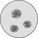 | It indicates the presence of reducing disaccharide namely lactose |
| Result: Reactions of lactose are thus studied—reducing disaccharide. | ||
Experiment | Observation | Inference |
|---|---|---|
Reactions of Disaccharides —Tests for Sucrose
Ex. No.6
Date:
Experiment | Observation | Inference |
|---|---|---|
| A reddish-purple or reddish-violet ring is seen at the junction of two layers | It is a general test for carbohydrate whether free or bound to substances such as proteins or lipids. Sucrose being a disaccharide gives positive reaction |
| No characteristic color is seen. Only yellow color of iodine is retained | It is a specific test for polysaccharide. Sucrose being a disaccharide does not answer this test |
| No characteristic color is seen | It is a reduction test indicating the presence of reducing sugar. Sucrose being a non-reducing sugar does not answer this test |
| No red precipitate is formed | It is a specific reduction test for monosaccharides. The given sample being a disaccharide does not answer this test |
| The precipitate which may be green, yellow or red color, depending on the amount of sugar, is obtained | Sucrose is a non-reducing sugar but after hydrolysis by concentrated HCl it is converted to glucose and fructose which are reducing sugars. This is a confirmatory test for sucrose |
| The sample turns into a cherry red colored complex | This test is positive for ketohexoses only. Fructose being a ketohexose answers this test. Shows the presence of fructose in the hydrolysate |
| Result: The reactions of sucrose (Non-reducing disaccharide) are thus studied. | ||
Experiment | Observation | Inference |
|---|---|---|
Reactions of Polysaccharides—Tests for Starch
Ex. No. 7
Date:
Starch
The most commonly available polysaccharide is starch, which is a mixture of amylose and amylopectin. The individual glucose unit in amylose are linked by 1,4-glycosidic linkages. Amylopectins have branching points contributed by alpha-1,6-glycosidic linkages. Starch is insoluble in cold water, but forms a colloidal solution in hot water. Starch has no detectable reducing property.
Experiment | Observation | Inference |
|---|---|---|
| A reddish-purple or violet ring is seen at the junction of two liquids | It is a general test for carbohydrate. Starch being a polysaccharide answers this test |
| A blue color is seen | It is a specific test for polysaccharide. Starch being a polysaccharide answers this test. Iodine forms colored complexes with polysaccharides |
| No characteristic reaction or colored precipitate is seen | It is a reduction test for reducing sugar. Starch being a non-reducing sugar does not answer this test |
| A precipitate which may be green, yellow or red may be obtained depending upon the amount of glucose present | Hydrochloric acid has hydrolyzed starch into its monomer glucose which gives positive result to Benedict's reagent |
| White precipitate is formed and no blue color is seen in the filtrate | A white precipitate is obtained which indicates that starch is precipitated by half saturation with ammonium sulfate. The filtrate does not give blue color with iodine as it does not contain starch |
| Result: The reactions of starch—a polysaccharide, are thus studied. | ||
Experiment | Observation | Inference |
|---|---|---|
Reactions of Polysaccharides—Tests for Dextrin
Ex. No. 8
Date:
Dextrin
They are formed as intermediate products during the course of hydrolysis of starch by dilute mineral acid and also by the reaction of amylases. These heterogeneous compounds are grouped into amylodextrin, erythrodextrin and achrodextrin based on the color produced by N/50 iodine. These three groups, which are in the order of decreasing molecular weight gives violet, red and no color in iodine reaction. Dextrins are partially soluble in water. They have more reducing activity.
Experiment | Observation | Inference |
|---|---|---|
| Reddish-purple ring is seen at the junction of the two liquids | It is a general test for carbohydrate. Dextrin being a carbohydrate, answers this test |
| Reddish-purple color is seen | It is a specific test for polysaccharide. Dextrin being a polysaccharide answers this test as iodine forms colored complexes with polysaccharides |
| A light green colored precipitate is obtained | Dextrin answers this test because it has free aldehyde group at the end of the chain of its molecule |
| Red colored precipitate is obtained | Dextrin on hydrolysis produces its monomeric units glucose which gives a positive reaction with Benedict's reagent |
| No precipitate is formed Red color is seen in the filtrate | Dextrin is not precipitated by half saturation with ammonium sulfate. So filtrate gives reddish-purple color on addition of Iodine as it contains dextrin |
| A reddish-purple color is seen in the filtrate | Dextrins are not precipitated even by full saturation with ammonium sulfate. So the filtrate gives reddish-purple color on addition of iodine as it contains dextrin |
| Result: The reactions of dextrin —a polysaccharide, are thus studied. | ||
Experiment | Observation | Inference |
|---|---|---|
SCHEME FOR IDENTIFICATION OF UNKNOWN CARBOHYDRATE—(SCHEME III)


Identification of an Unknown Carbohydrate
Ex. No. 9
Date:
Experiment | Observation | Inference |
|---|---|---|
| Purple ring is formed at the junction of 2 liquids No purple ring | Presence of carbohydrates Absence of carbohydrates |
| Blue color is formed No blue color | Presence of polysaccharide Absence of polysaccharide |
| White precipitate is formed No blue color in filtrate No white precipitate, filtrate gives reddish-purple color | Presence of starch Presence of dextrin |
| Red precipitate is formed No red precipitate | Presence of reducing sugar Presence of non-reducing sugar |
| Red precipitate | Presence of sucrose |
| Red precipitate | Presence of glucose |
| Red precipitate No red precipitate | Presence of monosaccharides Presence of reducing disaccharides |
| Cherry red color is formed No cherry color | Presence of fructose Absence of fructose |
| Blue color is formed No blue color | Presence of fructose Absence of fructose |
| Green color is obtained No green color | Presence of pentose Absence of pentose |
| Needle shaped crystals Sunflower shaped crystals Cotton ball shaped crystals | Glucose/fructose Presence of maltosazone Presence of lactosazone |
| Result: The unknown solution contains ............................................ which is ......................................................................... | ||
Exercise No. 10
Object—To study the acid hydrolysis of starch
Hydrolysis of starch by acid (demonstration)
- Set up a boiling water bath
- Arrange 10 test tubes in a rack, number them from 1 to 10
- To each tube numbered 1 to 5, add 5 mL distilled water
- To each tube numbered 6 to 10, add 2.5 mL Benedict's qualitative reagent
- Arrange another set of 5 test tubes separately. Number them as A,B,C,D and E. Add to each of these tube, 4 mL of 1% starch solution and 1 mL of 20% HCl. Mix
- Keep the tubes (A to E) in the boiling water bath and note the time as 0 minute
- After one and half minutes, take out tube A and cool under tap water (to inhibit further hydrolysis). Pour half of its contents into tube No. 1. To the other half add 20% sodium carbonate drop by drop till there is no effervescence (to neutralize the acid) and pour this solution into tube No. 6
- Similarly, proceed with the other tubes from the water bath at intervals of 5, 8, 12 and 20 minutes
- The contents of the tube B taken out at 5 minutes should be transferred in tubes 2 and 7
- The contents of the tube C taken out at 8 minutes should be transferred in tubes 3 and 8
- The contents of the tube D taken out at 12 minutes should be transferred into tubes 4 and 9
- The contents of the tube E taken out at 20 minutes should be transferred into tubes 5 and 10
- To each of the tubes 1 to 5, add 3 drops of N/50 iodine solution. Mix, note the color in each tube
- Finally arrange all the tubes 1 to 10 in the rack in their order of numbering
- Note the state of hydrolysis and relative reduction in each tube.
Observation | Inference | |||||
|---|---|---|---|---|---|---|
Time | Tube No. | Iodine test | Tube No. | Benedict's test | Stages of hydrolysis | Products formed |
1½ min | 1 | Blue | 6 | Blue | No Reduction | Solube starch |
5 min | 2 | Violet | 7 | Green | Slight reduction (+) | Amylodextrin + Maltose |
8 min | 3 | Reddish-violet | 8 | Red | More reduction (++) Maltose | Erythrodextrin + More |
12 min | 4 | No characteristic change (Yellow color of Iodine) | 9 | Red | Still more reduction (+++) | Achrodextrin + still more Maltose |
20 min | 5 | No characteristic change (yellow color of iodine) | 10 | Red | Maximum reduction (++++) | Maltose + Glucose |
| Note: Hydrolysis of starch by acid results in glucose as end product. Hydrolysis of starch by enzyme results in maltose as end product. | ||||||
| Result: The products yielded after the acid hydrolysis of starch are studied. | ||||||
RELEVANT QUESTIONS—CARBOHYDRATES
- How will you classify carbohydrates?
- Define carbohydrates.
-
- Monosaccharides
- Disaccharides
- Polysaccharides.
- Define—Aldoses and ketoses. Give example.
- What is meant by oligosaccharides? Give example.
- What are the monosaccharides present in:
- Maltose
- Lactose
- Sucrose
- Name the general test for carbohydrates. What is its principle?
- What is furfural?
- What is Molisch's reagent?
- What is meant by reducing sugar? Give example.
- Which are the tests that will answer positively for reducing sugars?
- What are the components of Benedict's reagent? What are their uses?
- What is the red precipitate formed in Benedict's test called?
- Why glucose is a reducing sugar?
- Why sucrose is a non-reducing sugar?
- What are the other names for sucrose?
- What are the components of Fehling's reagent?
- What are the advantages of Benedict's test over Fehling's test?
- Name the test which helps to differentiate monosaccharides from reducing disaccharides.
- What are the components of Barfoed's reagent?
- What is the precipitate formed in Barfoed's test?
- Name the tests which distinguish glucose from fructose.
- Name the ketohexoses.
- What are the components of:
- Seliwanoff's reagent
- Foulger's reagent.
- Why does glucose and fructose form the same osazone?
- What is the nature of glucosazone/fructosazone?
- How will you differentiate maltose and lactose?
- What are the components of osazone mixture?
- Why sucrose cannot form osazones?
- What is the confirmatory test for sucrose?
- Name the general test to detect polysaccharides.
- What is the principle of Iodine test?
- Why should we do Iodine test in acid medium?
- Name the hydrolytic products of starch by enzyme in the order of their formation.
- Name the test which differentiates starch from dextrin?
- How do you classify polysaccharides?
- How will you differentiate starch from dextrin?
- Which test is employed in the clinical laboratory to detect glucose in urine?
- How will you estimate the approximate concentration of glucose in urine by the color produced in Benedict's test?
PROTEINS
Proteins occupy the major portions of our body. It is a nitrogenous substance made up of carbon, hydrogen, oxygen and nitrogen. Some proteins contain sulfur and phosphorus.
AMINO ACID
All proteins are polymers of amino acids. There are 20 amino acids, which are genetically coded. According to their structure, they are classified as follows:
Aliphatic Amino Acids
- Mono-amino-mono-carboxylic amino acids:
- Unsubstituted simple amino acids—Glycine, Alanine
- Hydroxy amino acids—Serine, Threonine
- Branched chain amino acids—Valine, Leucine, Isoleucine
- Sulfur containing amino acids—Cysteine, Methionine
- Mono-amino-dicarboxylic amino acids and their amides (Acidic amino acids):
- Aspartic acid—Asparagine
- Glutamic acid—Glutamine
- Diamino-mono-carboxylic amino acids (Basic amino acids):
- Lysine
- Arginine
- Diamino-dicarboxylic amino acid:
- Cystine — Made up of 2 molecules of Cysteine (not genetically coded).
Aromatic Amino Acids (Benzene Ring)
Phenylalanine—Tyrosine (Hydroxyphenylalanine).
Heterocyclic Amino Acids
- With indole ring—Tryptophan
- With imidazole ring—Histidine.
Imino Acid
Proline.
PEPTIDES
Peptides are formed when two or more amino acids are linked.
Alpha carboxyl group of one amino acid reacts with alpha amino group of another amino acid to form a peptide bond or CO = NH bridge. As per the number of amino acids present, they are called as dipeptide (2 amino acids), tripeptide (3), etc. and more than 10 amino acids condense to form a polypeptide.
PROTEINS
Proteins are made up of peptide chains. They are classified as follows:
- As per their over all shape:
- Fibrous proteins, e.g. Collagen, Keratin
- Globular proteins, e.g. many enzymes, Globulin.
- As per their physical and chemical nature:
- Simple proteins—Made up of only amino acids, e.g. Albumin.
- Conjugated proteins—Proteins are conjugated to a non-protein prosthetic group, e.g. Hemoglobin (Heme—Prosthetic group).
- Derived proteins—Derived from hydrolysis or denaturation of proteins.
- Primary derived proteins (no change in peptide bonds), e.g. Proteans, Metaproteins.
- Secondary derived proteins (cleavage of peptide bonds), e.g. Proteoses, Peptones, Peptides.
When proteins are broken down, the following changes take place.
Proteins → metaproteins → primary proteoses → secondary proteoses → peptones → polypeptides → amino acids.
Primary proteoses: They are not coagulated by heat, but precipitated by half saturation with ammonium sulfate.
Secondary proteoses: They are not coagulated by heat but they can be precipitated only by full saturation with ammonium sulfate.
Peptones: They are not coagulated by heat. They cannot be precipitated by half and full saturation with ammonium sulfate.
The lower proteins—proteoses and peptones are not coagulated by heat. They require greater concentration of ammonium sulfate for precipitation, as the molecules of the broken down proteins are very small.
QUALITATIVE TESTS FOR PROTEINS (DEPENDING ON THE GENERAL PROPERTIES OF PROTEINS)
Precipitation Reactions of Proteins
Globular proteins form colloidal solutions, in which the particles have the diameter between 1 millimicron to about 200 millimicrons.
There are two types of colloids:
- Suspensoids.
- Emulsoids.
Suspensoids
The stability of suspensoids are due to the electrical charges over the surface of the molecule which prevent the formation of precipitation.
Emulsoids
There are two factors for the stability of an emulsoid:
- Electrical charges over the surface of the molecule.
- Hydration shell around the molecule.

Various Precipitation Methods of Proteins
A. By heavy metals
Lead acetate, Mercuric chloride, Mercuric nitrate
B. By negatively charged alkaloidal reagents
Picric acid, Trichloroacetic acid, Phosphotungstic acid, Sulfosalicylic acid, Tannic acid
C. By concentrated salt solutions (Salting out method)
Ammonium sulfate, Sodium sulfate
D. By bringing to isoelectric point by adjusting pH
Isoelectric point or isoelectric pH is the pH of a protein at which level it carries equal number of positive and negative charges and the net charge is zero. At this pH, the protein does not move either to the anode or to cathode in an electric field. Isoelectric point varies from protein to protein.
Isoelectric pH of albumin is 4.88 and that of casein is 4.60.
E. By heat and acid
F. By organic solvents like ethanol or acetone (not done)
A. Precipitation by Heavy Metals
Test No. 1
Reagent: 10% lead acetate solution.
Procedure: To 2 mL of protein solution, 5–10 drops of lead acetate solution are added and mixed. A white precipitate is formed.
Test No. 2
Reagent: Mercuric chloride or mercuric sulfate.
31Procedure: To 2 mL of protein solution, mercuric chloride or sulfate solution is added drop by drop. A white precipitate is formed.
Principle: Metal ions (lead, mercury) combine with the negatively charged groups on the protein to form the precipitate — metal proteinate. This principle is used in treating heavy metal poisoning by giving egg white (protein - albumin) orally which will precipitate the heavy metals.
B. Precipitation by Alkaloidal Solution
Test No. 1
Procedure: To 2 mL of protein solution 2–3 drops of sulfosalicylic acid are added and mixed. A white precipitate is formed.
Test No. 2
Procedure: To 2 mL of protein solution few drops of freshly prepared solution of tannic acid are added. A light brown or yellow precipitate is obtained.
Test No. 3
Procedure: To 2 mL of protein solution, 1 mL of picric acid is added and mixed. A yellow precipitate is formed.
Principle for test no 1 and 3: The negatively charged ions of the alkaloids—Sulphosalicylic acid, tannic acid and picric acid neutralize the positive charges on the protein causing denaturation which will end in precipitation.
Test No. 4
Procedure: To 2 mL protein solution, 3 drops of glacial acetic acid are added and mixed. Few drops of potassium ferricyanide are then added drop by drop to get a yellow precipitate.
Principle: Glacial acetic acid confers a positive charge on protein. When the negatively charged ferricyanide ions are added, the proteins get precipitated by neutralizing the charges.
C. Precipitation by Concentrated Salt Solution (Salting Out Method)
Reagents:
- Solid ammonium sulfate.
- Saturated solution of ammonium sulfate.
Procedure
Half Saturation Test: To 3 mL of protein solution, 3 mL of saturated solution of ammonium sulfate is added and mixed well.
A white precipitate is formed in the case of casein, gelatin and globulin. Albumin cannot be precipitated by half saturation.
Full Saturation Test: To 3 mL of protein solution, solid ammonium sulfate is added and mixed well till few undissolved ammonium sulfate crystals get settled at the bottom (saturated solution).
Albumin, gelatin and casein are precipitated by full saturation to give a white precipitate.
Peptone will not produce precipitate.
Principle: The colloidal protein solution has electric charge and the shell of hydration to keep it stable.
32Both these factors are removed by adding neutral salt like ammonium sulfate. Depending on the surface area of the protein, the amount of salt to be added will be varying. Smaller molecules like albumin have relatively large surface area and so it is precipitated by full saturation only. Casein and gelatin have smaller surface area and so they can be precipitated by half saturation and full saturation. Peptones are very small molecules and so cannot be precipitated even by half saturation.
D. Precipitation at Isoelectric pH (On Casein) (Casein has a pH of 4.6)
Reagents: Acetic acid, bromocresol green
Procedure: To 3 mL of casein solution, 3 drops of bromocresol green are added and mixed. A blue color is seen on the alkaline side. Then 2% of acetic acid is added drop by drop till the color changes to light green (pH 4.6). A white curdy precipitate is formed.
Principle: At isoelectric pH, the solubility of proteins is minimum as the molecules become electrically neutral at this pH.
E. Precipitation by Heat and Acid
Reagent: 1% acetic acid.
Procedure: Protein solution is taken up to three-fourth level of a test tube. The tube is inclined slightly and by holding it at the bottom, the upper portion of the solution is heated over a flame. An opalescent coagulum is formed at the heated portion only and there is no change at the nonheated bottom portion. 3–5 drops of 1% of acetic acid is added to get maximum white precipitate.
Principle: Proteins undergo denaturation by denaturing agents such as heat. Denaturation is a process in which the physical, chemical and biological properties of proteins are altered due to the breaking up of peptide bonds. There will be change in the conformation of the molecule. Denaturation is a reversible process and the denatured protein can be converted to its original form when the denaturing agents are removed. Coagulation, on the other hand, is an irreversible process.
II. Color Reactions of Proteins
Color reactions of proteins are produced by a particular amino acid or by a particular group of amino acids. These reactions are helpful in identifying the protein in a given solution and also to identify the particular amino acid.
A. General Color Reactions
1. Biuret Test
Reagents: 5% sodium hydroxide and 1% copper sulfate (Biuret reagent).
Procedure: To 2 mL of protein solution, 2 mL of 5% NaOH is added and mixed well. Then 2–3 drops of 1% copper sulphate solution is added. Protein gives a violet or purple colored complex.
Principle: This is a general test for proteins. Compounds which contain 2 or more peptide bonds will answer this. Dipeptide and free amino acids do not give biuret positive. The violet or purple color is due to the formation of the coordination complex between cupric ions and nitrogen atoms of the peptide bonds.
2. Ninhydrin Test (Demonstration Only)
Reagent: To 2 mL of protein solution 2–3 drops of ninhydrin are added and boiled. After cooling a bluish-purple colored complex is formed.
33Principle: This test is answered by proteins containing free alpha amino groups only. Alpha amino groups of the protein, react with ninhydrin to give a blue-violet colored complex called Ruhemann's purple. Proline and hydroxyproline will not answer this test as they do not have alpha amino group. Instead they will give yellow color only.
Alpha amino acid + Ninhydrin = Aldehyde + CO2 + NH3 + Hydrindantin
Hydrindantin + NH3 + Ninhydrin = Blue colored complex (Ruhemann's Purple).
B. Specific Color Reactions
1. Xanthoproteic Test (Based on the presence of Benzoid Radical)
Reagents: Concentrated nitric acid, 40% sodium hydroxide.
Procedure: To 3 mL of protein solution, 1 mL of concentrated nitric acid is added and heated for one minute. Due to denaturation, a yellow precipitate is formed. After cooling it under tap water, 2 mL of 40% sodium hydroxide solution is added which makes the precipitate to become orange in color.
Principle: Aromatic amino acids, which contain benzene ring, will answer this (phenylalanine, tyrosine and tryptophan). Nitration of benzene ring forms a yellow derivative which turns orange in alkaline medium.
2. Cole's Mercuric Nitrite Test (Modified Millon's Test) (Based on the presence of Hydroxy Benzene Radical)
Reagents: 10% mercuric sulfate, 10% sulfuric acid and 1% sodium nitrite.
Procedure: To 1 mL of protein solution, 1 mL of 10% mercuric sulfate in 10% sulfuric acid is added and boiled for half a minute. A yellow precipitate is formed. 3 drops of sodium nitrite are added to this and the precipitate is changed to red color.
Principle: This test is specific for hydroxyl group containing aromatic amino acid—tyrosine. The red color is due to mercury complex of nitrophenol derivative.
3. Aldehyde Test (Based on the presence of Indole Group Containing Amino Acid—Tryptophan)
Reagents: Dilute formalin (1 in 500), 10% mercuric sulfate in 10% sulfuric acid, concentrated sulfuric acid.
Procedure: To 3 mL of protein solution, 2 drops of 1/500 formalin and one drop of 10% mercuric sulfate in 10% sulfuric acid are added and mixed well. Then 3 mL of concentrated sulfuric acid is added through the sides of the tube. A purple or violet ring is formed at the junction of the 2 liquids.
Principle: Indole group of tryptophan combines with formaldehyde in the presence of sulphuric acid (Oxidizing Agent) and mercuric sulfate to give the violet or purple colored complex.
Note: Collagen and gelatin will not answer this due to the absence of tryptophan.
4. Sakaguchi Test (Based on the presence of Guanidine Group of Arginine)
Reagents: Alpha naphthol, 20% sodium hydroxide and bromine water.
Procedure: To 3 mL of protein solution, 5 drops of 20% sodium hydroxide and 4 drops of 1% alpha naphthol in alcohol are added and mixed. To this 10 drops of bromine water are added. A bright red color is formed.
Principle: This test is specific for guanidine group of arginine. In alkaline medium alpha naphthol combines with the guanidine group of arginine to form a complex which is oxidized by sodium hypobromite to produce a red color.
345. Sulfur Test (Based on the presence of Sulfur Group—Sulfur Containing Amino Acids—Except Methionine)
Reagents: 40% sodium hydroxide, 2% lead acetate.
Procedure: To 2 mL of protein solution, 2 mL of 40% sodium hydroxide is added and mixed and boiled for one minute. Then 5 drops of lead acetate solution are added. A black or brown precipitate is formed.
Principle: Sulfur containing amino acids — Cysteine and Cystine answer this but not Methionine. Sulfur present in the protein reacts with sodium hydroxide while boiling, and it is converted to inorganic form of sodium sulfide. This reacts with lead acetate to form a black precipitate of lead sulfide. Methionine does not answer this test as the sulfur is not released by alkaline digestion.
Note: Gelatin and casein do not answer this test due to the absence of cysteine.
6. Pauly's Test (Based on the presence of Imidazole Group of Histidine)
Reagents: 0.5% sulfanilic acid, 1% sodium nitrite and 10% sodium carbonate.
Procedure: To 1 mL of 0.5% sulfanilic acid and 1 mL of sodium nitrite solution, 2 mL of protein solution is added and mixed and cooled under tap water. Then 1 mL of 10% sodium carbonate is added to make the solution alkaline. An orange red complex is formed.
Principle: This test is specific for imidazole group of histidine. Diazotized sulfanilic acid reacts with the imidazole group of histidine to form this cherry red complex in alkaline medium.
7. Molisch's Test (Based on the presence of Carbohydrate Group in Glycoproteins such as Mucin and other Glycoproteins)
Procedure: To 2 mL of protein solution, 2 drops of 1% alpha naphthol in alcohol is added and then through the sides of the test tube, 2–3 mL of concentrated sulfuric acid is added to get a violet or purple ring at the junction of two liquids.
Principle: This test is a general test for carbohydrates, answered by carbohydrate containing glycoproteins such as mucin. Casein, gelatin albumin and peptones will not answer this as they are not glycoproteins.
Reactions of Proteins — Albumin
Ex. No:
Date:
Albumin
It is a simple heat coagulable protein. It is soluble in water. It is precipitated by full saturation with ammonium sulfate. It contains all amino acids.
Experiment | Observation | Inference |
|---|---|---|
I. Precipitation Reactions A. Precipitation by Salts of Heavy Metals | ||
| White precipitate is formed | This test indicates the presence of proteins. The metal cation combines with negatively charged group of proteins to cause precipitations of albumin |
| White precipitate is formed | This test indicates the presence of proteins. The metal cation combines with negatively charged group of proteins to cause precipitations of albumin |
B. Precipitation by Alkaloidal Reagents | ||
| White precipitate is formed | Sulfosalicylic acid has negatively charged ions, which neutralize the positively charged ions of albumin and cause denaturation resulting in precipitate formation |
| Light brown precipitate is formed | Tannic acid has negatively charged ions, which neutralize the positively charged ions of albumin and cause denaturation resulting in precipitate formation |
| Yellow precipitate is formed | Picric acid has negatively charged ions, which neutralize the positive charge of albumin causing denaturation resulting in precipitation |
C. Precipitation by Concentrated Salt Solution (Salting out method) 1. Half Saturation Test | ||
Take 3 mL of albumin solution in a test tube. Add equal volume of saturated ammonium sulfate solution. Mix well and allow to stand for 5 minutes. A white precipitate is formed. Filter it. To the filtrate add 3 mL of 40% sodium hydroxide and 2–3 drops of copper sulfate (Biuret reagent) | Filtrate gives violet color | |
2. Full Saturation Test | ||
Take 3 mL of albumin solution in a test tube. Add solid ammonium sulfate until little salt settles at the bottom. Wait for 5 minutes. A white precipitate is formed. Filter it. To the filtrate add 3 mL of 40% sodium hydroxide and 2–3 drops of 1% copper sulfate solution (Biuret reagent) | Filtrate gives blue color of copper sulphate | Albumin is completely precipitated by full saturation with ammonium sulfate. Hence, filtrate gives no color due to the precipitation of albumin |
D. Precipitation by Heat and Acid Heat Coagulation Test | ||
Fill 3/4th of test tube with albumin solution. Hold the test tube at its bottom. Incline it slightly and heat the upper portion of the solution over the flame. Add 3 drops of 1% acetic acid. (This test is used to confirm the presence of albumin in urine) | A white precipitate is formed | It indicates the presence of the coagulable protein albumin. Appearance of coagulation indicates denaturation of albumin and precipitation by acetic acid. Acetic acid gives the suitable pH to get maximum precipitate |
II. Color Reactions | ||
| A violet colored complex is formed | The color produced is due to denaturation and the formation of coordination complex between cupric ions and the nitrogen atoms of the peptide bonds |
| ||
| A bluish-purple colored complex is formed | Albumin contains free amino acids, which react with Ninhydrin to give a blue color. This test is not answered by proline and hydroxyproline |
| First a white precipitate is formed due to denaturation. It turns yellow on heating. On adding sodium hydroxide an orange yellow precipitate is formed | This test is specific for the benzene ring of the aromatic amino acids such as tyrosine and tryptophan. Based on the nitration of the benzene ring it forms a yellow derivative, which turns orange color. Phenylalanine may give a weak positive or negative reaction—due to the presence of unsubstituted phenyl group |
| A yellow precipitate is formed which becomes red after boiling and adding sodium nitrite solution | |
| A purple or violet colored ring is formed at the junction of the two liquids | This test is specific for the indole group of tryptophan. The indole ring of tryptophan acts with formaldehyde in the presence of oxidized agents to form a violet colored ring. This shows the presence of tryptophan in albumin |
| A bright red color is formed | This test is specific for guanidine group of arginine. In alkaline medium, alpha naphthol combines with the guanidine group of arginine to form a complex, which is oxidized by bromine water to produce red color. It shows the presence of arginine in albumin |
| A black or brownish precipitate is formed | This test indicates the presence of sulfur containing amino acids such as cysteine and cystine. On boiling with NaOH, the sulfur present in the amino acid is converted to inorganic form of sodium sulfide which gives black precipitate. Methionine does not answer the test as the sulfur group is not released by alkaline digestion. It shows the presence of cysteine and cystine in albumin |
| An orange red colored complex is formed | This reaction is specific for imidazole group of histidine. Diazotized sulfanilic acid reacts with imidazole group of histidine to give a cherry red color under alkaline condition. It shows the presence of histidine in albumin |
| No reddish-purple or violet ring is seen at the junction of the two liquids | This shows the absence of carbohydrate in albumin |
Result: The reactions of albumin are thus studied.
| ||
Experiment | Observation | Inference |
|---|---|---|
Reactions of Gelatin
Ex. No:
Date:
Gelatin is formed when collagen is hydrolyzed. It is not heat coagulable. It can be precipitated by half saturation.
Experiment | Observation | Inference |
|---|---|---|
I. Precipitation Reactions A. Precipitation by Heavy Metals | ||
| No precipitate is formed | Gelatin is not precipitated by the positively charged metal ions |
| No precipitate is formed | Gelatin is not precipitated by the positively charged metal ions |
B. Precipitation by Alkaloidal Reagents | ||
| White precipitate is formed | Sulfosalicylic acid has negatively charged ions, which neutralize the positive charge of gelatin to cause denaturation to produce precipitation of protein salicylate |
| Light brown precipitate is formed | Tannic acid has negatively charged ions which neutralize the positive charges on gelatin to cause denaturation to produce precipitation as protein tannate |
| Yellow precipitate is formed | Picric acid has negatively charged ions which neutralize positive charges on gelatin to cause denaturation to produce precipitation as protein picrate |
C. Precipitation by Concentrated Salt Solution (Salting out method) Saturation Test: | ||
Take 3 mL of gelatin solution in a test tube and add equal volume of saturated ammonium sulfate solution. Mix well and wait for 5 minutes. To the filtrate add 3 mL of 40% sodium hydroxide and 2–3 drops of 1% copper sulfate solution | Filtrate does not give violet color | Gelatin gets precipitated by half saturation method and so the filtrate does not give violet color with Biuret reagent as it does not contain gelatin |
D. Precipitation by Heat and Acid Heat Coagulation Test: | ||
Fill 3/4 of the test tube with gelatin solution. Hold the test tube at the bottom. Incline it slightly and heat the upper part of the solution over a flame. Add 3–5 drops of 1% acetic acid | No precipitate is formed | It indicates that gelatin is not a heat coagulable protein |
II. Color Reactions of Gelatin | ||
| A violet colored complex is formed | |
(Precaution: If copper sulfate is added in excess, violet color will not be formed) | ||
| A bluish-purple colored complex is formed | Gelatin contains free amino acids which react with Ninhydrin to give a blue color. This test is not answered by proline and hydroxyproline |
| A faint yellow color is obtained | Presence of trace amount of aromatic amino acids |
| A faint red color is obtained | Shows the presence of a trace amount of mercuric tyrosine in gelatin |
| No purple or violet colored ring is formed at the junction of the two liquids | This shows the absence of tryptophan in gelatin as this test is specific for the indole group of tryptophan |
| A bright red color is formed | This test is specific for guanidine group of arginine. In alkaline medium, alpha naphthol combines with the guanidine group of arginine to form a complex which is oxidized by bromine water to produce red color. It shows the presence of arginine in gelatin |
| No black or brownish precipitate is formed | This indicates the absence of sulfur containing amino acids — cysteine and cystine in gelatin |
| An orange red colored complex is formed | This test is specific for imidazole group of histidine. Diazotized sulfanilic acid reacts with imidazole group of histidine to give a cherry red color under alkaline conditions. It shows the presence of histidine in gelatin |
| No reddish-purple or violet ring is seen at the junction of the two liquids | This shows the absence of carbohydrates in gelatin |
Result: The reactions of gelatin are thus studied.
| ||
Reactions of peptone
Ex. No:
Date:
Peptone
Peptone is a secondary derived protein formed by the cleavage of peptide bonds. It is soluble in water. It is not heat coagulable. It is not precipitated by even full saturation with ammonium sulfate.
Experiment | Observation | Inference |
|---|---|---|
I. Precipitation Reactions A. Precipitation by Salts of Heavy Metals | ||
| A white precipitate is formed | This test indicates the presence of proteins. The metal cation combines with negatively charged group on peptones causing precipitations |
| A white precipitate is formed | This test indicates the presence of proteins. The metal cation combines with negatively charged group on peptones to cause precipitations |
B. Precipitation by Alkaloid Reagents | ||
| Brown precipitate is formed | Tannic acid has negatively charged ions which neutralize the positive charges on peptones causing denaturation resulting in precipitation |
| Yellow precipitate is formed | Picric acid has negatively charged ions which neutralize the positive charge of peptones causing denaturation resulting in precipitation |
| Yellow precipitate is obtained | Glacial acetic acid has negatively charged ions which neutralize the positive charge on peptones and cause denaturation resulting in precipitation. |
C. Precipitation by Concentrated Salt Solution | ||
| It gives rosy red color | Peptone is not precipitated by half saturation with ammonium sulfate. So, it gives rosy red color with Biuret reagent as it contains peptones |
| Rosy red color is formed | |
D. Precipitation by Heat and Acid | ||
| No white precipitate is formed | Peptones are not heat coagulable proteins |
I. Color Reactions | ||
| A rosy red or pink colored complex is formed | It is a general test for protein. This test is answered by compounds which contain 2 or more peptide bonds. The color produced is due to the formation of coordination complex of cupric ions and nitrogen atoms of the peptide bonds present in peptones |
| A bluish-purple colored precipitate is formed | Peptones give positive reaction. This test is answered by all amino acids containing free alpha amino group |
| A faint orange colored precipitate is formed | Peptones show faint reaction. This test is specific for benzene ring present in the aromatic amino acids such as tyrosine, and tryptophan |
| A red precipitate is formed | This test is specific for hydroxy phenyl group of tyrosine. The red precipitate is due to the mercury complex of nitrophenol derivative. It shows the presence of tyrosine in peptone |
| A violet or purple colored ring is seen at the junction of 2 layers | Peptones show positive reaction. This test is specific for indole group of tryptophan in peptones. The indole group combines with formaldehyde and oxidizing agent to form the violet ring. The presence of tryptophan in peptone is confirmed |
| A bright red color is seen | |
| A faint black or brown precipitate is formed | Peptones show faint positive reaction. It shows the presence of cysteine and cystine and not methionine. The sulfur present is liberated as sodium sulfide which react with lead acetate to form a black precipitate of lead sulfide. It shows that small amount of cysteine and cystine are present in peptones |
| Orange red colored complex is seen | Peptones show positive reaction. This test is specific for imidazole group of histidine. Diazotized sulfanilic acid reacts with imidazole group to form a cherry red colored complex under alkaline conditions. It shows the presence of histidine in peptones |
| No reddish-purple ring is seen at the junction of the two layers | Peptones show negative reaction due to the absence of carbohydrate |
Result: The reactions of peptones are thus studied.
| ||
Reactions of Casein
Ex. No:
Date:
Casein is the chief protein of milk. It is a conjugated protein (phosphoprotein). In milk, it is present as calcium caseinate. It is insoluble in water but soluble in dilute acids and alkali. It is not coagulable by heat. Its isoelectric pH is 4.6. It can be precipitated by half saturation with ammonium sulfate.
Experiment | Observation | Inference |
|---|---|---|
I. Precipitation Reactions | ||
| Filtrate does not give violet color | |
| A curdy white precipitate is formed | It indicates that casein is precipitated at isoelectric pH of 4.6 |
| A canary yellow or lemon yellow precipitate is formed | Indicates the presence of phosphorus in casein. On heating with sulfuric acid and nitric acid, casein is digested and phosphorus is liberated. It reacts with ammonium molybdate to form a canary yellow precipitate of ammonium phosphomolybdate |
Alternate test: To 5 mL of casein solution, 0.5 mL of 20% NaOH is added, heated and cooled. 0.5 mL of concentrated nitric acid is added and mixed. It is then filtered and to the filtrate, a pinch of solid ammonium molybdate is added and warmed | A canary yellow precipitate is formed | On boiling with NaOH, Organic phosphate is converted to inorganic phosphorus. On reacting with ammonium molybdate it is converted to ammonium phosphomolybdate |
II. Color Reactions | ||
| A violet or purple colored complex is formed | It is a general test for protein. This test is answered by compounds which contain 2 or more peptide bonds. The color produced is due to the formation of coordination complex of cupric ions and nitrogen atoms of the peptide bonds present in casein |
| A bluish-purple colored precipitate is formed | |
| An orange colored preci-pitate is formed | This test is specific for benzene ring present in the aromatic amino acids such as tyrosine and tryptophan present in casein |
| A red precipitate is formed | This test is specific for hydroxy phenyl group of tyrosine. The red precipitate is due to the mercury complex of nitrophenol derivative |
| A violet or purple colored ring is seen at the junction of 2 liquids | Casein shows positive reaction. This test is specific for indole group of tryptophan in casein. The indole group combines with formaldehyde in the presence of oxidizing agents to form a violet colored ring. The presence of tryptophan in casein is confirmed |
| A bright red color is seen | This test is specific for guanidine group of arginine. In alkaline medium alpha naphthol combines with guanidine group of arginine to form a complex which is oxidized by sodium hypobromite to produce a red color. It shows the presence of arginine in casein |
| No black or brown precipitate is formed | Casein shows negative reaction. It shows the absence of cysteine and cystine in casein |
| An orange red colored complex is seen | Casein shows positive reaction. This test is specific for imidazole group of histidine. Diazotized sulfanilic acid reacts with imidazole group to form a cherry red colored complex under alkaline conditions. It shows the presence of histidine in casein |
| No reddish-purple ring is seen at the junction of the two layers | Casein shows negative reaction due to the absence of carbohydrate |
Result: The reactions of casein are thus studied.
| ||
Analysis of Unknown Protein
Ex. No.
Date:
Experiment | Observation | Inference |
|---|---|---|
1. Biuret test | Violet color is produced | Presence of peptide bonds in protein |
2. Precipitation at isoelectric point | Curdy white precipitate No precipitate | Presence of casein Absence of casein |
3. Heat coagulation test | Formation of coagulum No coagulation | Presence of albumin Absence of albumin |
4. Half saturation test | Formation of violet color No violet color | Presence of albumin or peptone Presence of gelatin or casein |
5. Full saturation test | Formation of violet color No violet color | Presence of peptone Presence of albumin |
Color Reactions | ||
1. Biuret testss | Violet colored complex | Albumin, gelatin, peptone and casein answer this |
2. Ninhydrin test | Bluish-purple colored | All give positive result |
3. Xanthoproteic test | Precipitate orange colored | All give positive reaction Gelatin gives faint positive |
4. Cole's mercuric nitrite test | Red precipitate | All give positive reaction Gelatin gives faint positive |
5. Aldehyde test | Purple ring seen at the junction of 2 liquids No ring formation | Albumin, casein/peptones Gelatin |
6. Sulfur test | Black or brownish-black precipitate No black precipitate | Albumin or peptone Gelatin or casein |
7. Sakaguchi test | Bright red color | Albumin/Gelatin/Peptone/Casein |
8. Pauly's test | Orange red color | -do- |
9. Molisch's test | Reddish purple ring at the junction of 2 liquids No ring formation | Albumin Peptone/Gelatin/Casein |
| Result: The given protein is ...................... | ||
Experiment | Observation | Inference |
|---|---|---|
SCHEME FOR IDENTIFICATION OF AN UNKNOWN PROTEIN (SCHEME II)

RELEVANT QUESTIONS — PROTEINS
- Define proteins.
- How do you classify proteins? Give example.
- What are the stages of hydrolysis of proteins?
- Define suspensoid and emulsoid.
- What are the stabilizing factors for emulsoid?
- Name the heavy metals which are used to precipitate proteins.
- Name the alkaloidal reagents which are used to precipitate proteins.
- In clinical laboratory, how is the alkaloidal precipitation useful?
- What is the practical significance of heavy metal precipitation?
- What is the principle of precipitation reactions?
- What is denaturation?
- Name few denaturing agents.
- Is denaturation reversible?
- What is the clinical significance of heat coagulation test?
- What is isoelectric pH?
- What is the isoelectric pH of casein?
- Which is the general color reaction of protein?
- What is the principle of Biuret test?
- What is the practical clinical utility of this test?
- Which amino acids answer xanthoproteic test?
- Which amino acid answers Cole's mercuric nitrite test?
- What is the other name for this test?
- What is the red precipitate formed in this reaction?
- Name the amino acid with benzene ring or name the aromatic amino acids.
- Name the amino acid with indole ring.
- Which test is specific for indole group containing amino acid?
- Which amino acid answers Sakaguchi test?
- Name the sulfur containing amino acids.
- Name the amino acid which answers sulfur test.
- Why methionine does not answer this test?
- Name the amino acid which has the imidazole group.
- Name the test which is answered by the amino acid with imidazole group.
- Which color reactions are not answered by
- Casein.
- Gelatin?
- Which protein is not precipitated even by full saturation?
- Why does egg albumin answer Molisch's test?
NON-PROTEIN NITROGENOUS SUBSTANCES (NPN)
Non-protein nitrogenous substances include all substances containing nitrogen other than proteins. They are: urea, uric acid, creatine, creatinine and ammonia.
A. UREA
It is the end product of amino acid metabolism. It is synthesized in liver. The normal blood urea level is 15 to 40 mg per 100 mL and normal urinary urea is 15–30 g per day.
Reactions of Urea
1. Sodium Hypobromite Test
Reagents:
- 1% urea solution.
- Alkaline hypobromite solution—100 mL of 40% sodium hydroxide with 10 mL of bromine water.
Procedure: To 3 mL of urea solution, 3–5 drops of freshly prepared sodium hypobromite solution is added. A brisk effervescence is formed.
Principle: Sodium hypobromite acts on urea and decomposes it to give nitrogen, carbondioxide and water. Liberation of nitrogen gas produces brisk effervescence.
2. Specific Urease Test
Reagents:
- Urease suspension—(10 g of Horse gram powder is mixed with 100 mL of 30% ethanol).
- Phenolphthalein indicator.
Procedure: 3 mL of urease suspension is added to 3 mL of urea solution and then a drop of phenolphthalein indicator is added. Warm the tube with the palm of the hands. Wait for 5–10 minutes. A pink color is produced.
Principle: Horse gram contains the enzyme urease which acts on urea to form ammonium carbonate which changes the solution to pink color in the presence of the indicator.
B. URIC ACID
It is the product of catabolism of purines, in human body. It is synthesized in liver and excreted through urine.
Normal value in blood:
Male: 3–7 mg/100 mL
Female: 2.5–6.5 mg/100 mL
Normal level in urine
250–750 mg/day.
Reactions of Uric Acid
1. Benedict's Uric Acid Test
Reagents:
- Benedict's uric acid reagent: 100 g of sodium tungstate is dissolved in 150 mL of water. 51To this 16.3 mL of orthophosphoric acid and 16.8 mL of concentrated sulfuric acid are added and boiled for 2 hours using reflux condenser. After cooling the solution, solid sodium carbonate is added little by little with stirring
- 20% Sodium carbonate solution.
Procedure: To 3 mL of uric acid solution, 1 mL of Benedict's uric acid reagent and 1 mL of 20% sodium carbonate solution are added to get a deep blue color.
Principle: As uric acid is a reducing agent, it reduces phosphotungstic acid to tungsten blue in strong alkaline condition.
2. Schiff's Test
Reagents: Ammoniacal silver nitrate.
Procedure: A filter paper is moistened with ammoniacal silver nitrate. Then 2–3 drops of uric acid solution is added to that. A black color is produced.
Principle: Salt of silver is reduced by uric acid to metallic silver.
3. Murexide Test
Reagents:
- Concentrated nitric acid.
- Dilute ammonia (10%).
Procedure: In a China dish, 1 mL of uric acid is taken and few drops of concentrated nitric acid is added and heated over the flame gently. A reddish-yellow residue is formed. To this, 1–2 drops of dilute ammonia is added to get a purplish-red color.
Principle: Uric acid is oxidized by nitric acid to give the reddish-yellow purpuric acid which combines with ammonia to form ammonium purpurate or murexide, which is purple red in color.
C. CREATININE
Creatinine is the anhydride form of creatine phosphate. Creatine is synthesized from 3 amino acids—glycine, arginine and methionine.

Reaction of Creatinine
Jaffe's Test
Reagents:
- Saturated picric acid solution.
- 10% sodium hydroxide solution.
Procedure: 3 mL of creatinine solution is taken in one test tube and in another test tube 3 mL of distilled water is taken as control. To both the test tubes 1 mL of saturated picric acid solution and 5210 drops of 10% sodium hydroxide are added. A deep orange color is produced in the first tube containing creatinine and in the second tube containing water, yellow color is produced.
Principle: Creatinine reacts with picric acid to form creatinine picrate, which turns to deep orange color in alkaline medium.
Reactions of Non-protein Nitrogenous Substances
Ex. No.
Date:
Experiment | Observation | Inference |
|---|---|---|
I. Reactions of Urea | ||
| A brisk effervescence is observed in the tube containing urea | Alkaline hypobromite decom-poses urea to N2, CO2 and H2O. Brisk effervescence is due to liberation of N2, CO2 gas. This principle is used in the quantitative estimation of urea in urine |
| A pink color develops | Horse gram is the source of the enzyme urease which acts on urea to form ammonium carbonate which is identified by the indicator |
II. Reactions of Uric Acid | ||
| A deep blue color is seen | Uric acid in alkaline condition reduces phosphotungstic acid to tungsten blue |
| A black color develops | Uric acid reduces salts of silver nitrate to metallic silver |
| A purplish-red color develops on the addition of ammonia | Uric acid is oxidized by nitric acid to give purpuric acid which is reddish yellow in color. This combines with ammonia to form ammonium purpurate or murexide to form red color |
III. Reactions of Creatinine | ||
| A deep reddish-orange color develops in the tube containing creatinine and yellow color is seen in the control tube | Creatinine reacts with picric acid in the presence of alkali to form creatinine picrate which is reddish-orange in color |
| Result: The reactions of NPN are studied | ||
RELEVANT QUESTIONS
- Name the NPN substances.
- Define NPN.
- How and where is urea formed in the body?
- What is the normal urea level in blood and in 24 hours urine?
- Name the specific tests to detect:
- Urea
- Uric acid
- Creatinine.
- How is uric acid formed in the body?
- What is the normal value of uric acid in urine and in blood?
- What is the principle of Benedict's uric acid test?
- What is creatinine? How is it formed? What is its normal value in blood?
- What is the test to detect creatinine? What is the principle of the test?
- What is murexide?
SCHEME FOR IDENTIFICATION OF AN UNKNOWN SUBSTANCE OF BIOCHEMICAL IMPORTANCE (SCHEME I)

Experiment | Observation | Inference |
|---|---|---|
Experiment | Observation | Inference |
|---|---|---|
Scheme I: NPN—Urea, uric acid and creatinine
Scheme II: Proteins—Albumin, casein, gelatin and peptones
Scheme III: Carbohydrates—Glucose, fructose, maltose, sucrose, lactose, starch and dextrin.


LIPIDS
Lipids are heterogeneous group of substances, which are insoluble in water and soluble in organic solvents. Lipids are classified into:
- Simple lipids: Fats, waxes
- Complex lipids: Phospholipids, glycolipids
- Precursor and derived lipids: Fatty acids, glycerol, sterol, alcohols, etc.Uncharged lipids like acyl glycerol, cholesterol are called as neutral lipids.
Neutral fats of our body is triacylglycerol which is made up of 1 molecule of glycerol and 3 molecules of fatty acids — either saturated or unsaturated. The common fatty acids found in human body are palmitic and oleic acids.
QUALITATIVE TESTS FOR LIPIDS
1. Test for Unsaturation
Principle: When unsaturated fatty acids are treated with halogens like bromine, double bonds are saturated. Bromine adds across the double bonds, resulting in the disappearance of the yellow color.
Procedure: To 1 mL of palmitic acid (saturated acid) solution, bromine water is added drop by drop and mixed, to get yellow color.
To 1 mL of oleic acid (unsaturated fatty acid) solution in another test tube, bromine water is added similarly. But here the yellow color is disappeared.
2. Reactions for Cholesterol
Cholesterol has the cyclopentanoperhydrophenanthrene nucleus with 27 carbon atoms. It is present only in animal tissues. The derivatives of cholesterol are sex hormones, steroid hormones, bile acids and salts, and vitamin D.
a. Liebermann– Burchard Reaction
Principle: Cholesterol is dehydrated by sulfuric acid and acetic anhydride gradually and the color gets changed to red, blue and green.
Procedure: 2 mL of cholesterol in chloroform solution, is taken in a clean dry test tube and 10 drops of acetic anhydride and 3 drops of concentrated sulfuric acid are added and mixed. The solution turns to reddish-violet, blue and then emerald green. This reaction is used in the colorimetric estimation of cholesterol in blood.
b. Salkowski Reaction
Principle: Same as Liebermann-Burchard reaction.
Procedure: 2 mL of cholesterol in chloroform solution is taken in a clean dry test tube, and 2 mL of concentrated sulfuric acid is added along the sides of the test tube and mixed. A reddish-violet color is seen in the upper chloroform layer and a yellow color with green fluorescence is seen in the lower acid layer.
Reactions of Lipids
Ex. No.
Date:
Experiment | Observation | Inference |
|---|---|---|
1. Test for unsaturation | ||
| Pale yellow color of Bromine is retained | Indicates the presence of saturated fatty acid |
| Yellow color of Bromine, disappeared | Indicates the presence of unsaturated fatty acids. Bromine forms a dibromine with the double bonds of unsaturated fatty acid, and gets decolorized |
2. Color reaction tests for cholesterol | ||
| Solution turns purplish-red, then blue and finally emerald green in color | Indicates the presence of Cholesterol. The changes in color is due to dehydration by sulphuric acid and acetic anhydride |
| A reddish-violet color is seen in the upper chloroform layer and the lower acid layer becomes yellowish in color with green fluorescence | It indicates the presence of cholesterol |
| Result: The reactions of Lipids are thus studied. | ||
RELEVANT QUESTIONS
- How do you classify lipids?
- How do you classify fatty acids?
- What is the fat of our body called?
- Name the essential fatty acids.
- What are the functions of essential fatty acids?
- How will you detect essential fatty acids?
- How will you detect cholesterol?
- What is the principle of this test?
- What is the ring of cholesterol structure called as?
- How many carbon atoms are there in cholesterol?
- What are the derivatives of cholesterol?
- What is the normal cholesterol level in blood?
Experiment | Observation | Inference |
|---|---|---|
MILK
Milk is the secretion of lactating mammary gland. It is an emulsion of lipids in a solution of lactose, inorganic salts and proteins such as casein, lactalbumin and lactoglobulin. Milk is rich in vitamin A and riboflavin but poor in vitamin D. Minerals in milk include high calcium, phosphate, sodium, potassium and chloride. Milk is deficient in iron and copper. pH of fresh milk is 6.6 to 6.8. Specific gravity of milk is 1.030. Human milk has more lactose but less protein than cow's milk.
Constituents | Human milk g% | Cow's milk g% |
|---|---|---|
Proteins | 1.2 | 4.0 |
Fat | 3.6 | 3.5 |
Lactose | 6.8 | 4.6 |
Minerals | 0.2 | 0.75 |
ANALYSIS OF MILK
Ex. No:
Date:
Experiment | Observation | Inference |
|---|---|---|
1. Specific Gravity Measurement | ||
100 mL of milk is taken in a glass cylinder and the lactometer is allowed to float on it without touching the wall of the glass cylinder. The mark on the stem of the lactometer is noted | The mark on the stem coinciding the surface of the milk is ...... | This is the normal specific gravity of milk, which is between 1.028 to 1.034 |
2. Reaction of Milk | ||
A litmus paper or indicator paper is dipped in milk and the color is compared to the standard chart | pH of milk is ...... | pH of fresh milk is 6.4 to 6.8 |
3. Precipitation of Milk | ||
| A white precipitate is obtained | Casein with fat is precipitated |
| ||
4. A. Test with Precipitate | ||
i. Test for Casein | ||
| All color reactions are positive except sulfur test | |
| Canary yellow precipitate is obtained | Indicates the presence of casein, which is a phosphoprotein |
ii. Test for Fat | ||
A small portion of the precipitate is taken in a test tube and 3 mL of ether is added to dissolve it. The supernatant is decanted to a clean dry China dish and allowed to evaporate at room temperature | Oily spots are seen in the China dish | The fat precipitated along with casein are extracted with ether. This shows the presence of fat in milk |
B. Test with Filtrate | ||
i. Test for Heat Coagulable Proteins: | ||
5 mL of filtrate is taken in a test tube and the top portion is heated over the flame. Few drops of 1% acetic acid are added | Coagulum is seen in the heated portion | Indicates the presence of the heat coagulable proteins—Lactalbumin and lactoglobulins |
ii. Test for Lactose | ||
| Green or yellow or red precipitate is formed | Indicates the presence of reducing sugar in milk |
| Cottonball shaped lactozone crystals appear within 20 minutes |  Presence of lactose confirmed |
iii. Test for calcium and phosphorus: | ||
5 mL of filtrate is taken in a test tube and 5 drops of strong ammonia solution is added and mixed well to get a precipitate which is filtered and filtrate is discarded. Precipitate is dissolved in 5 mL of dilute acetic acid and divided into 2 parts | ||
| A white precipitate of calcium oxalate is formed | Shows the presence of calcium in milk |
| A canary yellow precipitate of phosphomolybdate is formed | Indicates the presence of phosphorus in milk |
| Result: Constituents of milk are thus analyzed. | ||
RELEVANT QUESTIONS
- What is the composition of milk?
- How does human milk differ from cow's milk?
- Name the vitamins and minerals present in milk.
- Name the sugar of milk.
- Name the milk proteins.
- What type of protein is casein?
GASTRIC JUICE
Gastric juice is secreted from the stomach by three types of cells, namely the chief cells, parietal cells, and mucous cells. It is highly acidic in nature. It is clear, odorless, pale yellow fluid having pH of 1–2. The important constituents of gastric juice are:
a. Hydrochloric acid: | Secreted by parietal cells. |
b. Enzymes: | Pepsin: Secreted by chief cells. |
Mucin: Secreted by mucous cells. |
Gastric juice contains 99% water and 1–2% solids. Solids include inorganic salts, mucin, and digestive enzymes like pepsin, renin and gastric lipase. The daily output varies from 2–4 liters. The abnormal constituents include lactic acid, blood, bile salt and bile pigment. Ryle's tube is to aspirate the gastric contents.
QUALITATIVE ANALYSIS OF GASTRIC JUICE
Ex. No.
Date:
Experiment | Observation | Inference |
|---|---|---|
A. Normal Constituents (Hydrochloric Acid) | ||
| Red color is seen | Indicates the presence of hydro-chloric acid |
B. Abnormal Constituents | ||
1. Iodine test for starch To 2 mL of gastric juice, add few drops of Iodine | Blue color is formed | Indicates the presence of starch due to the stasis of food in stomach as in duodenal ulcer or due to delayed emptying of stomach |
2. Benzidine test for blood To a pinch of benzidine powder, 1 mL of glacial acetic acid is added and dissolved. Then 1 mL of hydrogen peroxide is added to it and then 1 mL of gastric juice is added | Blue or green color develops which turns to brownish-black in color within few minutes | This shows the presence of blood in gastric juice which may be due to gastric ulcer or carcinoma of stomach or due to injury |
3. Test for bile
| Sulfur powder sinks to the bottom of the tube containing gastric juice and floats in water | Indicates the presence of bile salts in gastric juice. Bile could enter the stomach by regurgitation. Bile salts reduce the surface tension, and hence the sulfur powder sinks |
| A green color is developed | |
4. Test for lactic acid
| Violet color is changed to yellow color | Indicates the presence of lactic acid |
| Yellow color is developed in gastric juice. No color change in distilled water | Indicates the presence of lactic acid which is formed due to the fermentation of carbohydrates due to low HCl as found in cancer of stomach |
| Results: The gastric juice contains .......................... | ||
RELEVANT QUESTIONS
- What are the normal constituents of gastric juice?
- Name the types of gastric cells and the various secretions secreted by them.
- What is the pH of gastric juice?
- Which cells secrete HCl?
- What is the function of HCl?
- Name the enzymes present in the gastric juice.
- What is the daily output of gastric juice?
- What is the function of pepsin?
- What is the precursor form of pepsin?
- What are the possible abnormal constituents of gastric juice?
- In which diseases, blood will be present in gastric juice?
- If lactic acid is present in gastric juice, what pathology do you think of?
- In which conditions, bile will be present in gastric juice?
- Which indicator is used to detect the presence of HCl in gastric juice?
BILE
It is the chief secretion of liver, which is the largest gland in the body.
It is stored in the gallbladder and discharged into the duodenum on demand. It is a golden yellow, viscous fluid. Daily volume of secretion is 500–1200 mL. pH of hepatic duct bile is 7.8–8.6 and gall bladder bile is 7–7.4.
It is alkaline in nature.
COMPOSITION
- Water—97%
- Organic constituents—2%
- Inorganic constituents—1%; Bicarbonates, chloride, sodium, and potassium ions.
FACTORS AFFECTING BILE SECRETION
- Choleretics: Stimulate the secretion by liver, e.g. bile salts, hormones—secretin, vagal stimulation.
- Cholagogues: Stimulate the release of bile from gallbladder, e.g. cholecystokinin (This is stimulated by fatty acids, amino acids and calcium ions).
FUNCTIONS OF BILE
- Alkaline pH of bile neutralizes the acidity of the gastric juice.
- Acts as surfactants and detergents for the digestion of fats to form micelles.
- Excretes bilirubin which is the end product of heme metabolism.
- Excretes cholesterol to regulate the body cholesterol pool.
- Excretes drugs and metabolites after detoxication in liver.
ANALYSIS OF BILE
Ex. No.
Date:
Experiment | Observation | Inference |
|---|---|---|
1. pH: Test the pH with red litmus paper | litmus paper turns blue | pH of bile is alkaline |
2. Test for Bile Salts | ||
| Sulfur powder sinks to the bottom of the tube in bile and floats in water | Indicates the presence of bile salts. Surface tension of bile is lowered by bile salts and so sulfur powder sinks. (This is useful in detecting bile salts in urine) |
| A reddish-purple ring is seen at the junction of 2 liquids | Indicates the presence of bile salts. Sucrose is hydrolyzed and dehydrated by concentrated H2SO4 to form furfural derivative which condenses with bile salt to form the reddish-purple ring |
3. Tests for Bile Pigments | ||
| Green color develops in the precipitate | Shows the presence of bile pigments. The precipitate formed is barium sulfate which absorbs bile pigments. Ferric chloride present in the Fouchet's reagent oxidizes the bile pigments to give green colored pigment biliverdin |
| Green, blue and brown colored rings are obtained | Shows the presence of bile pigments. Bilirubin is oxidized by nitric acid to biliverdin (green), bilicyanin (blue), and bilifuscin (brown) |
| Greenish-white precipitate is formed which is not soluble in excess of acetic acid | Shows the presence of nucleo-proteins in bile. Excess of acid makes the precipitate insoluble by bile salts |
| Result: The reactions of bile are thus studied. | ||
RELEVANT QUESTIONS
- What is bile?
- What are its constituents?
- Name the bile salts.
- How do you classify bile salts?
- How are the bile salts synthesized?
- What is surface tension?
- What is the action of bile salts on surface tension?
- What are the uses of bile salts?
- Name the bile pigments.
- How are they formed?
- What is the role of bile pigments?
- In which disease bile salts and bile pigments are excreted more in urine?
- Name the tests to detect bile salts and bile pigments in urine.
HEMOGLOBIN
Hemoglobin is a conjugated protein consisting of globin, which is the protein part and heme, the prosthetic group. Heme is an iron porphyrin component.
- Oxyhemoglobin: Hemoglobin and 4 molecules of oxygen
- Carboxyhemoglobin: Hemoglobin and carbon monoxide
- Methemoglobin: Hemoglobin in which the iron (Fe++) is oxidized to Fe+++
- Hemochromogen: Denatured hemoglobin in which iron is in the Fe++ form.
These derivatives can be detected by viewing the solution through a simple device known as spectroscope.
SPECTROSCOPE
Spectroscope is a simple device that resolves white light into its seven component colors. It consists of a narrow slit through which light enters. A set of prisms resolves the light that can be viewed through an eyepiece. The wavelength region of light which the human eye can perceive, ranges from 400 to 700 nm. The wavelength of violet light is 400 nm and that of red light is 700 nm. When day light is viewed through the spectroscope, a few dark lines are seen. The two prominent lines are at 589 nm and 518 nm. They arise due to the absorption of light of particular wavelength by sodium and magnesium, respectively, present in the solar atmosphere. They are known as Fraunhofer's lines. When a solution of hemoglobin is viewed through a spectroscope, similar dark lines or bands are seen at definite wavelengths. They arise due to absorption of light by hemoglobin. The absorption maxima of these lines differ from one hemoglobin derivative to another, which is successfully used in differential identification of these compounds.

Study of Various Derivatives of Hemoglobin by Spectroscope
Ex. No.
Date:
Experiment | Observation | Inference |
|---|---|---|
1. Oxyhemoglobin | ||
1 in 200 dilution of blood is prepared, and taken in a clean test tube and examined with spectroscope | 2 bands are seen in the green region. The band closer to D line is narrow while other is broader. The reading of 2 bands are 576 and 540 nm, respectively (α and ß band) | Shows the presence of oxyhemoglobin  |
2. Reduced Hemoglobin | ||
To 1:100 diluted blood, a pinch of reducing agent sodium hydrosulfite is added and mixed. The solution is viewed through spectroscope | The contents turn purple. A single band in the green region is seen at 565 nm | Shows the presence of reduced Hb |
Now shake the tube vigorously | The purple color is changed to red | Due to the formation of oxyhemoglobin |
3. Methemoglobin | ||
To 5 mL of 1 in 50 diluted blood taken in a test tube, a small amount of potassium ferricyanide is added and mixed well | Red color changes to brown | Due to the formation of methemoglobin |
| A band is seen in the red region with its center at 630 nm. 2 faint bands are seen in green in the same place as oxyhemoglobin |  |
| The solution becomes purple. A single band of reduced Hb is seen | |
4. Preparation of Hemin Crystals (Demonstration Only) | ||
A drop of blood is spread on a glass slide to form a thin film and it is dried over a low flame. 2 drops of Nippe's fluid are added and a cover glass is placed in position. Then the slide is heated gently until gas bubbles are formed. 2 drops of Nippe's reagent are run underneath the glass and cooled. The glass slide is viewed under the microscope | Brown rhomboid crystals of hemin are seen  | Upon heating with acid, Hb is denatured and heme is oxidized to hematin which is then converted to hematin chloride which is called hemin |
| Result: The reactions of hemoglobin are thus studied. | ||

URINE
Urine is an excretory product of the body produced by the kidneys. Examination of urine may lead to the diagnosis of many metabolic and systemic diseases.
SPECIMEN COLLECTION
- Fresh mid-stream specimen of 10–20 mL is collected in a clean dry container.
- 24 hours urine collection—for total urinary proteins, calcium, and certain hormonal assays. This requires addition of preservatives such as 2N hydrochloric acid, concentrated sulfuric acid, crystals of thymol or 10% acetic acid, etc. depending upon the type of tests. Routine urinary examination may be carried out under the following three headings:
- Physical examination
- Chemical examination
- Microscopic examination (not needed).
I. Physical Examination
1. Appearance
- Freshly voided normal urine is clear and transparent.
- Abnormal urine may be turbid due to:
- Presence of pus cells in urinary tract infections (UTI).
- Increased excretion of phosphates in alkaline urine.
- Chyluria—milky white urine—presence of fat globules due to obstruction in the lymphatics of urinary tract as in filariasis.
2. Color
- Normal urine: Straw or amber-yellow in color due to the presence of the pigment Urochrome. Concentrated urine will be dark yellow in color due to low water intake. Yellow colored urine will be present in people who consume vitamin B complex. Certain food items also will change the color of normal urine.
- Abnormal urine:
- Deep yellow: Jaundiced due to the presence of bile pigments.
- Reddish: Hematuria—due to stones in the urinary tract, carcinoma of urinary bladder, injury to the urinary passage, stricture of the urethra.
- Reddish-brown: Hemoglobinuria — Incompatible blood transfusion
- Milky-white: Chyluria-filariasis
- Black on standing: Alkaptonuria — due to the presence of homogentisic acid (Inborn error of metabolism of tyrosine).
3. Volume
- Normal urine: 800–2000 mL/day. Day output is greater than night output.Factors influencing volume are:
- Quantity of fluids intake
- Quality of food taken
- Climate—output is low in hot climate due to excessive sweating
- Physical exercise.
- Abnormal urine:
- Polyuria: Increased volume. Due to:
- Diabetes mellitus
- Diabetes insipidus
- Later stages of chronic renal failure
- Drugs: Diuretics
- Oliguria: Decreased volume (less then 500 mL). Due to:
- Excess of fluid loss due to vomiting, diarrhea
- Acute nephritis
- Anuria: Total absence of urine. Due to:
- Shock
- Acute tubular necrosis
- Blood transfusion reaction
- Bilateral renal stones
The ratio of day urine to night urine is 2:1 or 3:1. The ratio may be altered in renal diseases.
4. Odor
Normal odor—Fresh urine aromatic odor.
Abnormal odor
- Putrid or ammoniacal odor: Bacterial decomposition
- Fruity odor: Diabetic ketoacidosis
- Mousy odor: Phenylketonuria.
5. Specific Gravity
Normal urine: 1.015–1.025.
High specific gravity
- Restricted water intake
- Presence of glucose in urine (Diabetes mellitus)
- Presence of albumin in urine (Albuminuria).
Low specific gravity
- Polyuria (except diabetes mellitus)
- High fluid intake
- Diabetes insipidus.
Specific gravity is measured with urinometer. Specific gravity is directly proportionate to the concentration of solutes excreted.
II. Chemical Characteristics
1. Acidity/pH
The pH of normal urine is 4.6–8 (average 6). Freshly voided urine is acidic. On standing, it may become alkaline due to the formation of ammonia from bacterial decomposition. pH of urine is influenced by the nature of diet.
High protein diet or low carbohydrate diet: Acidic urine.
Vegetables and fruits: Alkaline urine.
2. Constituents of Normal Urine
- Inorganic constituents: Chloride, calcium, phosphorus, inorganic sulfates, ammonia, sodium, potassium, magnesium.
- Organic constituents: Urea, uric acid, creatine, creatinine, ethereal sulfates (Also urobilinogen, hippuric acid, indican).
A. Inorganic constituents
- Chloride (Cl): Chief anion in urine.
- Normal: 10 to 12 g of chloride as NaCl/day.
Decreased Urinary Chloride
Increased Urinary Chloride
- Excessive intake of fluids
- Addison's disease.
- Calcium
Normal excretion | – 0.1 to 0.3 g/day |
High level | – Hyperparathyroidism |
– Hyperthyroidism | |
– Hypervitaminosis D | |
– Multiple myeloma. |
- Phosphates
Normal value: 0.8 to 1.3 g/day.
*(Presenting as inorganic as well as organic phosphates.)
Increased in | – Bone diseases |
– Rickets, osteomalacia, parathyroid dysfunction | |
Decreased in | – Diarrhea |
– Infections | |
– Nephritis | |
– Hypoparathyroidism | |
– Pregnancy. |
- Ammonia
(Normal excretion 0.7 g/day). Excreted as ammonium salts.
Increased in acidosis. Decreased in alkalosis.
- Sulfates
Three forms of sulfates:
- Inorganic sulfates (of Na and K) 80–85%
- Organic sulfates—ethereal sulfates 5%
- Neutral sulfur (organic sulfates) 15–20%.
They are derived from the metabolism of sulfur containing amino acids such as cysteine, cystine and methionine.
Normal value of total inorganic sulfates: 0.7 to 1 g/day.
Increased in: High protein diet.
Decreased in: Renal dysfunction.
B. Organic constituents
- Ethereal Sulfates (Organic) (5%)
Normal value: 0.06 to 0.12 g/day. It consists of Na and K salts of sulfuric acid, esters of phenols such as indoxyl, etc. They are formed during putrifaction of amino acids in the intestines.
Urinary sulfates | – Increased in |
– Inherited disorders—cystinuria, homocystinuria. | |
– Cyanide poisoning—thiocyanate |
- Urea
Major nitrogenous constituent of urine. It is produced from the breakdown of proteins.
Normal urinary urea | − 15–30 g/24 hours |
Increased in | – Increased protein catabolism |
– High protein diet | |
– Diabetes mellitus | |
Decreased in | – Low protein diet |
– Liver diseases | |
– Nephritis | |
– Acidosis |
- Uric Acid
It is the end product of purine catabolism in humans.
Normal value | – 0.5 to 1 g/day. |
Increased in | – Leukemias especially during cytotoxic drug therapy |
– Wilson's disease | |
– Administration of cortisone/ACTH. |
- Creatinine
It is the anhydride of creatine. Urinary creatinine is derived from muscle creatine. It is not influenced by the protein intake. Normal value (1–2 g/24 h).
Increased in—Fever, myopathy.
- Urobilinogen
Normal urine contains traces of urobilinogen, which is derived from bilirubin by the action of bacterial flora in the intestine, enters into circulation and then excreted by the kidneys.
Increased urobilinogen: Hemolytic jaundice, liver diseases.
Absence of urobilinogen: Obstructive jaundice.
NORMAL URINE
| : Straw colored : Clear : 6.0 : 1.015–1.025 : 1.5 liters |
Composition | |
| : 60 g/L : 1200 mL |
Inorganic constituents | |
| : 10–12 g/day : 0.1–0.3 g/day : 0.8–1.3 g/day : 0.7–1.0 g/day : 3.0 g/day : 0.6–0.7 g/day. |
Organic constituents | |
| : 15–30 g/day : 0.5 to 1.0 g/day : 1–2 g/day. |
Others | |
| : 0.6 g : 0.01 g. |
Analysis of Normal Urine
Ex. No.
Date:
(Each student should collect his/her urine in a 100 mL beaker and perform the following tests).
Experiment | Observation | Inference |
|---|---|---|
I. Physical Examination | ||
| Straw colored | Due to the presence of urochrome |
| Clear | Normal urine is clear and transparent |
| pH of the given urine is —(Acidic or alkaline) | pH of normal urine is 4.6 – 8.0(average 6.0) |
| Specific gravity of the given urine is — | Normal specific gravity of urine is 1.015 – 1.025 |
ii. Chemical Reactions | ||
A. Inorganic constituents | ||
| A white precipitate of silver chloride is formed | Shows the presence of chlorides |
| ||
| A white precipitate is obtained (Calcium oxalate) | It shows the presence of calcium in urine |
| A canary yellow precipitate is obtained. (Ammonium phosphomolybdate) | Indicates the presence of phosphorus in urine |
| Phenolphthalein indicator in the glass rod changes to pink color | |
| A white precipitate of barium sulfate is formed | This indicates the presence of inorganic sulfates in urine |
B. Organic constituents | ||
| A white precipitate of barium sulfate is obtained | The filtrate from the above experiment already contains excess BaCl2and HCl. Free sulfate is formed from organic sulphate by heating with HCl which reacts with barium chloride to form a white precipitate of barium sulfate |
| ||
| Brisk effervescence is produced | Shows the presence of urea in urine |
| A pink color develops | Presence of urea is confirmed |
| ||
| A deep blue color is formed | Shows the presence of uric acid in urine |
| A black color develops | Confirms the presence of uric acid which reduces silver nitrate to metallic silver |
| A deep orange color is developed in the test tube containing urine and yellow color in the test tube containing water | Orange color indicates the presence of creatinine in urine |
| A red color is seen when viewed through the mouth of the test tube | Urobilinogen reacts with p-dimethyl aminobenzaldehyde of the reagent to give red color. On standing, urobilinogen is oxidized to urobilin which does not answer this test |
| Result: The reactions of the constituents of normal urine are thus studied. | ||
Experiment | Observation | Inference |
|---|---|---|
ABNORMAL CHEMICAL CONSTITUENTS OF URINE
In diseased conditions, urine may contain certain abnormal constituents. Presence of these constituents in urine will help in the diagnosis of diseased conditions.
They may be:
1. REDUCING SUGARS — (MAINLY GLUCOSE)—GLYCOSURIA)
Glucose | : Diabetes mellitus, renal glycosuria |
Fructose | : Disorders of fructose metabolism |
: Essential fructosuria | |
: Hereditary fructose intolerance | |
Galactose | : Galactosemia |
Lactose | : Pregnancy, lactating woman |
Pentose | : Disorder of uronic acid pathway (Essential pentosuria) |
Non-sugars such as homogentisic acid (Alkaptonuria), vitamin C and some glucuronates will also give a positive result for Benedict's test.
Test for Reducing Sugar—Benedict's Test
To 5 mL of Benedict's reagent, 8 drops of urine are added and boiled and allowed to cool. Green or yellow or red precipitate is obtained depending upon the amount of reducing sugar present in it.
Color of the precipitate indicates the amount of glucose present in the urine of diabetes mellitus cases.
Light green turbidity | : 0.1–0.5% |
Green precipitate | : 0.5–1% |
Yellow precipitate | : 1–2% |
Red precipitate | : > 2% |
2. PROTEINS (MAINLY ALBUMIN)–ALBUMINURIA
The amount of protein excreted normally in 24 hours urine is insignificant and it is less than 20 to 80 mg per day. When proteins appear in detectable quantities in urine, it is called proteinuria (albuminuria). Normal glomeruli of kidneys do not permit molecules with molecular weight more than 60,000 to pass through. But when the glomeruli are damaged, they become more permeable and allow the leakage of proteins which will be present in urine. As albumin has smaller molecular weight, it passes through the glomeruli more easily. Bence Jones proteins will be present in urine in multiple myeloma.
Types of proteinuria and their causes:
a. | Functional proteinuria | |
b. | Organic proteinuria | |
i. Prerenal |
| |
ii. Renal |
| |
iii. Post-renal |
|
Tests for Proteins
- Heat coagulation test
Three-fourth of the test tube is filled with urine and the top portion of the tube is heated by holding the tube at the bottom. A turbidity is seen on the heated portion only. Two drops of 1% acetic acid are added. A cloudy white precipitate is seen at the top portion. Acetic acid is added to dissolve the phosphates.
- Sulfosalicylic acid test
To 2 mL of urine, few drops of 25% sulfosalicylic acid is added, to give white precipitate. Sulfosalicylic acid is an alkaloidal reagent and so it neutralizes the positively charged protein to produce precipitation.
- Heller's test
To 3 mL of urine, few mL of concentrated nitric acid is added to get a white ring at the junction of 2 fluids.
3. KETONE BODIES
The excretion of ketone bodies in urine is called ketonuria. This occurs in ketosis where there will be ketonemia and ketonuria. Ketone bodies are acetone, acetoacetic acid and beta hydroxybutyric acid. They are formed in excess when the glucose metabolism is slow (Diabetes mellitus) or when there is starvation and fat is used exclusively to give energy.
Test for Ketone Bodies
- Rothera's test (for acetone, and acetoacetic acid)
3 mL of urine is saturated with solid ammonium sulfate. Two drops of sodium nitroprusside are added followed by addition of 2 mL of strong ammonia along the sides of the tube to produce a purplish-pink ring at the junction of 2 liquids.
- Gerhardt's test (for acetoacetic acid)
To 5 mL of urine, 10 drops of 10% ferric chloride is added to get maximum precipitate of ferric phosphate. Filter and remove the precipitate. To the filtrate, excess of ferric chloride is added to get a purple/port-wine color.
4. BLOOD
- Hematuria: Passing of whole blood including erythrocytes in urine is called hematuria.Causes for hematuria- Injury to urinary tract or kidney.- Infection of urinary tract.- Benign or malignant carcinoma of kidney or urinary tract.- Enlargement of prostate due to rupture of engorged venous plexus.- Obstruction due to urinary stones.
- Hemoglobinuria: Excretion of free hemoglobin in urine. It is seen in incompatible blood transfusion, malaria, typhoid, hemolytic jaundice.
Test for Blood in Urine
Benzidine Test
To a knife point of benzidine, add 1 mL of glacial acetic acid and dissolve it. Then 1 mL of hydrogen peroxide is added and then 1 mL of urine is added and mixed well. Blue or green color develops which turns to brownish-black within few minutes. Hb in blood decomposes hydrogen peroxide and liberates oxygen which oxidizes benzidine to a colored compound.
5. BILE SALTS
Bile salts are sodium and potassium salts of glycocholic and taurocholic acids. They are derivatives of cholesterol. Normally, they do not enter general circulation and so are absent in normal urine. Bile salts are present in urine of patients having obstructive jaundice.
Test for Bile Salts
a. Hay's test
5 mL of urine and 5 mL of distilled water (Control) are taken in two test tubes each. Little quantity of sulfur powder is sprinkled over the surface of liquid in each tube. Sulfur powder sinks to the bottom of the tube containing urine as the bile salts decrease the surface tension. Sulfur powder floats on the tube containing water.
b. Pettenkofer's test
To 5 mL of urine, 5 drops of 5% sucrose solution are added. The tube is kept in an inclined position, and 2-3 mL of concentrated sulphuric acid is poured along the sides of the tube. A red ring is produced. The contents are mixed gently while keeping the tube under running water. The red color spreads throughout the liquid.
This is a less sensitive test than Hay's test.
6. BILE PIGMENTS
Bile pigments are bilirubin and biliverdin. They are produced by the breakdown of heme in the reticuloendothelial system. Bilirubin is in unconjugated form soon after it is produced from heme and it gets conjugated with UDP glucuronic acid in liver. Bile contains conjugated bilirubin, which is excreted into the intestines. In normal persons, bile pigments are not present in urine.
Test for Bile Pigments
a. Fouchet's test
To 3 mL of urine, few crystals of magnesium sulfate are added and dissolved by shaking. 2 mL of 10% barium chloride is added. A white precipitate of barium sulfate is formed. It is filtered by using filter paper. Unfold the filter paper and dry it. Add 1–2 drops of Fouchet's reagent to the precipitate in the filter paper, which oxidizes the bilirubin to give green colored pigment biliverdin.
b. Gmelin's test
5 mL of concentrated nitric acid is taken in a test tube. Urine is taken in a pipette and it is layered over the nitric acid. Green, blue or violet rings are seen at the junction of the liquids.
7. UROBILINOGEN
Urobilinogen is formed from bilirubin in the intestine by bacterial action. No urobilinogen is found in urine in obstructive jaundice. Urobilinogen is increased in hemolytic jaundice and in toxic jaundice.
Test for Urobilinogen
To 3 mL urine, few crystals of p-dimethyl aminobenzaldehyde (Ehrlich reagent) and 1 mL of hydrochloric acid are added and mixed. A cherry red color is seen if urobilinogen is present in large amount.
ANALYSIS OF ABNORMAL CONSTITUENTS OF URINE
Ex. No.
Date :
Experiment | Observation | Inference |
|---|---|---|
| ||
To 5 mL of Benedict's reagent, 8 drops of urine are added and boiled, and cooled | Green/yellow/red precipitate is obtained | Shows the presence of reducing sugars in urine |
| ||
Rothera's test | ||
3 mL of urine is taken in a test tube. This is saturated with ammonium sulfate powder until a little settles at the bottom. 2 drops of freshly prepared 5% solution of sodium nitroprusside are added and mixed. 2 mL of strong ammonia is added slowly along the sides of the tube and the tube is left in a rack for 5 minutes | Permanganate colored ring is seen at the junction of both liquids | |
| ||
| ||
Urine is taken up to 3/4th of a test tube and the upper portion is heated by holding the test tube at the bottom. 3–5 drops of 1% acetic acid is added | A white precipitate is formed | Indicates the presence of heat coagulable proteins (Albumin) |
| ||
To 2 mL of urine few drops of 25% sulfosalicylic acid is added | A white precipitate is formed | Indicates the presence of proteins in urine |
| ||
To 3 mL of urine few drops of concentrated HNO3is added | A white ring is formed at the junction of the 2 fluids | Indicates the presence of proteins in urine |
| ||
Benzidine test | ||
A small quantity of Benzidine powder is taken in a knife point and 1 mL of glacial acetic acid is added and mixed. 1 mL of hydrogen peroxide is added to that and mixed. 1 mL of urine is then added | A blue or green color develops first. Then it changes to black color within few minutes | Shows the presence of blood in urine |
| ||
Hay's test | ||
2 test tubes are taken. In the first tube 2 mL of urine and in the second tube 2 mL of distilled water are taken. A small quantity of sulfur powder is sprinkled over the surface of the liquid in each tube | Sulfur powder sinks in the tube containing urine | Indicates the presence of bile salts Bile salts reduce the surface tension, and hence the sulfur powder sinks to the bottom |
| ||
Fouchet's test | ||
To 3 mL of urine few crystals of magnesium sulfate are added and dissolved. Then 2 mL of 10% barium chloride is added and mixed. A white precipitate is formed. It is filtered and the precipitate in the filter paper is dried and 1–2 drops of Fouchet's reagent are added | A green color develops in the filter paper | Indicates the presence of bile pigments. The precipitate formed is barium sulfate which absorbs bile pigments. Ferric chloride present in Fouchet's reagent oxidizes bilirubin to green colored biliverdin |
| Result: The abnormal urine contains...................... | ||
RELEVANT QUESTIONS
Normal and Abnormal Urine
- Name the normal constituents of urine.
- What is the volume of normal daily urine?
- What is the normal color of urine?
- What is the normal specific gravity of urine?
- What is the pH of normal urine?
- What is the chief anion of urine?
- What is the normal level of calcium excreted in per day urine?
- In which diseases calcium will be excreted in large quantities in urine?
- What is the normal phosphate level in urine? In which disease the level will be increased?
- What is the level of normal excretion of ammonia?
- Name the NPN substances present in urine.
- How much of urea is excreted in per day urine?
- What is urea?
- How urea is formed and where is it synthesized?
- What is uric acid?
- How much of uric acid is excreted in per day urine?
- What is creatinine? How is it synthesized?
- What is creatine?
- What is normal excretory level of creatinine in urine?
- What is oliguria, polyuria and anuria?
- What is glycosuria? In which conditions it occurs?
- Name the test to identify reducing sugar in urine.
- What is proteinuria? In which diseases protein will be present in urine?
- Name the proteinuria in which light chains of immunoglobulins are excreted in urine? In which disease this proteinuria does occur?
- What is ketosis?
- In which diseases ketonuria does occur?
- Name the ketone bodies. How and where are they synthesized?
- Which test shows the presence of ketone bodies in urine?
- What is hematuria? Give some causes.
- What is hemoglobinuria? In which diseases there will be hemoglobinuria.
- Why do you add acetic acid in heat coagulation test?
- Name the condition in which bile salts and bile pigments are present in urine.
- What is jaundice? What are its types?
- What are the bile salts? How are they derived in human body?
- Name the bile pigments. From which substances are they derived?
- What are the differences between urobilins and urobilinogens?
Experiment | Observation | Inference |
|---|---|---|