

1.1 Absent Depressor Angularis Oris
Description
- There is facial asymmetry, especially on crying and facial symmetry at rest. Angle of mouth is deviated to one side on crying (to right side in Fig. 1.1).
- There is forehead wrinkling, nasolabial fold depth, and eye closure remain intact and equal on both side.
- Sucking is normal and there is no drooling. No other focal deficit. No history of birth trauma.
Etiopathogenesis
The etiology is probably multifactorial. There is congenital deficiency or absence of the depressor anguli oris muscle.
Clinical Features
Depressor anguli oris muscle draws the corner of mouth downward and everts the lower lip. While it can be an isolated phenomenon, coexisting major anomalies are seen in 10% with cardiovascular system and head and neck anomalies being the most common. Skeletal, genitourinary, gastrointestinal, central nervous system anomalies occur less frequently.


Investigations
To rule out associated anomalies, e.g. brainstem evoked response audiometry (BERA) for hearing, 2D echo for cardiac defect, etc. should be performed.
Differential Diagnosis
Facial palsy is characterized by asymmetrical forehead wrinkling, inability to close eye and loss of nasolabial fold depth on the affected side.
Treatment
No definitive treatment; parental assurance; treat comorbid conditions, if any.
Prognosis
This is a benign condition and mainly a cosmetic problem. It does not interfere with feeding or speech and improvement is seen within a month. It eventually disappears in the great majority of cases (as the muscle does not contribute significantly to facial expression in childhood/adulthood). Follow-up is required if coexisting anomalies exist.
1.2 Acrocyanosis
Description
- There is dusky discoloration of hands and feet. The tongue and lips appear pink, and the infant is alert and active with spontaneous respiration and cry (Fig. 1.2).
- There was no abnormality of pulse rate, volume, rhythm or feature to suggest cardiac disease.
Etiopathogenesis
Acrocyanosis is a functional peripheral vascular disorder often due to vasospasm of small cutaneous arteries, and arterioles along with compensatory dilatation in the capillary and postcapillary venules causing bluish discoloration. Usually, it is seen due to exposure to cold environment and physiologically during first hour of birth in both term and preterm.
Clinical Features
The condition is seen in delivery room immediately after birth and is transient and self-limiting. It may be seen in newborns with hypothermia. SpO2 is normal and above 95%. Pulse rate, volume and rhythm are normal.

Differential Diagnosis
Central cyanosis, vaso-occlusive disease.
Investigations
None.
Treatment
Re-assurance and ensuring normothermia.
Prognosis
A benign phenomenon.
Description
- There is fullness of breast on both the sides making them prominent (Figs 1.3A and B).
- It is painless with no local inflammation.
- The infant is asymptomatic.
Etiopathogenesis
Neonatal breast enlargement is a normal response to falling levels of maternal estrogen at the end of pregnancy, which trigger the release of prolactin from the newborn's pituitary.
Clinical Features
The condition is seen in both males, female and is self-limiting. At times, there may be milky discharge. It can persist for two months though palpable breast buds can persist into childhood.
Differential Diagnosis
Mastitis (local inflammation), breast abscess (local inflammation with pus discharge or accumulation).
Investigations
None.
Treatment
No treatment is necessary unless the area becomes red or tender. No massage or manipulation of the breast tissue should be carried out. Parents need to be reassured.

Prognosis
Benign condition.
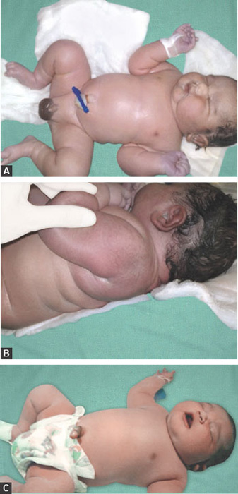
Description
- Note the firm, fluctuant, swelling of scalp, which increases in size after birth (Figs 1.4A to C).
- It is sharply limited by sutures.
- Does not transilluminate.
Etiopathogenesis
Repeated buffeting of the fetal skull against the maternal pelvis during a prolonged or difficult labor and mechanical trauma caused by use of forceps in delivery have been implicated in rupture of vessels that traverse from skull to periosteum.
Clinical Features
The swelling appears after birth. If isolated sign, the infant may be asymptomatic. It is known to exacerbate neonatal jaundice. Common associated complications are skull fracture and intracranial hemorrhage.
Investigations
Radiographs help in identifying the underlying skull fractures that can be seen in 5.4% of cephalhematomas. Magnetic resonance imaging (MRI) and/or computed tomography (CT) scan if suspect intracranial hemorrhage.
Differential Diagnosis
Differentiated from caput succedaneum and subgaleal bleed by its sharp periosteal limitations to one bone, the absence of overlying discoloration, the later initial appearance of the swelling, and the longer time before resolution.
Cranial meningocele is differentiated by pulsations, an increase in pressure during crying, and the demonstration of a bony defect.

Treatment
Therapy is not indicated for the uncomplicated cephalhematoma. Significant hyperbilirubinemia may result, necessitating phototherapy. Routine incision or aspiration of a cephalhematoma is contraindicated because of the risk for introducing infection.
Prognosis
Most cephalhematomas are resorbed within 2 weeks to 3 months, depending on their size.
1.5 Chest Indrawing
Description
- There is recession of the subcostal space and intercostal space. The sensorium is altered—drowsy or bouts of irritability (Fig. 1.5).
Etiopathogenesis
Chest indrawing is a sign of breathing difficulty and a marker of hypoxia. It is due to increased use of the chest muscles for breathing. In a normal breath, when an infant breaths in, the whole chest wall (upper and lower) and the abdomen moves out. With chest indrawing, the lower chest wall goes in while the upper chest and abdomen move out.
Clinical Features
It is a sign of increased work of breathing and is associated with hypoxia. It manifests as subcostal or intercostal indrawing. There may be audible sounds like grunt or stridor. It suggests moderate to severe lung disease and is a “danger sign”. It is best detected in a quiet infant.
Differential Diagnosis
Lung disease cardiac disease (history s/o heart disease, viz cyanosis, murmur, congestive cardiac failure, cardiomegaly or abnormal heart sound).
Investigations
X-ray chest; further tests based on clinical suspicion.
Treatment
Supportive care, oxygen, antibiotics and specific treatment of the underlying cause.
Prognosis
While prognosis depends on the diagnosis, it is generally good with prompt recognition and intervention.

Description
- The limb is elevated above the heart level. After applying digital pressure over the foot, there is blanching lasting more than 3 seconds suggesting delayed capillary refill time (CRT) (Fig. 1.6).
Etiopathogenesis
Capillary refill time is an indirect marker of circulatory stability. Normally, the capillaries refill instantly (<2 seconds) with adequacy of perfusion.
Clinical Features
Hypothermia, infection, shock lead to delayed CRT. One-time assessment and relying only on CRT for adequacy of perfusion is usually not conclusive. More the parameters deranged in addition to CRT (e.g. heart rate, pulse volume, central peripheral pulse volume, respiratory rate, skin temperature, core peripheral temperature mismatch, sensorium, blood pressure, urine output, pulse oximetry) more is the possibility of shock.
Differential Diagnosis
Hypothermia, sepsis.
Investigations
Tests are done depending on the cause suspected (e.g. sepsis screen, evaluation for congenital heart defect, etc.).

Treatment
Treatment is of the underlying cause. Correction of temperature (hypothermia), and early antibiotics, fluid resuscitation and inotropes for shock are indicated.
Prognosis
Prognosis is dependent on the underlying cause. Early recognition and prompt treatment has good outcome.
Description
- Symmetric, reticular mottling of the skin of the extremities and trunk was noted in the newborn on day 3 after birth (Fig. 1.7).
- Vital parameters are stable. The infant is asymptomatic.
- The lesions disappeared spontaneously after few hours.
Etiopathogenesis
It is caused by a vascular response to cold and usually resolves with warming. Sometimes, it may be a vasomotor phenomenon.
Clinical Features
The lesions appear spontaneously over the legs, arms or the trunk. They are transient and self-limiting. The infant is asymptomatic. There are no signs or symptoms. There are no associated anomalies.
Investigations
No specific investigations are required. It is a clinical diagnosis.
Differential Diagnosis
Physiologic cutis marmorata must be distinguished from cutis marmorata telangiectatica congenita, a vascular malformation in which the lesions do not resolve with warming. Cutis marmorata telangiectatica congenita is associated with extracutaneous findings.
Treatment
Resolves with warming. No treatment is required.
Prognosis
It is a benign condition.

Description
- There is bluish discoloration of mucous membrane and skin (Fig. 1.8). This is associated with low oxygen saturation (SpO2) and hypoxia (low PaO2).
Etiopathogenesis
Cyanosis is a reflection of the absolute concentration of reduced hemoglobin. It is detected when the amount of reduced hemoglobin in the blood exceeds 5 g%. It is not influenced by oxygen saturation or the ratio of reduced hemoglobin to oxyhemoglobin. While oxygenated hemoglobin is bright red, reduced hemoglobin is dark blue or purple in color, and is what produces the dusky or blue color of the skin and mucous membranes.
Clinical Features
Cyanosis is a marker of severity of illness and a “red flag” sign prompting urgent evaluation and stabilization. It is best visualized over mucous membranes. Infants with polycythemia may exhibit cyanosis at relatively high arterial saturations, while it is more difficult to discern cyanosis in a severely anemic infant unless the oxygen saturation is extremely low. Peripheral cyanosis, also known as acrocyanosis, is a bluish discoloration of hands and feet caused by peripheral vasoconstriction.
Differential Diagnosis
Acrocyanosis, polycythemia, cyanotic heart defect, upper airway malformation (e.g. atresia, stenosis or stricture), severe congenital or acquired lung disease (e.g. pulmonary hypoplasia, respiratory distress syndrome, meconium aspiration syndrome, pneumonia, air-leak syndrome, persistent pulmonary hypertension of the neonate), sepsis, hypoventilation, abnormal hemoglobin (e.g. congenital methemoglobinemia).
Investigations
Pulse oximeter; further tests depending upon clinical suspicion.
Treatment
Supportive care—assess, support and maintain airway, breathing and circulation, initiate oxygen and monitor the vitals—temperature, pulse rate, respiration, blood pressure, and oxygen saturation. Treat the underlying cause, viz partial exchange for polycythemia, prostaglandin infusion for critical cyanotic heart defect and definitive surgery, intercostals drain for air leak, etc.
Prognosis
While prognosis depends on the diagnosis, it is generally good with prompt recognition and intervention.

Description
- Note the whitish pearl-like structure, pinhead sized at the tip of the penis (Fig. 1.9).
- The infant is asymptomatic.
Etiopathogenesis
Epstein pearls are superficial epidermal inclusion cysts that contain laminated keratinized material and epithelial cells. This superficial skin developmental variation probably occurs with increasing maturity of the fetus, which could explain lack of this phenomenon in the preterm.
Clinical Features
The Epstein pearls occurs most frequently on the tip of the foreskin at the 6 o'clock position. It is also seen at the roof of the palate. It is a transient, self-limiting condition and disappears within a few days.
Differential Diagnosis
None.

Investigations
None.
Treatment
Assurance to parents.
Prognosis
Benign condition.
1.10 Fresh Stillbirth
Description
- There are no signs of life-spontaneous breathing or circulation or movements.
- Fresh stillborn baby does not show signs of maceration, i.e. peeling of skin or collapse of skull bones (Fig. 1.10).
Etiopathogenesis
Definition varies from country to country. Definition recommended by the WHO for international comparison is a baby born with no signs of life at or after 28 weeks of gestation. At least half of all stillbirths occur 10in the intrapartum period, representing the greatest time of risk. A fresh stillbirth has usually died during labor. Causes of fresh stillbirths include anoxia, birth trauma, instrumentation during delivery and rupture of the uterus. Anoxia may be acute or acute on chronic. Acute anoxia is due to prolonged labor, cord prolapse and cord compression. Chronic hypoxia states include pregnancy-induced hypertension, diabetes, systemic lupus erythematosus and other maternal systemic disease and infection. Premature and intrauterine growth restriction (IUGR) babies are more prone to asphyxia and trauma related deaths. Stillbirth rate is defined as the number of stillbirths per thousand of total births.
Investigations
A complete workup for maternal conditions leading to chronic fetal hypoxia states and strict intrapartum fetal monitoring by non-stress test and cardiotocography are necessary. Histopathological examination of the placenta and autopsy of the baby can help at times.
Clinical Features
The condition is not compatible with life. There are no signs of life—absent respiration, heart rate and movements to stimuli.
Differential Diagnosis
Sometimes live births with poor Apgar scores and severe fetal and prolonged acidosis may be confused with fresh stillbirths.
Treatment
A prompt delivery by cesarean section or assisted vaginal delivery, if the cervix is fully dilated, will save the baby. Once the fetal death is expected, vaginal delivery is preferred.
Prognosis
Prognosis depends on the etiology of the stillbirth. A sound antenatal workup and good intrapartum fetal monitoring may decrease the incidence of fresh stillbirths in future pregnancies.

Description
- Macrosomia (weight >90th percentile for gestational age).
- Large, plethoric baby with increased body fat in the abdominal and scapular regions (Fig. 1.11).
- Increased predisposition to hypoglycemia and congenital malformations.
Etiopathogenesis
One of the commonest causes of large for gestational age infant is maternal diabetes. Uncontrolled maternal diabetes, leads to fetal hyperglycemia stimulating the beta cells of pancreas resulting in fetal hyperinsulinemia. Because insulin is an anabolic hormone, the fetal hyperinsulinemia stimulates protein, lipid and glycogen synthesis causing increased fetal body fat, excessive adipose deposition, visceral organ hypertrophy, and acceleration of body mass accretion to cause macrosomia.
Clinical Features
The infant appears large and is asymptomatic. Problems in delivery room include asphyxia, birth trauma, respiratory distress and congenital anomalies (cardiomyopathy, caudal regression syndrome, anencephaly, hydrocephalus, spina bifida). These are prone to hypothermia, hypoglycemia, jaundice, hypocalcemia and polycythemia. Respiratory distress may be due to transient tachypnea, surfactant deficiency, asphyxia, birth injury, hypoglycemia, air leak syndrome, meconium aspiration or underlying heart defects.
Differential Diagnosis
Familial, constitutional, hydrops fetalis or genetic syndromes (e.g. Beckwith-Wiedemann syndrome, Simpson-Golabi-Behmel syndrome, Perlman syndrome, Sotos syndrome, Weaver syndrome, Costello syndrome).

Investigations
Screening for blood sugar, hematocrit, serum calcium, serum bilirubin and birth injury. Further tests are done based on clinical suspicion.
Treatment
Supervised feeding is initiated in the first hour of life to prevent hypoglycemia. All infant of diabetic mother are screened for hypoglycemia at birth, 30-min, 1-hour, 2-hour, 4-hour, 8-hour, 12-hour, 24-hour, 36-hour, 48-hour or when symptomatic by Dextrostix. Value less than 40 mg/dL identifies hypoglycemia and specific management based on glucose infusion rate is initiated. Anticipation and management of poor feeding, birth asphyxia, respiratory distress, polycythemia, jaundice, birth injury, hypocalcemia and hypomagnesemia is warranted.
Prognosis
Adverse outcomes are likely with prolonged or severe hypoglycemia and moderate to severe asphyxia requiring close neurodevelopmental follow-up. LGA (large-for-gestational-age) neonates continue to grow longer and heavier, and longitudinal data from developed countries convincingly associates macrosomia with long-term metabolic complications.
1.12 Intrauterine Growth Restriction
Description
- There is generalized loss of subcutaneous fat with a thin, wasted, “old man” look with a prominent head. There is loss of buccal fat, loose folds of skin in the nape of neck, axilla, interscapular area and groins (Figs 1.12A and B).
- Prominent sole creases, diminished breast bud formation and immature female genitalia are seen due to loss of subcutaneous fat.
- Weight is less than expected for the gestational age.
Etiopathogenesis
Growth restriction is reported in 3–10% of all pregnancies and in 20% of stillborn neonates. Maternal infections (malaria) and undernutrition are the major factors for growth restriction. Nearly one-third of intrauterine growth restrictions (IUGRs) are due to genetic causes, and two-thirds are related to the fetal environment. There is a strong link between IUGR, chromosomal abnormalities, and congenital malformations. IUGR occurs 10 times more frequently in twin pregnancies than in single gestations. Fetal growth that is less than normal for the population and for the expected growth of a specific neonate as documented on ultrasound. The term applies to the events in utero that cause fetal growth to be less than desired.
Clinical Features
Neonates with IUGR can be described as symmetrical or asymmetrical. If symmetrical growth restriction is present, the growth problems are proportional (less 13weight, length and head circumference). In asymmetric IUGR, weight and height are affected but head is spared.

The IUGR neonate is at risk for perinatal asphyxia, persistent pulmonary hypertension, respiratory distress, meconium aspiration, hypothermia, hypoglycemia, hyperglycemia, hypocalcemia, polycythemia and decreased immunity.
Investigations
Causative factors for growth restriction should be investigated antenatally.
Differential Diagnosis
Small-for-gestational-age (SGA) are infants with birth weight less than 2 SD below the mean or less than 10th percentile of a population-specific birth weight versus a gestational age plot.
Treatment
There are no effective therapies to reverse fetal growth restriction. Antenatal management is individualized and aimed at determining the ideal time and mode of 14delivery. Postnatal management involves supportive care, anticipating problems and specific treatment of the problem.
Prognosis
There is increased morbidity and mortality among premature IUGR neonates compared to term IUGR. Neurodevelopmental problems (e.g. cerebral palsy, learning deficits, short attention span, hyperactivity, learning difficulties and behavioral problems) are seen 5–10 times more often in IUGR neonates than AGA (appropriate for gestational age) neonates; the outcome depends on the severity and symmetry of the restriction. According to Barker's hypothesis, the SGA babies are more prone to develop diseases with the onset of adult age (diabetes, hypertension, ischemic heart disease, obesity and hypercholesterolemia).
1.13 Jaundice
Description
- Yellowish discoloration of skin and mucous membrane (Fig. 1.13).
- Cephalocaudal progression was noted, it first appeared on the face and then the trunk.
- There was no pallor, organomegaly, cephalhematoma or subgaleal bleed.
- The infant was active and feeding from breast.
Etiopathogenesis
Hyperbilirubinemia may result from an increase of unconjugated (indirect) or conjugated (direct) bilirubin. Hyperbilirubinemia is the result of an imbalance between bilirubin production and its elimination. In majority, early neonatal jaundice is due to rise in unconjugated (indirect) bilirubin. The etiological basis for progressive hyperbilirubinemia is usually multifactorial. Bilirubin is a potential neurotoxin causing bilirubin encephalopathy in some cases. Color of urine (dark colored staining the cloth) and stools (chalky white) suggests direct jaundice.
Clinical Features
Jaundice appears first on the face and then the trunk. Association of pallor and organomegaly is a feature of hemolysis. Mother blood group, peripheral smear and Coombs tests help to differentiate hemolytic from nonhemolytic causes. The infant's gestational age, birth weight, risk factors, aggravating factors, timing of onset of jaundice and rate of rise of bilirubin decide the management. Severe jaundice may affect the brain and the newborn may present with decreased activity, decreased tone, shrill cry, hypertonia, opisthotonus, neck retraction and seizures.

Investigations
Total and direct serum bilirubin, mother-infant blood group.
Treatment
Not all jaundice newborns need treatment. The decision making in jaundice management is based on gestation of baby, weight of baby, well or ill clinical status, age of onset (in hours) and type and severity of jaundice. Phototherapy is the standard treatment. Exchange transfusion is reserved for infants in whom bilirubin rises rapidly or fails to drop despite intensive phototherapy and for infants who show signs of acute bilirubin encephalopathy. The American Academy of Pediatrics guidelines (2005) are used for initiation of phototherapy and exchange transfusion.
Prognosis
Prognosis is excellent for uncomplicated newborn jaundice. Prognosis is guarded if jaundice is very high and/or baby shows signs of acute bilirubin encephalopathy.
1.14 Large for Gestational Age
Description
- The weight is more than 90 percentile for gestational age and the absolute weight was 4.2 kg.
- There is excessive fat deposition on body with relative sparing of head (normal head circumference). Note the “Buffalo hump” of fat on the nape of the neck, full, ruddy cheeks, full abdomen, rolls of fat on limbs (Figs 1.14A to C).
Etiopathogenesis
The common causes for large for gestational age (LGA) are familial, constitutional, infant of diabetic mother (IDM) and as a component of syndrome (Beckwith-Wiedemann syndrome, etc.). At times, the cause is idiopathic. The underlying problem in IDM is hyperinsulinemic state in utero due to poor diabetic control in mother leading to fetal hyperglycemia and excessive growth.
Differential Diagnosis
Familial, constitutional, transposition of great vessels, syndromic (Beckwith-Wiedemann syndrome).
Clinical Features
Large for gestation age infants have weight excessive for the gestation. The infant has a normal head size. The infants appear plethoric, chubby and have excessive thick skin folds. Hypoglycemia, hypocalcemia, hyperbilirubinemia, polycythemia are the common metabolic disturbances. Cardiac, gastrointestinal and neurologic malformations are seen with IDM but not with LGA infants.
Investigations
Check the diabetic status in mother if not already done (fasting, postprandial glucose and glycosylated hemoglobin), blood sugar and serum calcium monitoring is essential.
Management
Need supervised early breastfeeding. Continuous monitoring for glucose is needed for first 48 hours.
Prognosis
Prognosis is good in isolated LGA infants. With comorbid features, it depends on degree and severity of hypoglycemia.
Description
- The occipitofrontal circumference (OFC) or head circumference is more than two standard deviations above average for the child's age, sex and race (Fig. 1.15).
- There were no associated anomalies. The infant was asymptomatic.
Etiopathogenesis
The common causes of macrocephaly include familial, constitutional, hydrocephalus (increase in CSF volume due to obstruction, inflammation or infection), congenital infections (toxoplasmosis), megalencephaly (increase in brain volume as in Alexander's disease, Canavan's disease), cranial hyperostosis (bone overgrowth) and genetic disorders (e.g. neurofibromatosis, tuberous sclerosis). At times, it may arise as a sequelae to intraventricular hemorrhage, arachnoid cyst or subdural effusion. The associated complications and long-term morbidities include raised intracranial pressure, developmental delay, seizures and cognitive impairment.
Clinical Features
Large head should be seen in relation to other body parts. It large head associated with large body and organomegaly, one should consider Sotos syndrome. Large head with short limbs is characteristic of chondrodysplasia.
Differential Diagnosis
The various causes of hydrocephalus and megalencephaly are mentioned in etiopathogenesis, familial and constitutional large heads.

Investigations
An optimal measurement of the OFC and plotting the same on an appropriate growth chart for confirmation of macrocephaly. Serial measurement (weekly) to monitor the progress/regression of the head size, which may give a clue to the etiology and severity. Neuroimaging including serial cranial ultrasounds and MRI if required.
Treatment
There is no specific treatment for macrocephaly. Medical care for children with macrocephaly focuses on management of specific symptoms such as developmental delays and mental retardation and treatment of the primary diagnosis responsible for the macrocephaly.
Prognosis
For children with benign familial macrocephaly, the prognosis is excellent. In others, it is dependent upon the underlying cause.
Description
- Note the large tongue protruding beyond gum margins noticed at birth (Fig. 1.16).
- There is inability to close the mouth. It was difficult for the infant to breastfeed.
Etiopathogenesis
Congenital hypothyroidism is the most common cause of macroglossia. It may be due to an isolated anomaly (vascular—lymphatic malformation) or as an association with Beckwith-Wiedemann syndrome. Abnormal deposition of mucopolysaccharides as in mucopolysaccharidosis (MPS) may also cause macroglossia.
Clinical Features
Large tongue may be an isolated phenomenon or part of multisystem disorder. There is spectrum of manifestations from being asymptomatic to feeding, inability to close mouth or apnea depending upon the severity of lesion.
Investigations
Physical examination and family history help to identify underlying etiology. Chromosomal analysis, radiographs of spine and limbs, thyroid profile and thyroid scan, ultrasound of abdomen for visceromegaly, and urine glycosaminoglycan as indicated.
Differential Diagnosis
Beckwith-Wiedemann syndrome, isolated macroglossia, lingual lymphatic anomaly, hypothyroidism, MPS, transient neonatal diabetes, congenital isolated macroglossia (autosomal dominant), tongue hemangioma.
Treatment
Treatment of underlying condition.
Prognosis
The prognosis depends on the underlying cause.

Description
- A large pigmented lesion seen on the back and the right thigh. It is present at birth. There were no other malformations (Figs 1.17A and B).
Etiopathogenesis
This pigmented lesions result from delayed disappearance of dermal melanocytes. This is the most often encountered pigmented lesion in a newborn.
Clinical Features
It is seen commonly in lumbosacral area but may be found on leg, back and shoulder. Pigmentation is macular and gray-blue, lacks a sharp border, and might cover an area 10 cm or larger in diameter.
Investigations
Not required.
Differential Diagnosis
Congenital pigmented melanocytic nevi, Café au lait spots.
Treatment
No treatment is required.
Prognosis
Most of these lesions gradually disappear during the first few years of life, but aberrant lesions in unusual sites are more likely to persist.

Description
- A tooth is present at birth (Figs 1.18A to D).
Etiopathogenesis
Natal teeth most often develop on the lower gum, where the central incisor teeth will appear. They have little root structure. Histologic investigation reveals a failure of root formation despite eruption, a large vascular pulp, irregular genesis of dentin and a failure of cementum formation.
Clinical Features
It may be firmly fixed or loose, putting the infant at risk for aspiration. Usually, it is asymptomatic but may cause breast discomfort to the mother while feeding. They are different from neonatal teeth, which emerge through gingiva during the first 30 days after birth. Natal teeth are usually not well-formed, but they may cause irritation and injury to the infant's tongue when nursing. Natal teeth may also be uncomfortable for a nursing mother. It may rarely be associated with Ellis-van Creveld syndrome, Hallermann-Streiff syndrome, Pierre Robin syndrome, Pachyonychia congenita and Sotos syndrome.
Differential Diagnosis
Neonatal tooth, supernumerary teeth, cysts of the dental lamina, Bohn's nodules.
Investigations
None; X-ray tooth if surgery planned or diagnosis is doubtful.
Treatment
When well implanted, the teeth should be left in the arch. Teeth removal is indicated only when they interfere with feeding or when they are highly mobile, with the risk of aspiration. Smoothing of the incisal edge (to prevent potential discomfort during breastfeeding and ulcerations in the floor of the mouth) is also an option.

Prognosis
Good.
Description
- The newborn at birth appears uniformly pink. The posture has flexed upper and lower extremity with regular respiration (Fig. 1.19).
Etiopathogenesis
Transition from fetal to systemic circulation takes place at the time of birth leading to establishment of spontaneous respiration and cry. In 90% of newborns, this stress is well taken but in few, this may lead to distress and need for resuscitation. A normal newborn needs no intervention.
Clinical Features
A normal newborn is very active in the few hours of life. Color is pink, breathing is regular, skin is covered with vernix, umbilical cord is moist and peeling of skin may be seen in post-term infants. Temperature maintenance, feeding and infection control are the main concerns in this newborn.
Differential Diagnosis
None.
Investigations
Screening for metabolic disorder and hearing prior to discharge is desirable.
Treatment
Delayed cord clamping, early skin to skin contact, early and exclusive breastfeeding, assessment for risk factors, a thorough head to toe examination with special concern for congenital anomalies, assessment of weight, administration of vitamin K, parental assurance and counseling for hygiene, feeding, danger signs and vaccination prior to discharge.
Prognosis
Good.

Description
- The stool is yellowish green with pellets in it (Fig. 1.20).
Etiopathogenesis
Meconium is the black green stool, the newborn passes in the first few days of birth. As the newborn accepts milk feeds, the stool color changes to green and then to yellow. Transitional stool represents the change from meconium to the normal yellow, seedy stools that characterize infants feeding on milk only.
Clinical Features
The newborn stool normally shows variations in color, frequency and consistency, which varies with postnatal age. A well infant with weight gain is an assuring sign irrespective of the stool frequency. The stool pattern evolves over the first few days of life. After birth, a baby's first bowel movements are black and tarry called meconium, which last for couple of days. After few days, meconium is replaced with green-brown and then yellow-brown bowel stools. By about 5 days after birth, breastfed babies usually have seedy, loose bowel movements that are yellow to yellowish green or tan in color. The frequency of bowel movements varies widely from one baby to another.
Investigations
None.
Differential Diagnosis
Transitional stools, infective diarrhea.
Treatment
Assurance; continuation of exclusive breastfeeding.
Prognosis
Benign condition.

Description
- There is small reddish swelling with discharge of bowel content from an opening in the umbilicus (Figs 1.21A and B).
Etiopathogenesis
Vitellointestinal duct (VID), is an embryonic communication between the intraembryonic gut and yolk sac which is usually obliterated by the 7th week of intrauterine life. In about 2% of humans, this duct persists and gives rise to a group of anomalies—Meckel's diverticulum, vitelline cord, enteric cyst, umbilical sinus, enteric fistula or hemorrhagic umbilical mass.
Clinical Features
Presents as an umbilical nodule or polyp, bleeding from intestinal mucosa and intestinal small bowel prolapsed. There may be discharge from the umbilicus and the umbilicus is mostly moist. It may be associated with umbilical hernia, intestinal atresias, cardiac malformation, cleft lip and palate, and exomphalos.
Differential Diagnosis
- Umbilical granuloma (pink red, small, round, moist, erythematous, often pedunculated tissue protruding from the umbilicus).
- Patent urachus (a communication of bladder presents with urine in umbilical discharge).
- Unlike gastroschisis, the bowel protrudes through the umbilical ring rather than via a separate abdominal wall defect, and end up in a blind loop.
Investigations
Ultrasound examination of the abdomen for intra-abdominal collections/other congenital anomalies and water soluble 24contrast injecting through the opening to show the dye in the intestine.

Treatment
Antibiotics, primary closure of the VID if the patient arrives early without any complications. Where the defect is large, resection of the loop of intestine near the patent duct followed by primary anastomosis is the procedure of choice.
Prognosis
Delayed diagnosis may lead to peritonitis or intestinal perforation.
1.22 Pedal Edema
Description
There is fullness of dorsum of feet in preterm infant. There is no associated fullness over any other site of the body. The infant is not ill and the edema is transient, occurred on day 2 or 3 of age in the lower limbs (Figs 1.22A and B).
Etiopathogenesis
Typically, subcutaneous fluid collects on the dorsum of the feet and hands. It may occur in isolation or may be associated with generalized edema. The exact mechanisms remain unknown. About 30% of babies with Turner's syndrome have congenital lymphedema. Preterm infants may develop physiological edema due to immature kidneys and circulatory instability. Localized chronic cold injury may cause pitting edema with erythema of hands and feet. Capillary leak as in chikungunya fever may also cause pedal edema.
Clinical Features
Venous edema is typically pitting, dependent and bilateral. Lymphatic edema is non-pitting. Mechanical obstruction by amniotic bands may cause local edema. The systemic signs depend on the underlying cause.
Differential Diagnosis
Sclerema, subcutaneous fat necrosis, edema of prematurity, Milroy's disease, localized cold injury, chikungunya fever.

Investigations
For suspected edema of prematurity, no tests are required. Additional tests may be done depending on the cause.
Treatment
Treatment is directed to the underlying cause.
Prognosis
The condition is transient in preterm babies and resolves spontaneously. Peripheral lymphedema seen in neonates with Turner's syndrome usually resolves within a year. Residual lymphedema of the dorsum of the fingers may persist, along with swelling of the lower extremities, into adulthood.
1.23 Preauricular Tag
Description
- Note the skin tag near the tragus of the ear. It is soft, non-tender and pedunculated (Fig. 1.23).
- The infant is asymptomatic. There are no associated anomalies.
Etiopathogenesis
It is a common minor congenital anomaly of the first branchial arch or second branchial arch. It may appear as a skin tag or dimple. There is a known association with Goldenhar syndrome (oculo-auriculo-vertebral spectrum) and with Wildervanck syndrome.
Clinical Features
Skin tag is detected incidentally on routine evaluation. It may be single or multiple, unilateral or bilateral. It may be an isolated defect or part of syndrome. When an isolated defect, there are no signs or symptoms. It may be associated with cleft palate, cleft lip, ocular coloboma, hand or digit anomalies, mandibular hypoplasia or heart defects.
Investigations
None for isolated lesions. Presence of associated two or more minor anomalies or one major anomaly warrants renal ultrasound and hearing assessment.

Differential Diagnosis
Teratoma.
Treatment
Assurance; when part of syndrome, treatment is symptomatic; for cosmetic purpose, the skin tag may be removed.
Prognosis
A benign condition for an isolated defect.
Description
- Note the sternal indrawing with subcostal indrawing (Fig. 1.24).
- The infant is hypoxic.
Etiopathogenesis
Retractions may occur in several areas of the chest and are a sign of increased use of the chest muscles for breathing. Sternal indrawing is a clinical sign of respiratory distress. It is a mechanism to increase the intrathoracic negative pressure.
Clinical Features
It is associated with other chest signs like subcostal or intercostal indrawing and audible sounds like grunt or stridor. It may be seen normally in very preterm due to pliable chest wall. It serves as a soft marker sign for hypoxia.
Differential Diagnosis
Normal variant in preterm, pectus excavatum, respiratory distress (respiratory or cardiac cause) and sepsis.
Investigations
History and clinical examination gives a clue to possible etiology. Tests are done depending on the cause.
Treatment
Treatment is of the underlying cause. Supportive care involves correction of temperature (hypothermia), oxygenation, fluid therapy and antibiotics as indicated.
Prognosis
Prognosis is dependent on the underlying cause. Early recognition and prompt treatment has good outcomes.

Description
- Note the reddish discoloration over lateral margins of the sclera (Fig. 1.25).
- There is no eye discharge. The infant is asymptomatic.
Etiopathogenesis
It is proposed that in the majority of cases subconjunctival hemorrhage (SCHN) are a consequence of elevated venous pressure in the head and neck produced by compression of the fetal thorax and/or abdomen by uterine contractions. Tight umbilical cord around the neck probably represents an additional mechanism for SCHN. There is breakage of small blood vessels during pressure of delivery.
Clinical Features
The condition is asymptomatic. There are no signs or symptoms. It is self-limiting and transient in nature. In a minority of cases, it may also be indicative of child maltreatment. Therefore when SCHN is observed, it warrants a thorough and systematic assessment of the neonate.
Investigations
None.
Differential Diagnosis
Conjunctivitis, battered baby syndrome.
Treatment
None.
Prognosis
It is a benign condition.

Description
There is a tight piece of skin between the underside of their tongue and the floor of the mouth (Fig. 1.26).
Etiopathogenesis
It is a birth defect, the cause of which is not known. It may be familial in some cases. Normally, the tongue is loosely attached to the base of the mouth with a piece of skin called the lingual frenulum. If it is short and tight, it interferes with tongue movement leading to difficulty in feeding.
Clinical Features
Tongue-tie leads to inability to protrude the tongue past the edge of the lower gingiva or mandibular incisors. At times, the tongue may appear notched or heart-shaped when stuck out. Opinion varies regarding how frequently ankyloglossia truly causes problems. Babies with tongue-tie are not able to open their mouths wide enough to latch on to their mother's breast properly leading to sore nipples and inadequate breastfeeding and poor weight gain. In infancy, tooth decay and gingivitis may set in.
Differential Diagnosis
Bifid tongue, oral ranula, congenital furrowing, lingual thyroid.
Investigations
None.

Treatment
No consensus for management of short frenulum. Surgical intervention (frenotomy or frenuloplasty) is done only if the tongue-tie interferes with feeding. Speech difficulty (interfere with the ability to make certain sounds, such as “t,” “d,” “z,” “s,” “th” and “l,” and feeding difficulty of select food items may be seen in older age group and may require intervention.
Prognosis
Asymptomatic tongue-tie may cause no problems, as child gets older.
Description
- There is a single, soft, painless, non-pulsatile, swelling which appears to be fluid filled over the base of the umbilical cord (Fig. 1.27).
- The infant is asymptomatic. There is no local evidence of inflammation.
Etiopathogenesis
These are categorized as true cysts and false cyst. True cysts are lined with cells, and generally contain remnants of early embryonic structures. False cysts are fluid-filled sacs that can be related to a swelling of the Wharton's jelly.
Clinical Features
They may be isolated clinical phenomenon or associated with underlying malformations (20% cases). The cyst may be single (more common) or multiple. The cysts tend to resolve by the end of the first trimester if detected antenatal.
Differential Diagnosis
Pseudocysts, omphalomesenteric duct cysts, vascular disorders, abdominal wall defects, bladder exstrophy and urachal anomalies.
Investigations
Fetal karyotyping is indicated when other anomalies are found or when the cyst persists into the second trimester.

Treatment
Treatment of comorbid conditions, if any.
Prognosis
While single cysts in the first trimester are associated with favorable pregnancy outcome, the presence of multiple umbilical cord cysts, their persistence in the second and third trimester and their combination with other ultrasonographic abnormalities, is associated with increased risk of miscarriage, aneuploidy or other structural anomalies.
Description
- A pink red, small, round, moist, erythematous, often pedunculated tissue is seen protruding from the umbilicus which became apparent following the separation of the umbilical cord. There was clear umbilical discharge (Fig. 1.28). There is no local inflammation.
Etiopathogenesis
Umbilical granuloma is not a true congenital abnormality, but represents ongoing inflammation and granulation tissue formation, of an umbilicus that has yet to epithelialized. Bacterial colonization and low-grade infection may play a role in the pathogenesis.
Clinical Features
It is a granular polyp-like structure protrudes from the umbilicus. No contents are seen and there is no inflammation. The infant is asymptomatic. The lesion may ulcerate or bleed occasionally in case of friction.
Investigations
None.
Differential Diagnosis
- Umbilical polyp (a small remnant of intestinal or gastric mucosa in the umbilicus presents as a bright red nodule in the umbilical dimple)
- Patent vitellointestinal duct (a communication between the ileum and the umbilicus presents with feces or bile, often with a prolapse of the duct and adjacent ileum from the umbilicus)
- Patent urachus (a communication of bladder presents with urine in umbilical discharge).

Treatment
The common treatment is putting common salt over couple of days. Cauterization with 75% silver nitrate/copper sulfate, usually repeated 2–3 times may be considered. Rarely, if persistent, they need surgical removal.
Prognosis
Excellent; no residue.
Description
- There is bleeding per vagina, usually seen after 3–5 days after birth (Fig. 1.29).
- The infant is asymptomatic, and there is no rash or bleeding from any other site.
Etiopathogenesis
It occurs due to fall in level of sex hormones after birth when the baby is removed from placenta.
Clinical Features
Spontaneous vaginal bleed may occur episodically during first few weeks of life. There are no associated signs or systemic manifestations, and the infant is otherwise well. The condition is benign and resolves on its own.
Investigations
None.
Differential Diagnosis
Disseminated intravascular coagulation (sick, multiple bleeding sites).
Treatment
Assurance to the parents. Bleeding is mild and lasts for few days to week. Local hygiene of genitals is advised.
Prognosis
It is a self-limiting condition.

Description
There is a white, creamy, substance covering over the entire skin surface at birth (Fig. 1.30).
Etiopathogenesis
Vernix caseosa is laid down in the last trimester of pregnancy over the fetal skin produced in part by fetal sebaceous glands consisting of sebaceous gland secretions, lanugo, and desquamated epithelial cells. It consists of water (81%), lipid (9%) and proteins (10%). It facilitates extrauterine adaptation of skin in the first postnatal week. It facilitates passage through the birth canal, serves as a barrier to water loss, helps maintain temperature serves as moisturizer and anti-oxidant, and provides innate immunity. Absence of vernix caseosa is associated with the presence of physiological scaling of the newborn. Vernix caseosa peritonitis due to spillage of vernix has been reported as a complication in post cesarean mothers.
Clinical Features
This white creamy layer is present on the body at birth. This natural biofilm covering may at times be only confined to body folds. The closer to term the infant, the less the vernix; post-term infants have very little vernix. Preterm infants with less than 28 weeks of gestation lack the protective mantle of vernix. The coating progresses in cephalocaudal manner.

Investigations
None.
Differential Diagnosis
None.
Treatment
It is recommended to leave vernix intact on skin surface after birth. Removal of vernix is not necessary for hygienic reasons.
Prognosis
Emerging research suggests that vernix applied to burnt adult skin may enhance wound healing.
Description
There are rhythmic, wave-like muscular contractions over the abdomen (Figs 1.31A to C).
Etiopathogenesis
Peristalsis is normal mechanism for propulsion of food across the alimentary canal. Visible peristalsis occurs as a result of mechanical block in the alimentary canal signifying surgical emergency. Peristalsis waves temporarily increase in frequency and strength due to contraction of intestine to force the content past the obstruction. As a consequence, the wave of peristalsis become visible. The peristalsis wave may vanish suddenly due to increase in peristalsis overcoming the obstruction and the gastrointestinal tract become atonic.
Clinical Features
It appears episodic in nature. It is usually visible on stooping at the sides of the patient and inspecting the abdominal contour while the patient in supine position, immediately post-feed and in quiet condition. Pyloric obstruction is characterized by visible waves of peristalsis, which roll form left to right or from swollen epigastrium. Small and large bowel obstruction may also present with peristalsis waves visible in the upper abdomen in the early stages of bowel obstruction. It is associated with irritability, vomiting, dehydration, weight loss, acidotic breathing, hyperactive bowel sounds and distended abdomen. 34Visible peristalsis waves may also occur with normal stomach and those who are malnourished.

Differential Diagnosis
Pyloric stenosis, small bowel obstruction, large bowel obstruction, obstructed gastric outlet.
Investigations
Abdominal X-ray and barium studies.
Treatment
Nil by mouth, correction of hydration, electrolyte and acid-base imbalance, with surgical exploration once bowel obstruction is confirmed.
Prognosis
Detection in early stage has good outcomes. Late diagnosis may result in necrosis or gangrene of bowel requiring significant resection and anastomoses.
1.32 Lanugo
Description
- Note the fine, soft, unpigmented hairs over the shoulders, forehead, trunk and extremities at birth (Figs 1.32A to C).
Etiopathogenesis
Lanugo is fine, soft downy hair found on the body of the newborn baby.
Clinical Features
It is the first hair to be produced by the fetal hair follicles at about 24–25 weeks gestation and shed around 7 or 8 months of gestation. In some newborns, it may be present at birth and disappears within a few days or weeks. It is replaced by mature hair at about 33 to 36 weeks of gestational age. Lanugo functions as an anchor to hold the vernix caseosa on the skin. These hairs are transient and self-resolving.
Differential Diagnosis
None.
Investigations
None.
Treatment
Reassurance.
Prognosis
Benign phenomenon.
