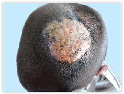
FIG. 1: Kerion note the boggy swellings.
FIG. 2: Boggy swelling misinterpreted as abscess and treated with antibiotic usage.
FIG. 3: Gray-patch type of tinea capitis.
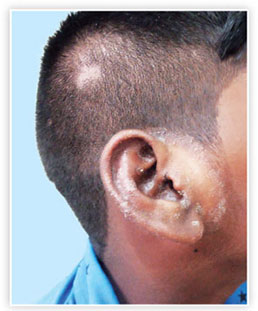
FIG. 4: Alopecia areata type of tinea capitis, note the involvement of the pinna.
FIG. 5: Multiple pustules in a child with tinea capitis.
FIG. 6: Noninflammatory type of tinea capitis in a child.
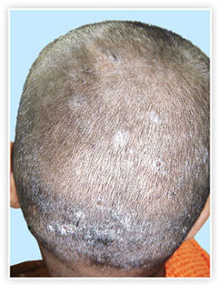
FIG. 7: Tinea capitis with intense scaling.
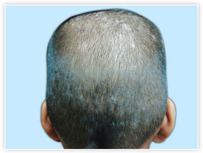
FIG. 8: Noninflammatory gray patch type of tinea capitis.
FIG. 10: Glabrous type of tinea capitis.
FIG. 11: Glabrous type of tinea capitis in an adult. Note extension on to the face.
FIG. 12: Tinea capitis in an adult mimicking psoriasis.
FIG. 13: Multiple patches of tinea capitis well seen after tonsuring.
FIG. 15: Glabrous type of tinea capitis. Note the involvement of face.
FIG. 17: Kerion with secondary bacterial infection.
FIG. 18: Kerion and inflammatory type of tinea capitis.
FIG. 19: Tinea capitis in brothers.