


[Year:2018] [Month:May-December] [Volume:11] [Number:2-3] [Pages:3] [Pages No:33 - 35]
DOI: 10.5005/jp-journals-10013-1343 | Open Access | How to cite |
Abstract
Aim: This study aimed to analyze the association of absolute eosinophil count, serum IgE, and spirometry with comorbid bronchial asthma in patients of allergic rhinitis. Materials and methods: This study involved 50 patients with signs and symptoms of allergic rhinitis who underwent clinical examination and various tests including spirometry and were followed up regularly. Patients found to have bronchial asthma or nasal polyposis were treated accordingly. Results: The study found the prevalence of bronchial asthma in patients with allergic rhinitis to be 58% and that the severity of bronchial asthma reduced significantly with less acute attacks and reduced hospitalizations with the effective treatment of allergic rhinitis (p = 0.064). Conclusion: This study showed that elevated AEC and serum IgE were significantly associated with coexisting allergic rhinitis and bronchial asthma and increased the chance of coexistence of the same. Spirometry is a useful tool for observing the response to treatment. Clinical significance: The findings of this study reinforce the unified airway concept and should therefore propel ENT clinicians to diagnose and tackle early bronchial asthma in patients of allergic rhinitis, thus reducing the overall morbidity.
[Year:2018] [Month:May-December] [Volume:11] [Number:2-3] [Pages:4] [Pages No:36 - 39]
DOI: 10.5005/jp-journals-10013-1342 | Open Access | How to cite |
Abstract
Introduction: Cerebrospinal fluid (CSF) rhinorrhea implies an abnormal communication between the subarachnoid space and the nasal cavity leading to drainage of CSF to the exterior. Aims and objectives: To study the clinical profile and factors associated with CSF rhinorrhea with outcomes of various treatment modalities at a tertiary healthcare center. Materials and methods: This study was conducted in the Department of ENT and Head-Neck Surgery, SSG Hospital and Government Medical College, Vadodara, Gujarat, from April 2014 to November 2016. Patients were selected from those attending the outpatient department (ENT/neurosurgery OPD), indoor patients (admitted at ENT ward 19/general surgery/neurosurgery wards), and emergency department of the hospital during the study period. Results: The age of patients ranged from 18 to 70 years with a mean age of 38.6 years; 12 (60%) were males and 8 (40%) were females. A total of 20 patients of CSF rhinorrhea were included in our study. Majority of rhinorrhea patients presented with watery nasal discharge as their primary complaint, which increased on straining or bending forward. The most common etiology was posttraumatic in 55% followed by iatrogenic in 25% and spontaneous in 20%. Treatment modalities used were: (A) conservative measures done in 50%, (B) endoscopic endonasal CSF leak repair in 35%; (C) repair by the craniotomy approach done in 15%. The CSF leak was successfully repaired by the endoscopic endonasal approach in six (86%) patients. About 71% of patients who were put on conservative measures responded favorably in the 1st week only and the rest 29% needed 2 weeks for stoppage of CSF rhinorrhea. Conclusion: Cerebrospinal fluid rhinorrhea has varied etiology. Mostly they are posttraumatic following road traffic accidents. Early identification of CSF rhinorrhea is important and can be safely managed conservatively in majority of cases.

Endoscopic Excision of Advanced-stage Angiofibroma: Predicting the Recurrence
[Year:2018] [Month:May-December] [Volume:11] [Number:2-3] [Pages:4] [Pages No:40 - 43]
DOI: 10.5005/jp-journals-10013-1348 | Open Access | How to cite |
Abstract
Objectives: To propose a novel scoring system for the prediction of recurrence disease after endoscopic excision in advanced stages of juvenile nasopharyngeal angiofibroma (JNA). Materials and methods: Retrospective case series review of 30 cases of advanced-stage JNA patients encountered within a period of three years (2016–2019). All the patients underwent a complete endoscopic excision which was preceded by embolization. Postoperatively, all the patients were followed up after 4 weeks, 3 months, and 6 months for recurrent disease. A scoring system was devised to reanalyze the risk of recurrent disease in all the patients who were a part of the study. Results: All the patients were male adolescents presenting with typical complaints of JNA like progressive unilateral nasal blockage, spontaneous painless epistaxis, and facial swelling. The patients who were recently diagnosed with JNA belonging to Fisch stages 3a, 3b, and 4 were only chosen for the study. Residual and recurrent cases were excluded. Preoperative contrast-enhanced computer tomography (CT) was done for all the patients. All the patients were preoperatively embolized, and the surgical technique used in all the patients was through a complete endoscopic approach. Conclusion: The endoscopic approach is possible for all stages of JNA. The endoscopic method reduced the postoperative morbidity to a great extent. The risk of recurrence could be predicted using the scoring system which was statistically significant.

[Year:2018] [Month:May-December] [Volume:11] [Number:2-3] [Pages:5] [Pages No:44 - 48]
DOI: 10.5005/jp-journals-10013-1341 | Open Access | How to cite |
Abstract
Introduction: Epistaxis is a common symptom in hypertensive patients. However, the relationship between hypertension and epistaxis is controversial and poorly understood. Objective: The present work was to study the histopathological changes underlying recurrent nasal bleedings in the patients with arterial hypertension (AH). Materials and methods: We have undertaken a prospective study based on the university clinic of Rostov-on-Don. Twenty-two hypertensive patients aged between 51 and 63 with recurrent epistaxis underwent surgical interventions due to severely deviated nasal septum hampering the search for the source of bleeding. Simultaneously nasal mucosae biopsies were taken in the bleeding point area. Tissue specimens were subjected to histological and ultrastructural investigations. Results: Histological and ultrastructural investigations of the biopsy samples revealed erythrocytic, hyaline, and fibrin thrombi in the vessels of the microcirculatory system, deendothelization, and destruction of the basement membrane alongside vascular subendothelium exposure. The above-mentioned changes in the nasal cavity mucosa lead to necrosis foci, which are the bleeding points. Conclusion: The cause of the nasal bleeding associated with AH is not a mechanical rupture of blood vessels but thrombosis and necrosis in the nasal mucosa. Clinical significance: Drug hemostatic treatment of hypertensive patients suffering from recurrent epistaxes is counterindicative due to possible serious thromboembolic complications (myocardial infarction, apoplexy, etc.). In case of a severely deviated septum hampering the search for the bleeding vessel, the treatment guidelines should include septoplasty.
[Year:2018] [Month:May-December] [Volume:11] [Number:2-3] [Pages:3] [Pages No:49 - 51]
DOI: 10.5005/jp-journals-10013-1340 | Open Access | How to cite |
Abstract
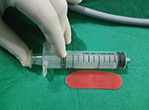
Background: Nasal packing and splints have been used to control bleeding in epistaxis and endonasal procedures using various materials for internal stabilization and as spacers to prevent synechiae and restenosis. Aims and objectives: The aim of this study was to compare the results of using wax plates as nasal splints along with non-absorbable sponge for anterior nasal packing following nasal surgeries. Materials and methods: A total of 60 patients were selected for various nasal surgeries involving the septum such as conventional and endoseptoplasty, septorhinoplasty, functional endoscopic sinus surgery with septoplasty, and posttraumatic nasal bone fracture correction. They were randomly classified into two groups: for group I patients (n = 30), post-surgery wax plate was used as an intranasal splint along with anterior nasal packing, whereas for those in group II (n = 30), only anterior nasal packing was used. Result: In this study, 28.33% (n = 17) of the patients were in the age group of 15–30 years, 41.66% (n = 25) were in the age group of 31–45 years, and 30% (n = 18) were in the age group of 46–60 years. In total, 60% (n = 36) of the patients were males and 40% (n = 24) were female. The number of synechiae cases noticed in group II (n = 9) was found to be statistically significant when compared to that of group I (n = 1), p value = 0.006 (<0.05). Conclusion: Wax plate is an ideal intranasal splint for nasal surgeries because of its low cost, easy availability, and low rate of synechia formation.

[Year:2018] [Month:May-December] [Volume:11] [Number:2-3] [Pages:3] [Pages No:52 - 54]
DOI: 10.5005/jp-journals-10013-1344 | Open Access | How to cite |
Abstract
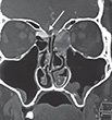
Proper identification of the site, size, multiple, and bilateral defects is very important to prevent recurrences after cerebrospinal fluid (CSF) leak repair. The utility of intrathecal fluorescein and a blue-light filter in this has been established before. We are reporting a case of a spontaneous CSF leak in an unusual site, which was identified completely with the help of intrathecal fluorescein and blue-light filter and successfully managed by the endoscopic approach.
Nasal Dorsum Swelling: A Rare Diagnosis of Necrobiotic Xanthogranuloma
[Year:2018] [Month:May-December] [Volume:11] [Number:2-3] [Pages:3] [Pages No:55 - 57]
DOI: 10.5005/jp-journals-10013-1347 | Open Access | How to cite |
Abstract
Necrobiotic xanthogranuloma (NXG) is a rare granulomatous disorder which presents as yellow plaques and nodules, commonly in the periorbital region. It is important to look for paraproteinemia by serum electrophoresis, as there is a 10% risk of developing multiple myeloma in these patients. This is the first report of NXG presenting as a nasal dorsum swelling. We wish to highlight the importance of histopathological diagnosis, serum electrophoresis and bone marrow biopsy in cases with similar clinical features.

[Year:2018] [Month:May-December] [Volume:11] [Number:2-3] [Pages:3] [Pages No:58 - 60]
DOI: 10.5005/jp-journals-10013-1346 | Open Access | How to cite |
Abstract
Background: Chronic rhinosinusitis (CRS) is a most common chronic debilitating disease that has great economical impact and can affect different age-groups. It significantly affects the physical and mental domains of the patient, leading to the deterioration in the quality of life. Purpose: Since medical treatment is not yet standardized for CRS because of scarcity of knowledge about its etiology, and postsurgery relapses are common, the purpose of the present case study was to explore the efficacy of osteopathic manipulative treatment (OMT) in a CRS patient to prevent the relapse. Case description: The author reported a 45-year-old female who presented with the complaints of facial pressure and pain, blocked nose, reduced sense of smell, pain around the eye, heaviness, ache in the head and neck on the right side with occasional postnasal drip and cough, unequal pressure in both the ears for the last couple of years, and always felt drain of energy. The patient had undergone pharmacological treatment for the same in the past, but her sinusitis persisted despite treatment. The X-ray (Water's view) of the paranasal sinus reveals opacification in the area of right maxillary sinus. The patient was treated with OMT technique twice a week for 4 weeks followed by the home exercise program. Results: The application of OMT revealed a significant change in the symptoms of the patient as self-reported and also improvement in the 22-item sinonasal questionnaire. Conclusion: Osteopathic manipulative treatment could be used as a conservative treatment strategy in patients with chronic sinusitis. However, its effectiveness can only be explored in randomized clinical trials in order to generalize the results to a larger population.

Airway Management in Arrhinia: A Case Report and Literature Review
[Year:2018] [Month:May-December] [Volume:11] [Number:2-3] [Pages:4] [Pages No:61 - 64]
DOI: 10.5005/jp-journals-10013-1345 | Open Access | How to cite |
Abstract
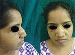
Aim: To describe optimal airway management in arrhinia. Background: Arrhinia is the congenital absence of the external nose, nasal cavities, and olfactory bulb, and this congenital anomaly is extremely rare. In newborns with this anomaly, airway management is urgent and crucial. There is no consensus on the airway management of these patients. Case description: A 22-year-old female visited our unit due to blindness and purulent discharge in the right eye of 5 years of evolution. She had arrhinia and was treated with an emergent tracheotomy at birth. Currently, she has purulent lacrimal ducts, leukoma in the right eye, hypertelorism, presence of nasofacial scar, absence of incisors, and neck with tracheocutaneous fistula. She had a previous history of six nasal reconstructive surgeries. She breathed through her mouth and tracheocutaneous fistula. She denied any airway difficulty. Discussion: We described an adult case with arrhinia. She breathed effortlessly through a tracheocutaneous fistula, and as a newborn a tracheotomy was necessary. An airway management consensus in arrhinia has not been described. We describe a thorough literature review on arrhinia and airway management. Conclusion: Arrhinia is a congenital malformation that carries the risk of respiratory difficulty. These patients require a multidisciplinary team to manage the newborn and choosing the appropriate alternative for securing the airway. Treatments described are orotracheal intubation, oropharyngeal tube, nasal reconstruction, and tracheotomy in patients who do not develop oral breathing. Tracheotomy is a definitive treatment in these patients. Clinical significance: There are various treatments for airway management of arrhinia. Clinicians should be aware of these treatment options.


© Jaypee Brothers Medical Publishers (P) LTD.

