

[Year:2017] [Month:July-December] [Volume:21] [Number:2] [Pages:1] [Pages No:0 - 0]
DOI: 10.5005/ijmb-21-2-iv | Open Access | How to cite |

A Comparative Study of Lipid Profile in Obese and Nonobese Men attending Master Health Checkup
[Year:2017] [Month:July-December] [Volume:21] [Number:2] [Pages:3] [Pages No:73 - 75]
DOI: 10.5005/jp-journals-10054-0024 | Open Access | How to cite |
Abstract
Obesity is emerging as an epidemic worldwide. Obesity is associated with a number of comorbid conditions, such as diabetes mellitus, hypertension, cancer, dyslipidemia, cardiovascular abnormalities, anemia, obstructive sleep apnea, and psychosocial abnormalities. This study aims at comparing the lipid profile levels of obese and nonobese men. This was a case—control study conducted at a tertiary care center. Totally, 80 men in the age group of 20 to 47 years attending the master health checkup were included in the study, out of which 40 men with normal body mass index (BMI) of 18 to 25 belonged to group I and 40 men with increased BMI of 30 and above belonged to group II. Lipid profile parameters, such as triglycerides (TGLs), total cholesterol, high-density lipoprotein (HDL) cholesterol, and low-density lipoprotein (LDL) cholesterol were estimated in them. The data were statistically analyzed using Statistical Package for the Social Sciences (SPSS) software version 15.0. Statistically significant difference was found in the total cholesterol levels with a p-value of 0.040 while the difference in LDL cholesterol was statistically highly significant with a p-value of 0.040. Among lipid profile parameters, only total cholesterol and LDL cholesterol showed significant difference between the obese and nonobese individuals. However, the other parameters like HDL cholesterol and TGLs did not show any significant difference. Babu SV, Jagadeesan AR, Ramalingam J. A Comparative Study of Lipid Profile in Obese and Nonobese Men attending Master Health Checkup. Indian J Med Biochem 2017;21(2):73-75.IntroductionAimMaterials and methodsResultsConclusionHow to cite this article
[Year:2017] [Month:July-December] [Volume:21] [Number:2] [Pages:5] [Pages No:76 - 80]
DOI: 10.5005/jp-journals-10054-0025 | Open Access | How to cite |
Abstract
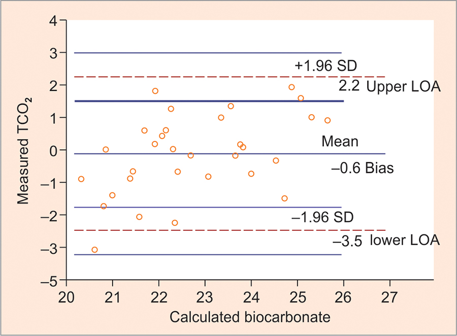
Measured total carbon dioxide (TCO2) from venous sample and calculated bicarbonate from arterial blood gas (ABG) have shown good agreement in some studies, while conflicting results have been obtained in few other studies. The objective of this study is to compare and assess the degree of agreement between the measured TCO2 and calculated bicarbonate and also whether they can be used interchangeably in our laboratory. We prospectively analyzed 89 ABG samples requested for calculated bicarbonate and then measured TCO2 from venous blood samples drawn simultaneously from the same participants between November 2016 and April 2017. Measured TCO2 results ranged from 5.7 to 39.9 mmol/L (mean 23.45 mmol/L), while calculated bicarbonate ranged from 9 to 40 mmol/L (mean 24.36 mmol/L). The values of TCO2 and bicarbonate correlated well (r = 0.95, p < 0.001), with the correlation given by the equation, y = 0.884 In majority of the cases, the calculated bicarbonate concentration from ABG showed a good correlation to the measured venous TCO2 concentration. Despite this excellent correlation, TCO2 did not show good agreement with calculated bicarbonate when Story and Poustie's criteria were applied, especially in cases of bicarbonate less than 20 mmol/L. Hence, clinicians should be aware of this discrepancy and be cautious when using measured TCO2 and calculated bicarbonate interchangeably in the assessment and management of acid—base disorders, especially in patients with metabolic acidosis. Mohan T, Kumar BV. Comparison of measured Serum Total Carbon Dioxide with calculated Bicarbonate calculated from Arterial Blood Gas Analysis. Indian J Med Biochem 2017;21(2):76-80.IntroductionMaterials and methodsResultsConclusionHow to cite this article

[Year:2017] [Month:July-December] [Volume:21] [Number:2] [Pages:5] [Pages No:81 - 85]
DOI: 10.5005/jp-journals-10054-0026 | Open Access | How to cite |
Abstract
Oral squamous cell carcinoma (OSCC) is a major health problem in Southeast Asia, including India. Areca nut chewing is a major health hazard in India, which has been implicated in the etiology of OSCC. Hypoxia-inducible factor-1 (HIF-1) is a major transcription factor involved in adaptation under hypoxic condition, a common finding in solid tumors. The present study was conducted to evaluate the effect of different habits including areca nut chewing on HIF-1 expression in patients with OSCC. It was a hospital-based observational case-control study. The study comprised 50 histologically proven cases of OSCC and 50 healthy controls. The HIF-1α level was measured by commercially available enzyme-linked immunosorbent assay (ELISA) in the blood samples. The data were analyzed using Statistical Package for the Social Sciences (SPSS) software version 20. The HIF-1α levels were found significantly higher in the patients with areca nut consumption in addition to other addictive habits. Isolated influence could not be discerned as there was only one patient who gave history of only areca nut chewing. Our findings prove that HIF-1α expression is upregulated by areca nut chewing, which leads to worse prognosis. This calls for widespread awareness programs regarding the deleterious effects of areca nut chewing among the general population. Prasad J, Goswami B, Agarwal K, Mehra P, Kumar S, Pahuja BK, Chauhan A, Ahirwar AK. Effect of Areca Nut Consumption on Hypoxia-inducible Factor-1 Alfa Expression in Patients with Oral Squamous Cell Carcinoma. Indian J Med Biochem 2017;21(2):81-85.IntroductionMaterials and methodsResultsConclusionHow to cite this article
[Year:2017] [Month:July-December] [Volume:21] [Number:2] [Pages:5] [Pages No:86 - 90]
DOI: 10.5005/jp-journals-10054-0027 | Open Access | How to cite |
Abstract
Perinatal asphyxia is one of the major causes of neonatal mortality and long-term morbidity. Although neonates with severe birth asphyxia are known to be at increased risk of early-onset hypocalcemia, the magnitude of the problem is not well documented. Magnesium plays a role in neuroprotection for neonates with hypoxic-ischemic encephalopathy (HIE). The objective of this study was to determine the prevalence of early-onset hypocalcemia and hypomagnesemia in severely asphyxiated neonates. This study was carried out on 75 newborns distributed as group I (50 asphyxiated neonates) and group II (25 healthy neonates). Serum calcium and serum magnesium was estimated within 24 hours after birth, followed by third and fifth day postbirth. Maximum number of cases (81.3%) were born by vaginal delivery. The mean value of serum calcium on days 1, 3, and 5 for group I was 7.004 ± 0.691, 7.482 ± 0.760, 8.184 ± 0.811 in contrast to group II: 8.788 ± 0.399, 9.476 ± 0.250, 9.992 ± 0.277 respectively. Whereas the mean value of serum magnesium for group I is reported as 1.545 ± 0.045, 1.496 ± 0.067, 1.556 ± 0.057 on days 1, 3, and 5, while that of group II was 1.518 ± 0.053, 1.597 ± 0.049, 1.66 ± 0.065 respectively. On HIE stage-wise comparison, abnormal calcium metabolism percentage increases with severity of asphyxia (46.6% abnormal in stage I, while 71.4% abnormal in stage III). Abnormal magnesium metabolism percentage also increases with severity of asphyxia (26.6% abnormal in stage I, while 71.4% abnormal in stage III) and this abnormality persists up to fifth day in stage III. Birth asphyxia is the most common and important cause of preventable cerebral injury occurring in the neonatal period. Serum calcium and magnesium level plays exceptionally imperative role for escaping HIE and other induced complications. Bhimte B, Vamne A. Metabolic Derangement in Birth Asphyxia due to Cellular Injury with Reference to Mineral Metabolism in Different Stages of Hypoxic-ischemic Encephalopathy in Central India. Indian J Med Biochem 2017;21(2):86-90.IntroductionMaterials and methodsResultsConclusionHow to cite this article
Prediabetes and Undiagnosed Diabetes Mellitus: The Hidden Danger
[Year:2017] [Month:July-December] [Volume:21] [Number:2] [Pages:5] [Pages No:91 - 95]
DOI: 10.5005/jp-journals-10054-0028 | Open Access | How to cite |
Abstract
Diabetes is considered as a major challenge to the public health system in India. Recent articles clearly mention that the hidden danger in the form of prediabetes and undiagnosed diabetes is greatly adding to the burden silently. Awareness regarding the same, particularly among youth, can help diagnose the condition very early and thus, initiate early management. With an aim to estimate the frequency of prediabetes and undiagnosed diabetes in the adult population, a camp was organized in our institute to screen the adults in our locality by estimating fasting plasma glucose (FPG) and glycosylated hemoglobin (HbA1c). A total of 246 individuals were selected for analysis after excluding the known diabetic cases. Height, weight, pulse, blood pressure (BP), waist circumference, and body mass index (BMI) were measured. Plasma fasting sugar and fasting serum lipid profile were analyzed. The HbA1c was estimated in hyperglycemic subjects. The frequency of hyperglycemia in the study population was found to be 28%. The total frequency of prediabetes was 18.3% and that of undiagnosed diabetes was 9.75%. The raised sugar could be significantly associated with age, waist circumference, BMI, hypertriglyceridemia, and cholesterol-to-high-density lipoprotein (Chol:HDL) ratio. Aging, greater BMI, hypertriglyceridemia, and raised low-density lipoprotein (LDL) depicted significant odds ratio (OR) to predict the risk factor for diabetes. The hidden burden of diabetes in our locality is quite high, which, if not taken care, would result in a public health catastrophe. Patel S, Nanda R, Abraham J, Sahoo S, Ganguly A, Mohapatra E. Prediabetes and Undiagnosed Diabetes Mellitus: The Hidden Danger. Indian J Med Biochem 2017;21(2):91-95.IntroductionObjectivesMaterials and methodsResultsConclusionHow to cite this article

[Year:2017] [Month:July-December] [Volume:21] [Number:2] [Pages:5] [Pages No:96 - 100]
DOI: 10.5005/jp-journals-10054-0029 | Open Access | How to cite |
Abstract
Maternal thyroid hormone level during pregnancy is a vital parameter for the health of mother as well as developing child. Fetal growth is affected by maternal thyroid levels. Various physiological changes like alterations of thyroxine binding globulins (TBGs), beta-human chorionic gonadotropin (β-hCG) level, and change of iodide metabolism affect maternal thyroid hormone levels. Therefore, reference intervals (RI) for thyroid hormones in pregnant population require to be established separately from general population. The RIs of serum triiodothyronine (T3), thyroxine (T4), and thyroid-stimulating hormone (TSH) were determined in healthy pregnant women by enzyme-linked immunosorbent assay (ELISA) technique after segregating them into three trimesters. This study was conducted in a 492-bedded zonal level hospital. The reference population was chosen from a study population of pregnant women by strict inclusion and exclusion criteria. The assays were done by the most commonly used economical ELISA method using standard kits. Tests were done using accurate and precise methods with proper quality control measures. The RIs were calculated from the central 95% of distribution of total T3, total T4, and TSH values located between 2.5 and 97.5 percentile values. The 0.90 confidence intervals (CIs) for the upper and lower reference limits were calculated. The values thus obtained were different from those provided by manufacturer kit literature. It is recommended to determine own laboratory-specific, method-specific, trimester-wise RI for maternal thyroid hormone status and use them for screening of pregnant mothers. Chakrabarty BK, Mitra B, Pal R, Hazra N. Specific Reference Intervals of Serum Triiodothyronine, Thyroxine, and Thyroid-stimulating Hormone in Normal Pregnant Indian Women as per Trimester. Indian J Med Biochem 2017;21(2):96-100.AimMaterials and methodsResultsConclusionHow to cite this article

Utility of Serum Paraoxonase Levels with reference to Severity of Organophosphorus Poisoning
[Year:2017] [Month:July-December] [Volume:21] [Number:2] [Pages:5] [Pages No:101 - 105]
DOI: 10.5005/jp-journals-10054-0030 | Open Access | How to cite |
Abstract
Organophosphorus (OP) compounds are widely used insecticides for agricultural and domestic purposes. Easy availability and less awareness regarding the toxicity caused by these compounds have resulted in high morbidity and mortality in India. Early diagnosis and initiation of treatment are required to reduce the mortality rate for which laboratory evaluation plays a vital role, in addition to various clinical scoring systems. A cross-sectional study was carried out for a period of 2 months. Forty clinically diagnosed acute OP poisoning cases admitted in emergency units formed the study subjects. Serum was used for the estimation of cholinesterase, for both basal and salt stimulated paraoxonase (PON) activity. Peradeniya organophosphorus poisoning (POP) scale was used as a tool to categorize patients into mild (0—3 score), moderate (4—7 score), and severe (8—11 score) poisoning. The mean age of the study participants was 31.9 ± 14.4 years. Seventy-five percent of the participants were males and 25% were females. Chlorpyrifos was the most common OP compound consumed by the study participants. There was a significant decrease in the serum cholinesterase activity (p = 0.001) and salt-stimulated PON activity (p = 0.016) as the severity increased. Serum cholinesterase and POP score showed statistically significant negative correlation (p = 0.003). There was a linear positive correlation between serum cholinesterase and serum PON activity, but the correlation was significant only with salt-stimulated PON activity (p = 0.005). The results suggest that subjects with higher levels of PON activity may have better detoxifying capacity toward OP poisoning. Rahul HV, Rani NA, Nusrath A. Utility of Serum Paraoxonase Levels with reference to Severity of Organophosphorus Poisoning. Indian J Med Biochem 2017;21(2):101-105.IntroductionMaterials and methodsResultsConclusionHow to cite this article
[Year:2017] [Month:July-December] [Volume:21] [Number:2] [Pages:6] [Pages No:106 - 111]
DOI: 10.5005/jp-journals-10054-0031 | Open Access | How to cite |
Abstract
Because of the varied presentation and associated high mortality, the identification of patients with acute myocardial infarction (MI) is very critical for patient management and has a bearing on the prognosis. The goal of present study was to correlate the diagnostic value of cardiac biomarkers in MI with survival and MI without survival. Diagnostic case—control study was conducted on 110 MI patients presenting to the Emergency Department within 12 hours of acute chest pain, and 120 healthy age- and sex-matched volunteers formed the control group. Serum ischemia-modified albumin (IMA), troponin I (TnI), creatine kinase-MB (CK-MB), lactate dehydrogenase (LDH), and aspartate transaminase (AST) were measured. Statistical software SYSTAT version 12 was used to analyze the data. The results were expressed in mean ± standard deviation. Comparisons of study groups and study groups with control groups were done by applying Z test. Correlation was tested by Student's Mean levels of serum IMA, TnI, CK-MB, LDH, and AST levels were significantly higher (p < 0.01) in patients with MI as compared with healthy controls. Serum levels of cardiac biomarkers were significantly elevated (p < 0.01) in MI patients without survival as compared with MI with survival. The serum levels of biomarkers were increased in MI without survival as compared with MI with survival. These study data prove that these changes might be helpful to obtain a comprehensive view of the infarct size and severity of vascular stenotic lesions. Patil SM, Bankar M, Padalkar R, Phatak A. Comparative Study of Potential Diagnostic Biomarkers in Myocardial Infarction with Survival and Myocardial Infarction without Survival. Indian J Med Biochem 2017;21(2):106-111.IntroductionMaterials and methodsResultsConclusionHow to cite this article

[Year:2017] [Month:July-December] [Volume:21] [Number:2] [Pages:5] [Pages No:112 - 116]
DOI: 10.5005/jp-journals-10054-0032 | Open Access | How to cite |
Abstract
Diabetes is a common endocrinal disorder. Abnormal lipid and magnesium levels are observed in diabetes in many studies. The current study was done with an aim to find the relationship between lipid with magnesium and diabetes mellitus in Gujarati population. The cross-sectional study included 60 diabetics and 50 healthy subjects. Each subject was interviewed, examined, and investigated for serum lipid profile and magnesium. A 12-hour overnight fasting was recommended. The statistical analysis of data obtained was done by Student's t-test and calculation of Pearson correlation coefficient. Routine biochemical investigations showed a significant rise (p < 0.01) of fasting plasma glucose, triglycerides (TGs), total cholesterol (TC), low-density lipoprotein-cholesterol (LDL-C), and very low-density lipoprotein-cholesterol (VLDL-C) in diabetics, in comparison with controls (p < 0.01). Among diabetics, males have significantly higher (p < 0.01) TC, TG, and LDL-C while significantly lower high-density lipoprotein-cholesterol (HDL-C). Significant inverse correlation of magnesium with TC (r = −0.18), TGs (r = −0.14), LDL-C (r = −0.27), fasting blood sugar (FBS; r = −0.12) and direct correlation with HDL-C (r = 026) were observed in cases. The major highlights of the current study are lower magnesium levels in cases compared with controls. In the current study, serum magnesium level has been found to be inversely related to cholesterol, TG, and LDL-C levels, while it is directly associated with HDL-C level. Hence, it could play a role in controlling the risk of coronary artery disease (CAD)-associated morbidities in future. Sendhav SS, Kakaiya A, Chatterjee B. Evaluation of Serum Magnesium Level along with Lipid Profile in a Gujarati Population diagnosed with Diabetes Mellitus. Indian J Med Biochem 2017;21(2):112-116.Aims and objectivesMaterials and methodsResultConclusionHow to cite this article

[Year:2017] [Month:July-December] [Volume:21] [Number:2] [Pages:7] [Pages No:117 - 123]
DOI: 10.5005/jp-journals-10054-0033 | Open Access | How to cite |
Abstract
Lymphatic filariasis is a mosquito-borne disease affecting nearly 120 million people across the world. Filarial antigen detection is a good indicator for mapping new filarial cases and for evaluation of filarial elimination programs as compared with the low sensitivity associated with the direct evidence of microfilaria (Mf) in blood samples. To overcome low sensitivity and night-time blood collection method for parasite detection in filariasis cases, the sandwich enzyme-linked immunosorbent assay (ELISA) was standardized for detection of circulating filarial antigen using monospecific polyclonal antibodies raised against recombinant filarial antigen rWbL2. In the present study, the specific antibodies raised against novel recombinant antigens rWbL2 were explored to develop suitable filarial antigen assays. It was possible to come out with a filarial antigen assay that could detect WbL2 or its equivalent antigen with 40% sensitivity (by using mouse anti-WbL2 antibody as capturing antibody), 60% sensitivity (using FSIgG human filarial serum immunoglobulin G as capturing antibody), and 100% specificity. These assays show promise to detect and monitor active filarial infection and thus prove to have potential as a useful diagnostic and monitoring tool in the elimination program. Gandhe MB, Gandhe SM. Generation of Monospecific Polyclonal Antibodies to Recombinant Filarial Antigen rWbL2 and Evaluation of Its Immunodiagnostic Utility in Filariasis. Indian J Med Biochem 2017;21(2):117-123.IntroductionHow to cite this article
Glucose Fluctuations and Activation of Oxidative Stress in Patients with Type II Diabetes Mellitus
[Year:2017] [Month:July-December] [Volume:21] [Number:2] [Pages:3] [Pages No:124 - 126]
DOI: 10.5005/jp-journals-10054-0034 | Open Access | How to cite |
Abstract
In recent years, the oxidative stress (OS)-induced free radicals have been implicated in the pathology of diabetes mellitus (DM). Persistently high glucose levels can lead to the generation of higher amounts of free radicals. The purpose of the present study was to evaluate the role of hyperglycemia [by measuring variables: Glycated hemoglobin (HbA1c), fasting plasma glucose (FPG)] in the induction of OS [(by analyzing the OS marker: Malondialdehyde (MDA)] in type II DM. This observational study was conducted among 50 type II DM patients without complications and 50 type II DM patients with complications in S.B.K.S. Medical Institute and Research Centre, Waghodiya, Gujarat, India. Correlations between variables were tested using the Pearson rho correlation test. Chi-squared (χ The MDA values correlated significantly with HbA1c and FPG values in type II diabetic subjects with complications (r = +0.29, p = 0.04; r = +0.47, p = 0.0006). Glucose fluctuations may activate OS in DM. Assessment of glycemic control marker HbA1c and lipid peroxidation marker MDA is useful in DM patients for detection of risk of diabetic complications at an early stage of the disease. Nayak MS, Sharma R, Patani SS. Glucose Fluctuations and Activation of Oxidative Stress in Patients with Type II Diabetes Mellitus. Indian J Med Biochem 2017;21(2):124-126.IntroductionMaterials and methodsResultsConclusionHow to cite this article

Study of Insulin Resistance in Women with Preeclampsia
[Year:2017] [Month:July-December] [Volume:21] [Number:2] [Pages:4] [Pages No:127 - 130]
DOI: 10.5005/jp-journals-10054-0035 | Open Access | How to cite |
Abstract
The root cause of preeclampsia is placental ischemia due to impaired trophoblastic invasion in the uterine spiral arterioles. Ischemic placenta liberates various inflammatory mediators that cause widespread endothelial dysfunction leading to insulin resistance (IR). Increased IR in pregnant females can further lead to high occurrence of maternal and fetal complications. To compare and evaluate the role of measuring IR among women with preeclampsia and normal pregnancy. A total of 35 women with preeclampsia and 35 women with normal pregnancy were included in the study as cases and controls, respectively. Fasting plasma glucose (FPG) and fasting plasma insulin (FI) were measured and IR indices, such as FPG to FI ratio (FGIR), quantitative insulin sensitivity check index (QUICKI), and log FI were calculated. Unpaired Student's t-test was used for comparison. Statistical analysis was done using Statistical Package for the Social Sciences (SPSS) version 17.0. The mean FI and log FI were significantly higher while QUICKI and FGIR were significantly lower in cases when compared with controls (p < 0.001). As disease advances, IR increases. There is increased risk of maternal and fetal complications in presence of increased IR. Screening of all hypertensive pregnancies for IR and timely intervention may help to improve outcome. Sonagra AD, Deba Z, Makandar A, Biradar SM. Study of Insulin Resistance in Women with Preeclampsia. Indian J Med Biochem 2017;21(2):127-130.IntroductionObjectivesMaterials and methodsResultsConclusionHow to cite this article

Effect of Gestational Age and Birth Weight on Serum Creatinine in the First Week of Newborn Life
[Year:2017] [Month:July-December] [Volume:21] [Number:2] [Pages:5] [Pages No:131 - 135]
DOI: 10.5005/jp-journals-10054-0036 | Open Access | How to cite |
Abstract
Serum creatinine concentration in health is essentially a function of muscle mass. We designed a prospective study to know the influence of gestational age and birth weight on creatinine in the first week of life. A total of 218 neonates were enrolled during a 2-year study period (May 2014—April 2016) at Kamineni Hospitals, Hyderabad, India. The study group was categorized based on gestational age [very preterm (VPT), preterm (PT), and term] and birth weight [very low birth weight (VLBW), low birth weight (LBW), and normal birth weight (NBW)]. The serum creatinine was assayed on the day of delivery in the mother and on days 0, 3, 5, and 7 in the neonates. The method of estimation of serum creatinine was by the modified kinetic Jaffe's reaction. Serum creatinine was high in the VPT neonates (28—33 weeks of gestational age) and VLBW neonates (<1.5 kg) on day 0. An increase in mean serum creatinine was recorded on day 3 in VPT and VLBW neonates. Mean neonatal serum creatinine in all the subgroups based either on gestational age or birth weight decreased on day 7. The serum creatinine in the first week of a newborn should be interpreted cautiously as the kidneys are in the process of maturation. High serum creatinine in neonates with insignificant muscle mass is a reflection of maternal serum creatinine coupled with an immature kidney. Ayyala VL, Devabathina N, Gurappagari P. Effect of Gestational Age and Birth Weight on Serum Creatinine in the First Week of Newborn Life. Indian J Med Biochem 2017;21(2):131-135.IntroductionMaterials and methodsResultsConclusionHow to cite this article

[Year:2017] [Month:July-December] [Volume:21] [Number:2] [Pages:6] [Pages No:136 - 141]
DOI: 10.5005/jp-journals-10054-0037 | Open Access | How to cite |
Abstract
Symptoms of porphyria usually overlap with other clinical conditions, thereby leading to misdiagnosis and inappropriate treatment, especially when patient presents with uncommon features. An accurate diagnosis of porphyria can be made only when enzyme defects can be detected, which is costly and not easily available. Classically, Ehrlich's test used to screen porphyria has certain disadvantages too. Hence, we planned this study. To develop comparatively simpler diagnostic tests feasible at tertiary care centers to work up porphyria cases. A suspected case of porphyria was screened using Ehrlich's test. Thereafter, patient's urine and serum samples were analyzed by ultraviolet (UV) absorption spectral scan and fluorescence emission spectral scan using multimode microplate reader and compared against normal controls to validate the results. The UV absorption spectral scan revealed a small peak at 410 nm for patient's urine sample, which intensified on acidification. The UV absorption spectral scan for patient's serum also showed absorbance peak at 405 nm Novel methods like UV absorption spectral scan and fluorescence emission spectral scan using patient's urine and serum samples can be developed as diagnostic tests considering their practicality and affordability. Further, an algorithm formulated based on clinical features and basic lab tests can also identify the type of porphyria. Nimesh A, Agarwal V, Garg S, Mehndiratta M. Ultraviolet Absorption Spectral Scan and Fluorescence Emission Spectral Scan Analysis: Potential Tests with Diagnostic Utility in Porphyria. Indian J Med Biochem 2017;21(2):136-141.IntroductionAimMaterials and methodsResultsConclusion and clinical significanceHow to cite this article

[Year:2017] [Month:July-December] [Volume:21] [Number:2] [Pages:5] [Pages No:142 - 146]
DOI: 10.5005/jp-journals-10054-0038 | Open Access | How to cite |
Abstract
Namitha D, Nusrath A, Rajeswari A, Rani NA, Shilpashree YD. Apolipoprotein A-I and Apolipoprotein B: Better Indicators of Dyslipidemia in Diabetic Retinopathy Patients? Indian J Med Biochem 2017;21(2):142-146.How to cite this article
[Year:2017] [Month:July-December] [Volume:21] [Number:2] [Pages:4] [Pages No:147 - 150]
DOI: 10.5005/jp-journals-10054-0039 | Open Access | How to cite |
Abstract
Preeclampsia, a pregnancy-specific disorder, is a global health problem. The major mineral calcium has been proposed to play an important role in the pathogenesis of pre-eclampsia. The present study was aimed to compare the level of serum calcium in normal pregnant women and in preeclampsia and determine the association of serum calcium with severity of the disease. This study included 60 pregnant women diagnosed with preeclampsia as cases and 60 healthy pregnant women as controls in the third trimester of gestation. The concentration of total serum calcium was measured in both groups. Serum calcium level was significantly decreased in preeclamptic women than in normal pregnant women. Serum calcium showed significant negative correlation with systolic and diastolic blood pressure. Hypocalcemia found in preeclamptic women in our study suggests that serum calcium may have a role in the etiopathogenesis of this disorder. Routine estimation of serum calcium may be useful as a diagnostic marker in preeclampsia. Aghade SM, Bavikar JS. Comparative Study of Serum Calcium in Preeclampsia and Normal Pregnancy at Government Medical College and Hospital, Aurangabad City, India. Indian J Med Biochem 2017;21(2):147-150.IntroductionStudy designResultsConclusionHow to cite this article
[Year:2017] [Month:July-December] [Volume:21] [Number:2] [Pages:6] [Pages No:151 - 156]
DOI: 10.5005/jp-journals-10054-0040 | Open Access | How to cite |
Abstract
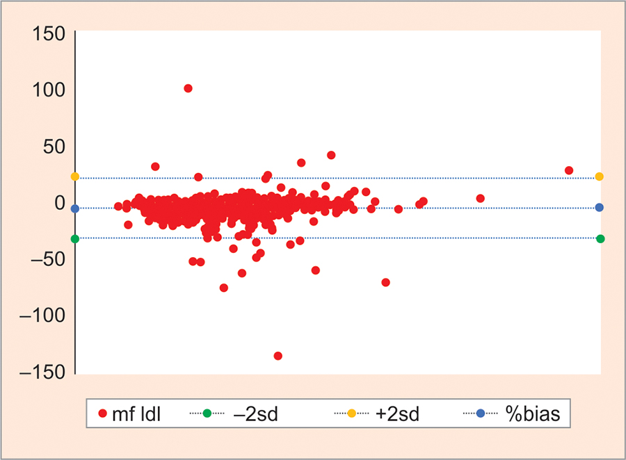
Recent recommendations of the Adult Treatment Panel and the Adolescents Treatment Panel of the National Cholesterol Education Program make the low-density lipoprotein cholesterol (LDL-C) levels in serum the basis of classification and management of hypercholesterolemia. This makes accurate reporting of LDL-C decisive in the management of coronary heart disease (CHD). Direct measurement of LDL by homogeneous method is accurate but reagent is costly. Therefore, we have to compare different calculated LDL values with direct LDL (D-LDL) values. The aim of this study was (1) to decide if LDL-C level was underestimated/overestimated after it was calculated using the formulae compared with direct measurement of LDL-C and (2) to choose the best calculated method that compares maximum with the direct method. We measured total cholesterol (TC), triglycerides (TG), high-density lipoprotein (HDL), D-LDL by direct homogeneous method in 500 fasting samples. Simultaneously, Friedewald's (F-LDL-C), modified Friedewald's (MF-LDL-C), and Anandaraja's (A-LDL-C) formulas were also used for calculation of LDL-C. A good correlation was found between D-LDL-C as compared with F-LDL-C, MF-LDL-C, and A-LDL-C. Pearson's coefficient of correlation between MF-LDL-C and D-LDL-C was 0.94, which was moderately higher than other calculated methods. Pearson's coefficient of correlation between A-LDL-C and D-LDL-C was 0.92 and F-LDL-C and D-LDL was 0.93. In conclusion, among the three LDL-C formulas, the Friedewald formula and Anandaraja' s formulas give a higher percentage of error compared with the modified Friedewald formula Therefore, modified Friedewald's formula is better than the other two formulae for calculating LDL-C in a more cost-effective manner and can be used in large population studies. Kanani DN, Mishra A. Comparison of Different Estimated Formulas with Direct Estimation of Low-density Lipoprotein Cholesterol. Indian J Med Biochem 2017;21(2):151-156.IntroductionAimMaterials and methodsResultsConclusionHow to cite this article

Study of Parathyroid Hormone as an Independent Risk Marker of Heart Failure
[Year:2017] [Month:July-December] [Volume:21] [Number:2] [Pages:5] [Pages No:157 - 161]
DOI: 10.5005/jp-journals-10054-0041 | Open Access | How to cite |
Abstract
Heart failure (HF) is a clinical syndrome characterized by cardiac pump failure with signs and symptoms arising from salt and water retention. Heart failure is associated with considerable mortality and morbidity. Identification of modifiable risk factors may reduce incidence of HF and its complications. The aim of our study is to assess parathyroid hormone (PTH) as a risk marker for HF and its association with severity of HF. In this cross-sectional study, 120 subjects with HF were recruited and they were compared with 60 age- and sex-matched controls. Along with the routine parameters, N-terminal pro B-type natriuretic peptide (NT-proBNP), intact PTH, and vitamin D were estimated. The study group was divided into quartiles depending on PTH value. The median PTH (81.5 pg/mL) and NT-proBNP (3753 pg/mL) in HF patients are found to be significantly higher (p < 0.0001) when compared with control subjects. The median vitamin D concentration (18 ng/mL) though low in cases is not statistically significant when compared with controls. Demographic, clinical, and laboratory characteristics are compared across the quartiles of PTH. Highest number of New York Heart Association (NYHA) class IV HF cases are found in highest quartiles of PTH. Logistic regression analysis demonstrated that high concentration of PTH [odds ratio of 1.1113; 95% confidence interval (CI) 1.07—1.14; p < 0.0001] and low levels of vitamin D (odds ratio of 1.053; 95% CI 1.0079—1.1009) are significantly associated with HF. This study has demonstrated that higher concentration of PTH is associated with severe form of HF. Vitamin D deficiency is also seen in the study group. Khan SA, Iyyapu KM, Sai Baba KSS, Yerram S. Study of Parathyroid Hormone as an Independent Risk Marker of Heart Failure. Indian J Med Biochem 2017;21(2):157-161.IntroductionMaterials and methodsResultsConclusionHow to cite this article

Effect of Sucralose on Glucose Uptake in Rat L6 Myotubes
[Year:2017] [Month:July-December] [Volume:21] [Number:2] [Pages:4] [Pages No:162 - 165]
DOI: 10.5005/jp-journals-10054-0042 | Open Access | How to cite |
Abstract
With growing awareness of the link between diet and health and the problem of obesity, public concern over sugar levels in the diet is forcing a worldwide trend toward cutting down on sugar by using artificial sweeteners (AS). To study the effect of increasing concentrations of sucralose (an AS) on glucose uptake in rat L6 myotubes. The L6 cell line from American type cell culture (ATCC) was grown in Dulbecco's Modified Eagle's Medium (DMEM) and differentiated into myotubes. The wells were exposed to either 0, 1 nM, 1 μM, or 1 mM of sucralose alone or with 10 nM insulin for 24 hours. Glucose uptake was studied after this period. Significant decrease was seen between the insulin-stimulated basal glucose uptake and insulin-stimulated glucose uptake across all the concentrations of sucralose treatment. Increased concentration of sucralose appears to decrease glucose uptake even on insulin stimulation. It may not be beneficial to use sucralose in certain groups of people who have insulin resistance or are prone to it. Prakash SN, Shanthakumari J, Devanath A. Effect of Sucralose on Glucose Uptake in Rat L6 Myotubes. Indian J Med Biochem 2017;21(2):162-165.IntroductionAimMaterials and methodsResultsConclusionClinical significanceHow to cite this article
Role of Active Vitamin D3 in Immunity
[Year:2017] [Month:July-December] [Volume:21] [Number:2] [Pages:10] [Pages No:166 - 175]
DOI: 10.5005/jp-journals-10054-0043 | Open Access | How to cite |
Abstract
The active vitamin D3—1,25 dihydroxy cholecalciferol—is the key player in calcium and phosphorus metabolism and skeletal growth and functions. However, recent new developments have revealed its role in other tissues as well, referred to as the nonclassical actions of vitamin D. Not only the endocrinal effects, evidence indicates that vitamin D3 also has autocrine and paracrine functions due to its extrarenal synthesis by many cells, including the immune cells. All cells of the immune system have vitamin D receptors and show wide-ranging effects to it. It impacts both the innate and adaptive immune systems and the overall influence points to anti-infective, anti-inflammatory, immunosuppressive, and regulatory roles. It shows a significant role in chronic inflammatory and autoimmune diseases as well in susceptibility to infections. In this review, newer developments on the role of vitamin D in immunity and the underlying mechanism are discussed with possible future reflections. Verma R, Singh S, Singh B, Goswami B, Gupta SK. Role of Active Vitamin D3 in Immunity. Indian J Med Biochem 2017;21(2):166-175.IntroductionHow to cite this article


© Jaypee Brothers Medical Publishers (P) LTD.

