


Do We Observe While Learning and Teaching?
[Year:2014] [Month:July-December] [Volume:4] [Number:2] [Pages:2] [Pages No:1 - 2]
DOI: 10.5005/jsd-4-2-1 | Open Access | How to cite |

Expression of Bax in Oral Lichen Planus – An Immunohistochemical Study
[Year:2014] [Month:July-December] [Volume:4] [Number:2] [Pages:6] [Pages No:3 - 8]
Keywords: Oral Lichen Planus, Apoptosis, Bax, Immunohistochemistry
DOI: 10.5005/jsd-4-2-3 | Open Access | How to cite |
Abstract
Background: Oral lichen planus (OLP) affect a minor percentage of the general population, but the morbity caused by this disease is a cause of concern. Apoptosis is known to occur during the course of OLP. Aims and Objectives: To find the expression of Bax using immunohistochemistry and To find its role in the pathogenesis of oral lichen planus. Methodology: The present study was done using Immunohistochemical procedure on the 21 samples procured from the archival material. The pro-apoptotic marker Baxwas demonstrated and analysed using peroxidase – antiperoxidaseimmunostainingmethod. Results: The results were analysed using the positivity of the immunostaining of the samples. Further the positivity was graded in terms of their staining intensity, distribution and location of stain uptake. The results were subjected to statistical analysis and analysed using a non-parametric analysis, Mann Whitney test and was found to be statistically significant with p value of 0.000. Conclusion: The results obtained in this study suggest that the process of apoptosis occurs in OLP. Hence inhibition of apoptosis in the patients could reduce the severity of the lesions and thus representing new specific targets for treatment of Lichen Planus.

Estimation of Malondialdehyde Level in Oral Submucous Fibrosis
[Year:2014] [Month:July-December] [Volume:4] [Number:2] [Pages:5] [Pages No:9 - 13]
Keywords: Oral submucous fibrosis, Malondialdehyde
DOI: 10.5005/jsd-4-2-9 | Open Access | How to cite |
Abstract
Background: Oral submucous fibrosis are the common Potentially Malignant Disorders prevailing in India. The primary etiological factor include arecanut which contain numerous Reactive Oxygen Species (ROS). Malondialdehyde (MDA) is the end product of lipid peroxidation and it is mutagenic and tumorigenic. Aims and Objectives: To estimate the serum Malondialdehyde level in Oral submucous fibrosis. Methodology: The control group comprised twenty normal individuals (Group 1). The experimental group comprised twenty patients with oral submucous fibrosis (Group 2). Blood samples were obtained and evaluated for serum Malondialdehyde and antioxidants level. Serum Malondialdehyde level was estimated using TBARS assay by Spectrophotometric method. Results: The present study revealed a statistically significant increase in serum MDA level in OSMF patients in comparison with corresponding normal individuals. Conclusion: While the oxidant (Malondialdehyde) levels are increased, indicating the potential role in premalignancy status, it is important to see from a larger sample if these results are reproducable and if so can it be sensitively used in detection of these potentially malignant disorders (Oral submucous fibrosis).

[Year:2014] [Month:July-December] [Volume:4] [Number:2] [Pages:6] [Pages No:14 - 19]
Keywords: Root canal morphology, Dental pulp, Mandibular central incisors, Buccolingual width
DOI: 10.5005/jsd-4-2-14 | Open Access | How to cite |
Abstract
Background information: Mandibular incisors are associated with high failure rate in endodontic therapy because of presence of two canals. There is no clinical diagnostic method to identify the number of canals in the mandibular incisors. Aim: To assess the correlation between incidence of two canals in mandibular incisors and its bucco-lingual width measured at the cervical third of crown. The secondary aims were to assess the percentage of different levels of canal bifurcation and the discrepancy in radiographic measurement of tooth width. Methodology: 70 extracted human mandibular incisors were included in the study. Using Vernier caliper, the bucco-lingual width at the cervical aspect of each mandibular incisor was measured. RVG image was taken through the mesiodistal plane to assess the number of canals, their level of bifurcation and also the buccolingual width of crown at cervical aspect. Canal configuration was categorised according to Vertucci's classification. Result: From the observations of this study, it is inferred that the incidence of Vertucci's type III canal configuration was 44.28%. Type III canal was found when the bucco-lingual width at the cervical third of crown was greater than 5.68 mm (from ROC curve) with 97% sensitivity and specificity. The bifurcation level of type III canals was 93% at middle – apical third, 3% at coronal, and 3 % at apical third. The spearman's correlation between radiographic measure of buccolingual width and its actual measure was 0.82. Conclusion: Within the limitations of this study, it can be concluded that if the bucco-lingual width at the cervical aspect of a mandibular incisor is greater than 5.68mm, then two canals (type III) can be found in 97% of the mandibular incisors among the South Indian population. The incidence of type III canals was 44.28%; the bifurcation level was mostly at middle third of root.
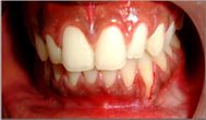
Double Papilla Flap for Root Coverage - A Case Report
[Year:2014] [Month:July-December] [Volume:4] [Number:2] [Pages:5] [Pages No:20 - 24]
Keywords: Double-papilla flap, Gingival recession, Root coverage
DOI: 10.5005/jsd-4-2-20 | Open Access | How to cite |
Abstract
Gingival recession is defined as the displacement of the gingival margin apical to the cementoenamel junction (CEJ). Root coverage is being achieved by a variety of techniques. Double-papilla flap technique was introduced by Cohen and Ross for the treatment of gingival recession. This case report describes three cases of class 2 gingival recession associated with dentinal hypersensitivity treated using double-papilla flap technique.

Treatment of Intrabony Defects by Using Platelet Rich Plasma Combined With Bone Graft: A Case Report
[Year:2014] [Month:July-December] [Volume:4] [Number:2] [Pages:5] [Pages No:25 - 29]
Keywords: Periodontitis, Periodontal regeneration, Intrabony defects, PRP, DBM
DOI: 10.5005/jsd-4-2-25 | Open Access | How to cite |
Abstract
Background: Regeneration of periodontium using autologous materialshave been attempted as it addresses safety issues and ensures easy availability. Among the autologous options available, use of platelet concentrates are promising due to ease of procurement, handling and low cost. Platelet-rich plasma (PRP) is an autologous product that is derived from whole blood through the process of gradient density centrifugation. We hereby report a case of chronic periodontitis with intra bony defects in relation 46 and 47 treated with PRP combined with bone grafts with a six month follow up. Methodology: 5ml of patient's venous blood was collectedand PRP obtained after centrifugation. The platelet concentrate obtained was used in combination with bone graft in intra bony defect in relation to 46 and 47. Results: A reduction in the Probing pocket depth (PPD) from 7mm (Pre-operative) to 3mm and CAL (clinical attachment level) from 9 mm (Pre-operative) to 5 mm at 6 month recall was observed respectively. Conclusion: Significant improvement in clinical parameters such as PPD, CAL indicates success of regenerative therapy using PRP with bone grafts.

Giant Sialolithiasis - A Case Report and Review
[Year:2014] [Month:July-December] [Volume:4] [Number:2] [Pages:4] [Pages No:30 - 33]
Keywords: Giant sialolith, salivary gland, soft tissue laser
DOI: 10.5005/jsd-4-2-30 | Open Access | How to cite |
Abstract
Sialolithiasis or salivary gland duct calculus or salivary stones are the most common pathologies of the salivary gland. Sialolithiasis accounts for more than 50% of diseases of the major salivary glands and is the most common cause of acute and chronic infections. Sialoliths are deposits obstructing the ducts of major or minor salivary glands or its parenchyma. Salivary stones larger than 15 mm are classified as giant sialoliths. The prevalence of sialoliths varies by location. About 85% of sialoliths occur in the submandibular gland and 5-10% occurs in the parotid gland. In about 5% of cases, the sublingual gland or a minor salivary gland is affected. Sialolith in the parotid gland is less common when compared to submandibular gland. This case report describes a case of giant sialolith of submandibular salivary gland.

[Year:2014] [Month:July-December] [Volume:4] [Number:2] [Pages:6] [Pages No:34 - 39]
Keywords: Pharyngeal Obturator, Velopharyngeal insufficiency, Hyper-nasality, Speech intelligibility
DOI: 10.5005/jsd-4-2-34 | Open Access | How to cite |
Abstract
Maxillary obturator prostheses restore the congenital or acquired defects of the soft palate and allow adequate closure of palatopharyngeal sphincter. Our patient, a 30-year-old female with a diagnosis of soft palate defect presented with complaints of speech difficulty and hyper-nasality. There was history of surgery for ameloblastoma of hard and soft palate one month ago. Intra oral examination revealed a soft palate defect. Since the patient had full dentition, pharyngeal obturator with clasp retention was given. Adequate velopharyngeal closure was detected after the patient was examined during drinking water in the upward head position. Moreover no nasal reflux was observed. A speech pathologist confirmed that the hyper-nasality was reduced immediately and advised for speech training classes. The production of oral and nasal consonants and the speech was noticeably improved after perceptual speech evaluation. We therefore emphasize the integration of an interdisciplinary team in order to increase the efficacy of rehabilitation ant the quality of life of these patients.

Management of Fractured Endodontic Instruments In Root Canal: A Review
[Year:2014] [Month:July-December] [Volume:4] [Number:2] [Pages:9] [Pages No:40 - 48]
Keywords: Endodontic instruments, Rotary NITI instruments, Separated instrument removal, Fractured endodontic instruments
DOI: 10.5005/jsd-4-2-40 | Open Access | How to cite |
Abstract
With the increased practice of rotary endodontics in recent years, separated rotary nickel-titanium (NiTi) files in root canals is the most commonly reported mishap, causing lot of stress and anxiety among clinicians and patients. No clear guidelines can be drawn from the literature available because there are either too few studies of the effects of broken files on prognosis, or the few studies that have been performed involved so few patients. The prognosis is also dependent on file location, prior condition of the pulp, presence or absence of periapical lesion and many other factors. Each case is different. This paper offers a flowchart to help the general dentists and non-endodontists decide which strategy is best when faced with a broken file in root canal.

Lesion Sterilization and Tissue Repair (LSTR): A Review
[Year:2014] [Month:July-December] [Volume:4] [Number:2] [Pages:7] [Pages No:49 - 55]
Keywords: Lesion Sterilization and Tissue Repair, Triple-Antibiotic Paste, 3-Mix Paste, Regenerative Endodontics, Non-Instrumentation Endodontics, Revascularization
DOI: 10.5005/jsd-4-2-49 | Open Access | How to cite |
Abstract
Residual infection in root canal system has always been an area of penumbra for a treating dentist. Lesion sterilization and tissue repair (LSTR) therapy is an endodontic treatment procedure that involves non-instrumentation or minimal instrumentation followed by placement of antibiotic mixture to disinfect root canal systems. It is evolving as an alternate treatment procedure in comparison to traditional pulpectomies/root canal treatment and extractions for treatment of nonvital or pulpless teeth. This article reviews the rationale of the technique, evolution, indications, uses and clinical procedures required in performing the technique.

Craniofacial Abnormalities and Syndromes
[Year:2014] [Month:July-December] [Volume:4] [Number:2] [Pages:6] [Pages No:56 - 61]
Keywords: craniofacial abnormalities, diagnosis, syndromes
DOI: 10.5005/jsd-4-2-56 | Open Access | How to cite |
Abstract
Symmetry and proportion plays an important role in cosmetic part of face and golden ratio depicts a strong correlation between mathematical proportion of face and appearance. This can be affected by multiple factors including syndromes which contribute a significant role in craniofacial abnormalities. This paper intends to bring out the craniofacial manifestations of syndromes which play an essential role in diagnosis and more interestingly serve as a window to overall systemic health exploring the associated systemic manifestations.


© Jaypee Brothers Medical Publishers (P) LTD.

