


[Year:2019] [Month:July-August] [Volume:10] [Number:4] [Pages:2] [Pages No:251 - 252]
DOI: 10.5005/jp-journals-10015-1660 | Open Access | How to cite |

[Year:2019] [Month:July-August] [Volume:10] [Number:4] [Pages:6] [Pages No:253 - 258]
Keywords: Aptitude tests, Oral implantology, Questionnaire, Resident program, Validation
DOI: 10.5005/jp-journals-10015-1645 | Open Access | How to cite |
Abstract
Aim: The aim of the present study was to develop and validate the psychometric properties of the questionnaire (POCTOIR-30) for the evaluation of the perception of clinical training and future goals of residents of oral implantology. Materials and methods: The development of the questionnaire followed the process of five phases: (a) preparation of the instrument, (b) validation of the content, (c) validity of the construct, (d) evaluation of internal consistency and reliability, (e) evaluation of the level of knowledge, for which an instrument on implant training of residency programs was used as a reference. To evaluate the sensitivity and validity of the questionnaire, a pilot sample of 20 residents between 20 years and 50 years of a residency program in oral implantology was used. Results: The factor analysis of the data and the adjustment statistics process resulted in the quality care of 4 factors and 30 questions that were included in the present questionnaire (POCTOIR), which covers: logistics and infrastructure (five items), training in oral implantology (10 items); information on the postgraduate program (seven items) and accessibility and sustainability of care (ten items) and future goals after postgraduate studies (five items). Content validity was measured using the index that combines the ease of calculation and the evaluation of results at the statistical level (κ 0.833). The reliability of the instrument was evaluated to measure the degree of internal correlation (Cronbach's α 0.738). Finally, the reliability was evaluated to measure the constancy of the responses repeatedly with the same subjects (intraclass correlation 0.74). Conclusion: This study showed that the POCTOIR-30 instrument has adequate levels of validity and reliability to be used in residents of areas related to oral implantology in new research. Clinical significance: The impact of the academic training of the residents in oral implantology significantly influences their performance at the time of performing the surgical and prosthetic protocols. Therefore, this research validates statistically the perception of academic training and future goals of postgraduate residents in order to quantify and apply improvement strategies during their years of study.

Assessing the Use of Glycine Powder Air Polishing in Periodontal Maintenance Therapy
[Year:2019] [Month:July-August] [Volume:10] [Number:4] [Pages:5] [Pages No:259 - 263]
Keywords: Dental polishing, Glycine, Periodontitis
DOI: 10.5005/jp-journals-10015-1650 | Open Access | How to cite |
Abstract
Aim: The aim of this study is to evaluate the clinical efficacy of subgingival glycine powder air polishing in periodontitis during PMT. Materials and methods: Forty PMT patients were recruited from subjects referred to the Department of Periodontology, Faculty of Dentistry, Damascus University. AgP group comprised twenty patients diagnosed with generalized aggressive periodontitis, whereas ChP group included 20 patients diagnosed with generalized chronic periodontitis. Using a split mouth study design, subgingival plaque was removed by means of glycine powder air polishing GPAP or curettes MCD. Clinical periodontal parameters were recorded at baseline and at 3 months after treatment. A visual analog scale was used to evaluate inconvenience during therapy. Results: The two studied groups showed improvements in all clinical indices after 3 months (p = 0.001). There were no significant statistical differences between GPAP and MCD in both of AgP and ChP groups (p > 0.05). GPAP therapy was significantly less painful than MCD in AgP and ChP groups (p = 0.018, p = 0.038, respectively). Conclusion: Subgingival glycine air polishing was effective in treatment periodontitis during maintenance care and perceived to be more acceptable by the patients than conventional curettes. Clinical significance: Subgingival glycine air polishing has been shown to remove biofilm in periodontal pockets without causing damage to the root surfaces or soft tissues; therefore, it was suggested to be used as an alternative to repeated use of conventional curettes in periodontal maintenance therapy (PMT).

Flexural Strength of Monolithic Zirconia after Different Surface Treatments
[Year:2019] [Month:July-August] [Volume:10] [Number:4] [Pages:6] [Pages No:264 - 269]
Keywords: Ceramics, Crowns, Dental materials, Dental polishing, Dental porcelain, Surface properties
DOI: 10.5005/jp-journals-10015-1653 | Open Access | How to cite |
Abstract
Aim: The purpose of this study was to evaluate the effect of grinding, over-glazing, regrinding, and polishing on flexural strength of monolithic zirconia. Materials and methods: In this in vitro study, 50 bar-shaped zirconia specimens were fabricated and divided into 5 groups (n = 10) of no-treatment control group (C), grinding (G), grinding + glazing (GGl), grinding + glazing + grinding (GGlG), and grinding + glazing + grinding + polishing (GGlGP). A universal testing machine was used to measure the flexural strength. Data were analyzed using the Tukey's HSD test and ANOVA (p < 0.05). Results: There were significant differences between group C and other groups (p < 0.001). Group C showed the highest mean flexural strength among all groups. There was also a significant difference between groups GGlG and GGlGP (p = 0.031), and the latter group showed higher mean flexural strength. There was no significant difference between other groups (p > 0.001). Conclusion: Regrinding does not have a significant effect on flexural strength of monolithic zirconia compared with grinding. Glazing slightly decreased the flexural strength of ground zirconia surfaces, which was not statistically significant. Polishing improved the flexural strength of ground zirconia surfaces; however, their mean flexural strength was significantly lower than that of the control group. Clinical significance: Grinding may lead to significant strength reduction and decrease the durability of zirconia restorations. Therefore, polishing of ground surfaces is suggested while glazing has an adverse effect on the strength.

[Year:2019] [Month:July-August] [Volume:10] [Number:4] [Pages:5] [Pages No:270 - 274]
Keywords: Facial esthetic, Orthognathic surgery, Postoperative complication, Quality of life
DOI: 10.5005/jp-journals-10015-1646 | Open Access | How to cite |
Abstract
Introduction: Most patients undergo orthognathic surgery (OS) to improve their beauty and correct functional imbalances. OS is also performed in the case of temporomandibular joint disorders to reduce facial pain and enhance the quality of life of patients. Aim: The aim of the study was to evaluate the quality of life of patients who undergo OS in terms of esthetic appearances, chewing, and postoperative complications and to assess the percentage of satisfaction among patients in terms of facial appearances, friends’ and relatives’ opinions, and functional performance of the oral cavity. Materials and methods: Data of 73 patients who underwent OS in Kingdom of Saudi Arabia were collected from their medical records. A brief questionnaire was prepared on the basis of previous studies with some modifications. Data were collected and a questionnaire was answered via a telephone interview with these patients. Questions were related to the patients’ basic demographic information and their postoperative complications, such as swelling, pain, chewing difficulty, and snoring. The second part was introduced as a questionnaire designed by the researchers to measure the patients’ satisfaction regarding OS. This questionnaire was related to the appearance of the face from the side and front views and to the level of attractiveness perceived by patients themselves and their relatives and friends after OS. This questionnaire also included improvements in oral functions, such as chewing. The overall performance of the oral cavity was assessed. Data were analyzed using SPSS. Results: A total of 50 patients completed the telephone interview and the examination. The highest number of subjects was in the young age group of 19–25 years. Of these patients, 31 (62%) showed a positive result on the snoring outcomes, and 25 (50%) complained of pain and swelling. Furthermore, 33 (66%) and 27 (54%) patients reported improvements in chewing and facial esthetic outcomes, respectively. More than 50% of the patients were satisfied in their facial appearance from the side and front views. Patients’ perception of their attractiveness and their friends’ and family's opinion about their facial appearance recorded were between 60% and 62%. Other questions dealing with improvements in eating, chewing, and overall oral cavity performance gained 42–18%. All variables showed significant differences at the level of p value more than 0.050. Conclusion: Overall, the majority of the OS patients showed positive results in terms of chewing, snoring, facial esthetics, pain, and swelling during their recovery time. Patients are mostly satisfied with the overall facial attractiveness results as well as the opinions of their relatives and friends. Clinical significance: OS is performed to enhance the appearance and function of the oral cavity, which may end with a positive effect of the patient's oral function, self-esteem, and social interaction.

[Year:2019] [Month:July-August] [Volume:10] [Number:4] [Pages:5] [Pages No:275 - 279]
Keywords: CD4 count, Highly active antiretroviral therapy, Human immunodeficiency virus, Systemic comorbidity
DOI: 10.5005/jp-journals-10015-1642 | Open Access | How to cite |
Abstract
Aims: Oral and systemic comorbidities are common in human immunodeficiency virus (HIV)/acquired immunodeficiency syndrome (AIDS) and are considered to be important predictors of the disease. Cluster of differentiation 4 (CD4) count serves as an important marker for the progression of HIV to AIDS. Our objective was to correlate the oral and systemic comorbidities associated with HIV infection with CD4 count in patients on highly active antiretroviral therapy (HAART). Materials and methods: This was an observational study among 110 HIV-diagnosed patients. The oral and systemic comorbidities were noted and compared to their CD4 counts. A Chi-square analysis was carried out to see the association of oral manifestations. Results: Among the study subjects, 50 (45.5%) participants had a CD4 count of >500 cells/μL, 46 (41.8%) patients had a CD4 count of 200–499 cells/μL, whereas 14 (12.7%) had <200 cells/μL. The major oral manifestations observed were dental caries (n = 30, 60%), periodontitis (n = 25, 50%), and lipoatrophy (n = 25, 50%) in patients with CD4 >500 cells/μL; dental caries (n = 28, 60.90%), intraoral pigmentation (n = 23, 50%), and periodontitis (n = 20, 43.50%) in patients with a CD4 count between 200 and 499; and dental caries (n = 9, 64.30%), periodontitis (n = 7, 50%), and candidiasis 6 (42.90%) among subjects with CD4 counts <200. The most common systemic comorbidity observed was tuberculosis (p < 0.001) and pneumonia (p < 0.003). Conclusion: Early intervention strategies in diagnosis and management for HIV-infected individuals have shown promising results. With the advent of HAART, the quality of life has significantly improved. Clinical significance: The prevalence of oral and systemic comorbidity among HIV-infected patients have declined since the advent of HAART. Oral and general physicians should be able to identify and treat the patients at the earliest, which in turn could reduce the morbidity and mortality rates among those infected with HIV.

[Year:2019] [Month:July-August] [Volume:10] [Number:4] [Pages:5] [Pages No:280 - 284]
Keywords: Crowns, Fabrication place, Full veneer crown, Gingival health, Gingival index, Gingival margins
DOI: 10.5005/jp-journals-10015-1643 | Open Access | How to cite |
Abstract
Aim: The aim of this study was to assess the effect of different marginal locations and fabrication places on the gingival health of patients treated with full veneer crown restorations, at Al-Riyadh province, Kingdom of Saudi Arabia. Materials and methods: A cross-sectional study was performed to assess the gingival health of patients with full veneer crown restorations. Thirty-nine patients with 45 full veneer crowns have been examined in two places: College of Dentistry (Prince Sattam Bin Abdulaziz University) and East Riyadh Specialist Dental Center; both places are in Al-Riyadh province, Kingdom of Saudi Arabia. The gingival index by Loe and Sillness has been used to assess gingival health. Patients have been examined from December 2016 to March 2017. Results: A tooth restored with full veneer crowns showed a high gingival index (1.10) than the contralateral unrestored tooth in the same patients. There were significant differences between subgingival margin (1.14), supragingival (0.69), and equigingival (0.73) margin on gingival health. There were significant differences between full veneer crowns fabricated at government clinics (0.81) and full veneer crowns fabricated at private clinics (1.10) on gingival health. There were no significant differences on gingival health between patients with different educational levels. Conclusion: Subgingival margin of full veneer crowns has a higher gingival index than the equigingival and supragingival margin. Full veneer crowns fabricated at governmental clinics have a lower gingival index compared with full veneer crowns fabricated at private clinics, with no positive relationship between the educational level and gingival health around the crown. Clinical significance: The location of the finish line as well as the fabrication place will significantly impact the gingival health of the final full veneer restorations.

[Year:2019] [Month:July-August] [Volume:10] [Number:4] [Pages:6] [Pages No:285 - 290]
Keywords: Bacterial contamination, Cling films, Cross-infection control, Light-curing unit, Sleeves, Wrapping
DOI: 10.5005/jp-journals-10015-1647 | Open Access | How to cite |
Abstract
Aim: To compare and assess the level of infection control provided by a cling film against a sleeve and their impact on the light intensity level of dental light-emitting diode (LED) light-curing units (LCUs). Materials and methods: A sleeve and a cling film of proprietary brands were compared on their reduction of light output and bacterial colonies on agar plates. Including a control group, 120 samples of analog radiometer readings were obtained. A total of 90 samples, including 10 each for positive and negative controls, were obtained in a laboratory setting via swabbing of light-guiding tips placed intraorally. These swabbings were inoculated on 5% sheep blood agar in a biological cabinet and cultured for 48 hours at 37°C; the inoculated surfaces were photographed and analyzed for area of coverage by bacterial colonies. The data obtained were subjected to ANOVA and Mann–Whitney U tests. Results: There is neither statistically significant reduction in output nor difference in output between either barriers (p > 0.05). There is statistically significant reduction in bacterial colonies on the inoculated surface by both barriers compared to no barrier (p < 0.01), but there is no statistically significant difference between the two barriers (p > 0.05). Conclusion: Both barriers do not significantly affect light output and are equally efficacious as cross-contamination barriers, and the choice lies with the operator. Clinical significance: Use of barriers is very important to prevent cross-infection control. The results of our study help the clinician select appropriate measures to prevent cross infection while using LCUs across the patients.
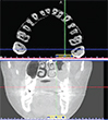
[Year:2019] [Month:July-August] [Volume:10] [Number:4] [Pages:4] [Pages No:291 - 294]
Keywords: Cone-beam computed tomography, Dental operating microscope, Maxillary first molar, MB2 canal
DOI: 10.5005/jp-journals-10015-1644 | Open Access | How to cite |
Abstract
Aim: To assess and compare the internal morphology of the maxillary first molar and the incidence of the fourth root canal, especially mesiobuccal (MB2) using three diagnostic methods, clinically with or without dental operating microscopy (DOM) and cone-beam computed tomography (CBCT). Materials and methods: A total of 336 maxillary first molar teeth were examined and distributed into the following groups: group I (n = 112), the teeth were treated by dental interns without any means of magnification. Group II (n = 112), the teeth were treated by an endodontist with the use of the a dental operating microscope (DOM). Group III (n = 112), the CBCT images were selected and examined carefully. Results: The second mesiobuccal root canal within the maxillary first molar was present in groups I, II and III with the incidence of 27%, 46% and 60%, respectively. Conclusion: Within the limitations of this study, it can be concluded that the maxillary first molar teeth showed significant variations of their root canals. CBCT and dental operating microscope significantly facilitated the location and identification of the second root canal of the mesiobuccal root of maxillary first molar. Clinical significance: Knowledge of root canal anatomy is an important factor for a successful endodontic outcome.

Efficacy of Virgin Coconut Oil and Chlorhexidine as an Oral Antimicrobial: A Comparative Pilot Study
[Year:2019] [Month:July-August] [Volume:10] [Number:4] [Pages:6] [Pages No:295 - 300]
Keywords: Coconut oil, Gingival index, Plaque index, Streptococcus mutans
DOI: 10.5005/jp-journals-10015-1652 | Open Access | How to cite |
Abstract
Aim: The present study aimed to evaluate the efficacy of oil pulling therapy using virgin coconut oil (VCO) in reducing S. mutans counts, plaque, and gingival indices, and to compare it with the gold standard chlorhexidine. Materials and methods: Twenty subjects (study) using VCO and 20 subjects using chlorhexidine (control) visiting the outpatient department of periodontics in the institute were chosen for the study. The gingival and plaque indices (baseline) in both groups were recorded following which unstimulated whole saliva samples were collected by spit method and sent for microbial count. They were then provided either of the VCO/mouthwash to swish once daily. Three weeks post-intervention, the recording of indices was repeated for both groups along with the microbial count. Results: The mean values of gingival and plaque indices pre- and post-intervention showed a statistically significant reduction in the control population compared with the study group, while there was no statistically significant reduction in the bacterial count seen. The difference in the scores of plaque index pre- and post-intervention was more in control group while the difference in the gingival index was similar in both groups, but statistically insignificant. Conclusion: Virgin coconut oil may not be as effective as chlorhexidine in reducing plaque while it may be as effective as chlorhexidine in reducing gingival index. Clinical significance: In comparison with newer chemical oral hygiene aids, coconut oil could still be used as a traditional adjuvant to reduce gingivitis in addition to routine brushing.

[Year:2019] [Month:July-August] [Volume:10] [Number:4] [Pages:5] [Pages No:301 - 305]
Keywords: Composite resins, Dental amalgam, Fracture strength, Pulpotomy, Tricalcium silicate cement
DOI: 10.5005/jp-journals-10015-1655 | Open Access | How to cite |
Abstract
Aim: Biodentine is a biocompatible, bioactive material with dentin regeneration potential that is known as the future material of choice in primary tooth pulp therapy. Biodentine (BD) is also designed as a dentine substitute in direct posterior restorations. So, the aim of this study is to evaluate the fracture resistance of BD pulpotomized primary molars, restored with different restorative techniques. Materials and methods: A total of 36 extracted primary second molar teeth were selected. Standardized class I access cavity preparation was done and then the teeth were randomly divided into three experimental groups of 12 in each. Group I (n = 12): BD and glass ionomer as liners with composite resin as restoration, group II (n = 12): BD as both liner and restoration; and group III (n = 12): BD and glass ionomer as liners with amalgam as restoration. After water storage and thermocycling, static fracture resistance was tested. Data (in Newtons) were analyzed using one-way ANOVA (α = 0.05). Results: Statistically significant difference was observed among groups of the study (p value = 0.000). Composite group showed the maximum fracture resistance and amalgam group exhibited the least (2371.67 N vs 1912.17 N). Application of composite and BD respectively led to higher numbers of restorable fractures (75%). Conclusion: In pulpotomized primary molars using biodentine, composite restoration shows the best fracture resistance followed by BD and amalgam restorations. Clinical significance: In order to improve the outcome of endodontic treatment in primary molars, biodentine can be used successfully as both pulpotomy and restorative material to achieve less time-consuming treatment in children.

[Year:2019] [Month:July-August] [Volume:10] [Number:4] [Pages:4] [Pages No:306 - 309]
Keywords: Canal preparation, Centering, Cone-beam computed tomography, Root canal therapy
DOI: 10.5005/jp-journals-10015-1654 | Open Access | How to cite |
Abstract
Aim: Centering ability of an instrument is the ability of the instrument to act centrally inside the canal without deflections. This property is of significance in assessment of any endodontic file because endodontic accidents due to instrumentation, commonly apical transportation can be avoided in case of a perfectly centered instrument. Therefore, the aim of the present study was to compare three different endodontic files, K File, Hand ProTaper, and Rotary ProTaper for their centering ability using cone-beam computed tomography (CBCT). Materials and methods: On 30 extracted mandibular premolars, 3 reference lines were created from the apex. Preoperative CBCT images were made and analyzed. D1 and D2 are the centering ratios measured buccolingually and mesiodistally, following which the samples were randomly allotted to one of the three groups (K File, Hand ProTaper, and Rotary ProTaper) for instrumentation. Postoperative images were obtained and canal dimensions were assessed. The differences were calculated and the centering ability was determined using the centering ratio formula. Comparison was done using ANOVA test. Results: The mean working length of all the samples was 20.8 mm. The average preoperative D1 and D2 values obtained were 0.0067 and 0.0117, respectively. Following instrumentation, the obtained D1 and D2 values in group I, group II, and group III were 0.0048 and 1.07, 0.783 and 1.24, and 0.785 and 0.96, respectively. Intergroup comparison showed insignificant p value (p > 0.05). Conclusion: K File, Hand ProTaper, and Rotary ProTaper were equally efficient to act centrally in straight canals. Clinical significance: Centering ability of an instrument is of significance in avoiding accidents such as canal transportation. K File, Hand ProTaper, and Rotary ProTaper were found to be equally efficient to act centrally in straight canals.

[Year:2019] [Month:July-August] [Volume:10] [Number:4] [Pages:6] [Pages No:310 - 315]
Keywords: Dental implant, Le Fort osteotomy, Mandibular fractures, Maxillofacial surgery, Vascular grafting
DOI: 10.5005/jp-journals-10015-1656 | Open Access | How to cite |
Abstract
Background: Facial trauma may lead to severe fractures and loss of bone and dental structures. Depending on the magnitude of the trauma, whole segments of the jaws may be lost due to comminution, exposure of internal structures, or infection. The treatment of such lesions can be challenging, and success is often hindered by unsatisfactory esthetic, phonetic, and functional outcomes. The aim of this case report was to describe a multidisciplinary approach for implant rehabilitation in a complex case of trauma in the maxillofacial region in a young patient. Case description: A 22-year-old female patient was referred with extensive loss of soft and hard tissues in the mandible as well as loss of several teeth due to facial trauma. The patient was submitted to innumerous surgical reconstructive procedures throughout years of treatment, which included vascularized soft and hard tissue grafts, installation of dental implants, Le Fort I osteotomy for maxillary repositioning, and subsequent implant-supported prosthetic rehabilitation. Conclusion: A 24-year follow-up of comprehensive surgical and prosthodontic reconstruction of a facial injury with several fractures due to motorcycle accidents using the multidisciplinary approach was described. The multidisciplinary approach employed in this case demonstrated successful outcomes for the reconstruction of the maxillofacial region after extensive facial trauma. After six years postoperatively no complications with the rehabilitation treatment has been detected to the present date. Clinical significance: The multidisciplinary treatment planning employed was able to restore masticatory function and facial esthetics, enhancing patient quality of life and satisfaction.

[Year:2019] [Month:July-August] [Volume:10] [Number:4] [Pages:5] [Pages No:316 - 320]
Keywords: Clear cell odontogenic carcinoma, Mandibular neoplasm, Special stains
DOI: 10.5005/jp-journals-10015-1648 | Open Access | How to cite |
Abstract
Aim: To resolve the diagnostic dilemma of clear cell odontogenic carcinoma (CCOCa) using various special stains. Background: Clear cell odontogenic carcinoma is a rare malignant odontogenic tumor with a female predilection, occurring predominantly in the mandible. It is an aggressive tumor with frequent recurrences (55%) and high metastatic potential (31%). Case description: Herein, we report a case of an unusual extensive mandibular tumor mass in 49-year-old male patient who posed diagnostic challenges due to similar microscopic features of oral squamous cell carcinoma (OSCC), clear cell mucoepidermoid carcinoma (CCMEC), hyalinizing clear cell carcinoma (CCC), epithelial myoepithelial carcinoma (EMC), clear cell variant of calcifying epithelial odontogenic tumor (CCCEOT), and clear cell odontogenic ghost cell tumor (CCOGCT). Conclusion: The use of appropriate special stains like mucicarmine, Van Gieson, and Periodic Acid Schiff with and without diastase paved the way toward confirmatory diagnosis. Clinical significance: Good knowledge and right practice of various special staining techniques in histopathology provides a rapid and reproducible result with better diagnosis.

Tasty Herb that Heals: A Review of Cosmos caudatus (Ulam Raja) and its Potential Uses in Dentistry
[Year:2019] [Month:July-August] [Volume:10] [Number:4] [Pages:4] [Pages No:321 - 324]
Keywords: Cosmos caudatus, Pharmacological use, Traditional medicine, Ulam raja
DOI: 10.5005/jp-journals-10015-1651 | Open Access | How to cite |
Abstract
Aim: In recent years, there has been an increase in research on herbal derivatives with regards to their medicinal use. This review aims to summarize the scientific reports on one such herb, Cosmos caudatus. Our primary focus is to highlight its pharmacological properties and their potential applications in the field of dentistry. Background: The vascular plant biodiversity of Southeast Asia is second to that of South America, with many of these plants being used for their traditional medicinal benefits. One such herb that is popular in Malaysia is C. caudatus, locally referred to as ulam raja. It is an edible herb, commonly served in the form of a salad and is also used in traditional medicine practices to boost blood circulation, increase strength of bones, reduce fever, treat infections, and improve skin texture. Recent scientific studies have reported numerous pharmacological benefits of C. caudatus ranging from being an antioxidant agent to having bone-protective properties. These properties are attributed to a rich source of biochemical compounds found in the herb. A PubMed and Science Direct database search was conducted revealing 25 papers relevant to our review of C. caudatus, describing its traditional uses, pharmacological uses, botanical description, and phytochemical description. Review results: Published studies have reported that C. caudatus has potent antioxidant properties. It has shown both antibacterial and antifungal activities. Extracts of C. caudatus are capable of acting as an antihypertensive and antidiabetic agent, in both human and animal models. It also plays a role as a bone-protective and bone-proliferative agent. Conclusion: C. caudatus has the potential of being greatly beneficial in treating various dental diseases due to its pharmacological properties. Clinical significance: There is need for further research into this abundant natural resource in order to explore its potential uses in clinical dentistry.


© Jaypee Brothers Medical Publishers (P) LTD.

