
Subscribe to be the first to know about Best Deals and Exclusive Offers!
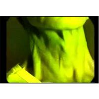
The video recordings of the internal jugular pulsations as well as that of the precordial pulsations from a 65 years old Italian woman are shown in this video. Patient had history of a previous mitral commissurotomy for significant mitral stenosis when she was about 40 years of age. She had good improvement in her symptoms. She essentially presented with exertional fatigue and peripheral edema. The mitral valve area by Echo-Doppler studies was 2.2 cm2. The internal jugular pulsations were recorded with simultaneous arterial pulse Doppler. The early part of the squishing noise of the arterial Doppler flow signals is systolic in time. The pulsations over the apex and the adjoining lateral chest wall are recorded with simultaneous heart sounds for timing purposes. The most likely cause of the findings in the jugulars and the precordium is: (A) Mitral stenosis with secondary pulmonary hypertension and right ventricular (RV) failure, (B) Significant mitral regurgitation with secondary pulmonary hypertension and RV failure, (C) Congestive heart failure with pulmonary hypertension, (D) Severe tricuspid valvular regurgitation with large “V” wave and dominant right ventricle with lateral retraction. This patient actually had done well following her mitral commissurotomy with improvement in her symptoms and findings. The residual mitral stenosis was quite minimal. She had no significant diastolic rumble at the apex. She had a pansystolic murmur over the precordium. This was almost all due to tricuspid regurgitation. She only had mild mitral regurgitation. The apex was formed by an active RV which was not sustained and there was a large area of lateral chest wall retraction which is clearly visible on the video. The Jugular pulsations reflect the severe tricuspid regurgitation with a large CV wave followed by a single y descent. The tricuspid regurgitation was primary and related to the rheumatic involvement. The pulmonary artery pressures were not elevated.


© 2019 Jaypee Brothers Medical Publishers (P) LTD. | All Rights Reserved

 Refer to Friend
 Recommend To Librarian
