
Subscribe to be the first to know about Best Deals and Exclusive Offers!
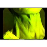
The video recording of the internal jugular venous pulsations from an 80 years old gentleman. The Doppler arterial flow signals from the radial artery have been recorded for timing. Patient had been operated at age 63 for severe aortic stenosis and received a mechanical aortic valve prosthesis. At age 78 he required a permanent pacemaker insertion for symptoms of pre-syncope associated with significant pauses demonstrated on the ambulatory monitoring. His risk factors include hypertension and dyslipidemia. He has mild exertional dyspnea with negative perfusion stress test for ischemia. All of the following statements are correct except: (A) The jugular contour shows a single descent, (B) The descent seen is a y descent, (C) This patient is unlikely to have significant tricuspid regurgitation, (D) Jugular contour shows intermittent cannon “a wave” due to a paced ventricular rhythm with A-V dissociation. Patient had had previous cardiac surgery and has received a pacemaker. The rhythm is clearly regular. The patient was in electronically paced ventricular rhythm. The early part of the squishing noise of the arterial Doppler flow signals is systolic in time. The descent in the jugulars is clearly diastolic and out of phase with the systolic Doppler flow signal. There is no prominent rise of any wave noted. The wave preceding the single y descent is usually the v wave. The v wave rise is not prominent. In addition, patient is lying almost flat and the venous pressure is not significantly elevated. Although there could be some tricuspid regurgitation because of the pacemaker wire across the tricuspid valve, it is unlikely to be anything but mild in this patient since the v wave rise is not prominent and the venous pressure is not elevated.


© 2019 Jaypee Brothers Medical Publishers (P) LTD. | All Rights Reserved

 Refer to Friend
 Recommend To Librarian
